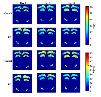
Non-invasive and quantifiable diagnosis of Parkinson's disease (PD) is an ongoing challenge for researchers. In this study, we compared two different chemical exchange saturation transfer (CEST) imaging quantification metrics in substantia nigra (SN), cortex and hippocampus of acute 1-methyl-4-phenyl-1,2,3,6-tetrahydropyridine (MPTP)-induced PD mice at three different time points including before MPTP administration, and the 3rd and 10th day after MPTP administration. Area of amine (Areaamine), magnetization transfer ratio yielding Rex (MTRrex) and apparent exchange-dependent relaxation (AREX) in SN derived from 5-pool Lorentzian fitting and inverse Z-spectrum analysis showed a statistically significant decrease after successful modeling, while there were no significant alterations in T1, T2, and magnetization transfer ratio based on asymmetry analysis (MTRasym). The results suggested that the former quantification is superior to that based on Z-spectrum asymmetry in vivo due to the exclusion of direct saturation (DS) and magnetic transfer (MT) effects and may reflect the destroying SN of PD.
[1] DUGUID J R, DE LA PAZ R, DEGROOT J. Magnetic resonance imaging of the midbrain in Parkinson's disease[J]. Ann Neurol, 1986, 20(6):744-747.
[2] BAUDREXEL S, WITTE T, SEIFRIED C, et al. Resting state fMRI reveals increased subthalamic nucleus-motor cortex connectivity in Parkinson's disease[J]. Neuroimage, 2011, 55(4):1728-1738.
[3] PRAKASH B D, SITOH Y Y, TAN L C, et al. Asymmetrical diffusion tensor imaging indices of the rostral substantia nigra in Parkinson's disease[J]. Parkinsonism Relat Disord, 2012, 18(9):1029-1033.
[4] LANGKAMMER C, PIRPAMER L, SEILER S, et al. Quantitative susceptibility mapping in parkinson's disease[J]. PLoS One, 2016, 11(9):e0162460.
[5] AU W L, ADAMS J R, TROIANO A, et al. Neuroimaging in Parkinson's disease[J]. J Neural Transm Suppl, 2006, (70):241-248.
[6] TAMBASCO N, PELLICCIOLI G P, CHIARINI P, et al. Magnetization transfer changes of grey and white matter in Parkinson's disease[J]. Neuroradiology, 2003, 45(4):224-230.
[7] WANG M Y, HONG X H, CHANG C F, et al. Simultaneous detection and separation of hyperacute intracerebral hemorrhage and cerebral ischemia using amide proton transfer MRI[J]. Magn Reson Med, 2015, 74(1):42-50.
[8] ZAISS M, WINDSCHUH J, GOERKE S, et al. Downfield-NOE-suppressed amide-CEST-MRI at 7 Tesla provides a unique contrast in human glioblastoma[J]. Magn Reson Med, 2017, 77(1):196-208.
[9] ZAISS M, WINDSCHUH J, PAECH D, et al. Relaxation-compensated CEST-MRI of the human brain at 7 T:Unbiased insight into NOE and amide signal changes in human glioblastoma[J]. Neuroimage, 2015, 112:180-188.
[10] TAO Q, YI P W, WEI G J, et al. Recent progress on the method, principle and application of pH imaging based on chemical exchange saturation transfer mechanism[J]. Chinese J Magn Reson, 2018, 35(4):505-519. 陶泉, 易佩伟, 魏国境, 等. 基于CEST机制的pH成像方法、原理和应用[J]. 波谱学杂志, 2018, 35(4):505-519.
[11] ZHANG M, LU J H, CAI C B, et al. Effects of lipids signals on nuclear overhauser enhancement contrast imaging at 7 T[J]. Chinese J Magn Reson, 2015, 32(4):606-617. 张苗, 卢建华, 蔡聪波, 等. 7 T下脂肪对基于NOE的磁共振对比成像的影响[J]. 波谱学杂志, 2015, 32(4):606-617
[12] LI C M, PENG S, WANG R, et al. Chemical exchange saturation transfer MR imaging of Parkinson's disease at 3 Tesla[J]. Eur Radiol, 2014, 24(10):2631-2639.
[13] LI C M, WANG R, CHEN H B, et al. Chemical exchange saturation transfer mr imaging is superior to diffusion-tensor imaging in the diagnosis and severity evaluation of Parkinson's disease:A study on substantia nigra and striatum[J]. Front Aging Neurosci, 2015, 7:198.
[14] BAGGA P, CRESCENZI R, KRISHNAMOORTHY G, et al. Mapping the alterations in glutamate with GluCEST MRI in a mouse model of dopamine deficiency[J]. J Neurochem, 2016, 139(3):432-439.
[15] ZAISS M, XU J Z, GOERKE S, et al. Inverse Z-spectrum analysis for spillover-, MT-, and T1-corrected steady-state pulsed CEST-MRI-application to pH-weighted MRI of acute stroke[J]. NMR Biomed, 2014, 27(3):240-252.
[16] WINDSCHUH J, ZAISS M, MEISSNER J E, et al. Correction of B1-inhomogeneities for relaxation-compensated CEST imaging at 7 T[J]. NMR Biomed, 2015, 28(5):529-537.
[17] SAEED U, COMPAGNONE J, AVIV R I, et al. Imaging biomarkers in Parkinson's disease and Parkinsonian syndromes:current and emerging concepts[J]. Transl Neurodegener, 2017, 6:8.
[18] VAN ZIJL P C M, LAM W W, XU J D, et al. Magnetization transfer contrast and chemical exchange saturation transfer MRI. Features and analysis of the field-dependent saturation spectrum[J]. Neuroimage, 2017, 168:222-241.
[19] ZAISS M, BACHERT P. Chemical exchange saturation transfer (CEST) and MR Z-spectroscopy in vivo:a review of theoretical approaches and methods[J]. Phys Med Biol, 2013, 58(22):R221-R269.
[20] GUAN J J, FENG Y Q. Quantitative magnetic resonance imaging of brain iron deposition:comparison between quantitative susceptibility mapping and transverse relaxation rate (R2*) mapping[J]. Journal of Southern Medical University, 2018, 38(3):305-311. 关基景, 冯衍秋. 脑铁沉积的MR定量分析方法:定量磁化率成像与横向弛豫率成像比较[J]. 南方医科大学学报, 2018, 38(3):305-311.
[21] GROGER A, KOLB R, SCHAFER R, et al. Dopamine reduction in the substantia nigra of Parkinson's disease patients confirmed by in vivo magnetic resonance spectroscopic imaging[J]. PLoS One, 2014, 9(1):e84081.
[22] CAI K J, SINGH A, POPTANI H, et al. CEST signal at 2ppm (CEST@2ppm) from Z-spectral fitting correlates with creatine distribution in brain tumor[J]. NMR Biomed, 2015, 28(1):1-8.
[23] ZU Z L, LOUIE E A, LIN E C, et al. Chemical exchange rotation transfer imaging of intermediate-exchanging amines at 2 ppm[J]. NMR Biomed, 2017, 30(10). doi:10.1002/nbm.3756.

