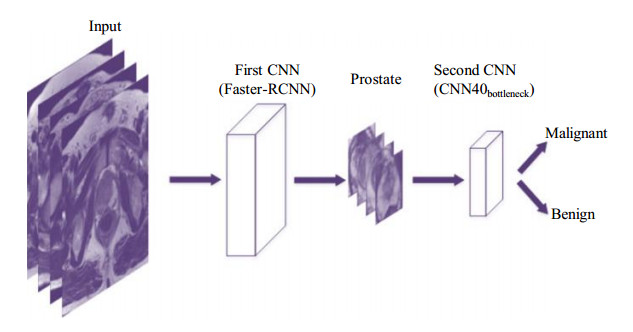
针对深度学习训练成本高,以及基于磁共振图像的前列腺癌临床诊断需要大量医学常识且极为耗时的问题,本文提出了一种基于级联卷积神经网络(Convolutional Neural Network,CNN)和磁共振图像的前列腺癌(Prostate Cancer,PCa)自动分类诊断方法,该网络以Faster-RCNN作为前网络,对前列腺区域进行提取分割,用于排除前列腺附近组织器官的干扰;以基于ResNet改进的网络结构CNN40bottleneck作为后网络,用于对前列腺区域病变进行分类.后网络由瓶颈结构串联组成,其中使用批量标准化(Batch Normalization,BN)、全局平均池化(Global Average Pooling,GAP)进行优化.实验结果证明,本文方法对前列腺癌诊断结果较好,而且缩减了训练时间和参数量,有效降低了训练成本.
刘可文
,
刘紫龙
,
汪香玉
,
陈黎
,
李钊
,
吴光耀
,
刘朝阳
. 基于级联卷积神经网络的前列腺磁共振图像分类[J]. 波谱学杂志, 2020
, 37(2)
: 152
-161
.
DOI: 10.11938/cjmr20192717
Interpreting magnetic resonance imaging (MRI) data by radiologists is time consuming and demands special expertise. Diagnosis of prostate cancer (PCa) with deep learning can also be time consuming and data storage consuming. This work presents an automated method for PCa detection based on cascaded convolutional neural network (CNN), including pre-network and post-network. The pre-network is based on a Faster-RCNN and trained with prostate images in order to separate the prostate from nearby tissues; the ResNet-based post-network is for PCa diagnosis, which is connected by bottlenecks and improved by applying batch normalization (BN) and global average pooling (GAP). The experimental results demonstrated that the cascaded CNN proposed had a good classification results on the in-house datasets, with less training time and computation resources.
[1] SIEGEL R L, MILLER K D, DVM A J. Cancer statistics, 2018[J]. Ca:A Cancer Journal for Clinicians, 2018, 68(1):11.
[2] HAN S J, ZHANG S W, CHEN W Q, et al. Analysis of the status and trends of prostate cancer incidence in China[J]. Chinese Clinical Oncology, 2013, 18(4):330-334. 韩苏军, 张思维, 陈万青, 等. 中国前列腺癌发病现状和流行趋势分析[J]. 临床肿瘤学杂志, 2013, 18(4):330-334.
[3] YANG J Y, YANG M Z, WEI W. Epidemiological study on the occurrence and development of prostate cancer[J]. J Clin Urology, 2017, 9:74-78. 杨进益, 杨明州, 魏伟. 前列腺癌发生发展的流行病学研究进展[J]. 临床泌尿外科杂志, 2017, 9:74-78.
[4] MADDAMS J, UTLEY M, MØLLER H. Projections of cancer prevalence in the United Kingdom, 2010-2040[J]. Brit J Cancer, 2012, 107(7):1195-1202.
[5] PENG Y, JIANG Y, YANG C, et al. Quantitative analysis of multiparametric prostate MR images:differentiation between prostate cancer and normal tissue and correlation with Gleason score——a computer-aided diagnosis development study[J]. Radiology, 2013, 267(3):787-796.
[6] ROOIJ M D, HAMOEN E H J, FÜTTERER J J, et al. Accuracy of multiparametric MRI for prostate cancer detection:A meta-analysis[J]. AJR Am J Roentgenol, 2014, 202(2):343-351.
[7] FEHR D, VEERARAGHAVAN H, WIBMER A, et al. Automatic classification of prostate cancer Gleason scores from multiparametric magnetic resonance images[J]. Proc Natl Acad Sci U S A, 2015, 112(46):6265-73.
[8] PENG Y, JIANG Y, YANG C, et al. Quantitative analysis of multiparametric prostate mr images:differentiation between prostate cancer and normal tissue and correlation with gleason score—a computer-aided diagnosis development study[J]. Radiology, 2013, 267(3):787-796.
[9] HASEGAWA A, LO S C B, FREEDMAN M T, et al. Convolution neural-network-based detection of lung structures[J]. Proceedings of SPIE-The International Society for Optical Engineering, 1994:654-662.
[10] LO S B, LOU S A, LIN J S, et al. Artificial convolution neural network techniques and applications for lung nodule detection[J]. IEEE Trans Med Imaging, 1995, 14(4):711-718.
[11] SAHINER B, CHAN H P, PETRICK N, et al. Classification of mass and normal breast tissue:a convolution neural network classifier with spatial domain and texture images[J]. IEEE Trans Med Imaging, 1996, 15(5):598-610.
[12] YANG X, LIU C Y, WANG Z W, et al. Co-trained convolutional neural networks for automated detection of prostate cancer in multi-parametric MRI[J]. Med Image Anal, 2017, 42:212-227.
[13] WANG Z W, LIU C Y, CHENG D P, et al. Automated detection of clinically significant prostate cancer in mp-MRI images based on an end-to-end deep neural network[J]. IEEE Trans Med Imaging, 2018, 37(5):1127-1139.
[14] SIMONYAN K, ZISSERMAN A. Very deep convolutional networks for large-scale image recognition[J]. Computer Science, 2014. arXiv:1409.1556.
[15] REN S, HE K, GIRSHICK R, et al. Faster R-CNN:towards real-time object detection with region proposal networks[C]//International Conference on Neural Information Processing Systems. 2015.
[16] HE K M, ZHANG X Y, REN S Q, et al. Deep residual learning for image recognition[C]//Proceedings of the IEEE Conference on Computer Vision and Pattern Recognition. IEEE, 2016:770-778.
[17] LIN M, CHEN Q, YAN S C. Network in network[J]. Computer Science, 2013:196-204.
[18] IOFFE S, SZEGEDY C. Batch normalization:accelerating deep network training by reducing internal covariate shift[C]//International Conference on International Conference on Machine Learning. JMLR.org, 2015.
[19] SZEGEDY C, LIU W, JIA Y, et al. Going deeper with convolutions[C]//Proceedings of the IEEE Conference on Computer Vision and Pattern Recognition. 2015:1-9.
[20] SZEGEDY C, VANHOUCKE V, IOFFE S, et al. Rethinking the inception architecture for computer vision[C]//Proceedings of the IEEE Conference on Computer Vision and Pattern Recognition. 2016.
[21] SZEGEDY C, IOFFE S, VANHOUCKE V, et al. Inception-v4, Inception-ResNet and the impact of residual connections on learning[C]//Thirty-First AAAI Conference on Artificial Intelligence. 2017:711-718.
[22] GEERT L, OSCAR D, JELLE B, et al. Prostatex challenge data. The cancer imaging archive[OL]. https://doi.org/10.7937/K9TCIA.2017.MURS5CL.
[23] SCHAEFER S, MCPHAIL T, WARREN J. Image deformation using moving least squares[C]//ACM Siggraph. 2006:711-718.