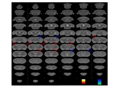
本文探究了西洋参-石菖蒲(X-S)药液对糖尿病认知障碍(DACD)大鼠学习记忆相关脑区的影响.腹腔注射链脲佐菌素(STZ)诱导糖尿病大鼠模型,并将动物分为糖尿病组和X-S组,STZ注射7天后给予X-S药,每日1次,连续113天;于STZ注射80天后采用Morris水迷宫方法筛选DACD和非认知障碍(DNCD)大鼠;120天后运用磁共振成像(MRI)技术扫描大鼠全脑,利用感兴趣区(ROI)法和基于体素形态学(VBM)法对各组大鼠大脑灰质和白质进行体积和密度的分析;并通过苏木精-伊红(HE)染色观察大鼠海马神经元的形态结构.ROI分析法显示:与DACD组相比,X-S组大鼠左侧颞叶联合皮质体积减小(p<0.05);VBM分析法显示:与DACD组相比,X-S组大鼠海马CA1、CA3区及其它脑区的体积和密度或增高或降低(p<0.005).HE染色结果显示:与DACD组相比,X-S组大鼠海马CA1、CA3区的细胞固缩及排列紊乱减轻.结果表明X-S药液对DACD大鼠与学习记忆相关的海马及其它脑区存在双向调节,从而发挥学习记忆能力的改善作用.
This study explored the effects of Panax quinquefolius L.-Acorus Tatarinowii (X-S) on learning and memory behaviors and related brain regions in rats with diabetes-associated cognitive decline (DACD). Type 1 diabetes mellitus (DM) was induced by intraperitoneal injection of streptozotocin (STZ). The DM rats were randomly divided into the DM group and X-S group. Starting at day 7 after the STZ induction, X-S was administered once a day for 113 consecutive days. Morris water maze test was performed to screen the DM rats with and without cognitive impairment at 80 days after the STZ induction. The volume and concentration of gray matter and white matter in the whole brain were analyzed from magnetic resonance imaging (MRI) data acquired at day 120 after the STZ induction, with a region of interest (ROI)-based method and voxel-based morphometry (VBM). Hippocampal neurons were stained with hematoxylin and eosin (HE). The results of ROI-based analysis showed that:the volume of left temporal association cortex of the X-S rats decreased significantly (p<0.05), compared with the DACD rats. The VBM results showed that:the volume or concentration of CA1, CA3 and other brain regions increased or decreased in the X-S rats (p<0.005), compared with the DACD rats. The HE staining results showed that:X-S treatment alleviated cell pyknosis of the CA1 and CA3 neurons. It was concluded that X-S treatment has both positive and negative effects on the hippocampus and other brain areas related to learning and memory in the DACD rats.
[1] CHO N H, SHAW J E, KARURANGA S, et al. IDF diabetes atlas:Global estimates of diabetes prevalence for 2017 and projections for 2045[J]. Diabetes Res Clin Pract, 2018, 138:271-281.
[2] MIJNHOUT G S, SCHELTENS P, DIAMANT M, et al. Diabetic encephalopathy:A concept in need of a definition[J]. Diabetologia, 2006, 49(6):1447-1448.
[3] SIMA A A. Encephalopathies:the emerging diabetic complications[J]. Acta Diabetol, 2010, 47(4):279-293.
[4] 中华医学会糖尿病学分会. 中国2型糖尿病防治指南[M]. 北京:北京大学医学出版社, 2014.
[5] OHARA T. Epidemiology of diabetes and risk of dementia[J]. Brain Nerve, 2016, 68(7):719-727.
[6] 赵瑛, 谢海龙, 王冬雪, 等. 一种防治糖尿病认知障碍的药物组合物及其制备方法:中国, 201611190858.4[P]. 2017-05-31.
[7] HUANG W, CAO Z Y. STZ-induced progressive brain atrophy studied by magnetic resonance imaging and histochemical staining[J]. Chinese J Magn Reson, 2015, 32(3):439-449. 黄微, 曹子玉. STZ诱导大鼠1型糖尿病进行性脑萎缩的磁共振成像及组织化学研究[J]. 波谱学杂志, 2015, 32(3):439-449.
[8] 刘月婷. 糖尿病致认知损害大鼠脑脊液代谢标志物的筛选[D]. 哈尔滨:哈尔滨商业大学, 2016.
[9] 毕田田. 糖尿病致认知障碍大鼠脑脊液差异代谢物的动态变化[D]. 哈尔滨:哈尔滨商业大学, 2019.
[10] 高伟莹. 糖尿病认知损害大鼠海马组织蛋白质组学的研究[D]. 哈尔滨:哈尔滨商业大学, 2020.
[11] LI C, YANG Q F, ZHOU L N, et al. Atrophy of entorhinal cortex in evaluation of cognitive performance in patients with diabetes mellitus[J]. Chin J Med Imaging Technol, 2018, 34(1):25-29.
[12] 苏力担卡扎·仇曼, 何铁英, 林海, 等. 链脲佐菌素建立1型糖尿病大鼠模型的最优剂量研究[J]. 中国普外基础与临床杂志, 2016, 23(1):84-85.
[13] BEDNARIK P, MOHEET A A, GROHN H, et al. Type 1 diabetes and impaired awareness of hypoglycemia are associated with reduced brain gray matter volumes[J]. Front Neurosci, 2017, 11:529.
[14] 谢海龙. 基于代谢组学的西洋参-石菖蒲对糖尿病认知损害大鼠干预作用研究[D]. 哈尔滨:哈尔滨商业大学, 2019.
[15] WEINSTEIN G, MAILLARD P, HIMALI J J, et al. Glucose indices are associated with cognitive and structural brain measures in young adults[J]. Neurology, 2015, 84(23):2329-2337.
[16] 赵瑛, 刘琳, 刘月婷, 等. 一种基于水迷宫的动物学习记忆的个体化评价方法[P]. CN 106070014 B, 2020-06-11.
[17] PRESSLER M, NOPOULOS P, HO B C, et al. Insular cortex abnormalities in schizophrenia:Relationship to symptoms and typical neuroleptic exposure[J]. Biol Psychiatry, 2005, 57(4):394-398.
[18] GLASER N, LITTLE C, LO W, et al. Treatment with the KCa3.1 inhibitor TRAM-34 during diabetic ketoacidosis reduces inflammatory changes in the brain[J]. Pediatr Diabetes, 2017,18(5):356-366.
[19] LO W, O'DONNELL M, TANCREDI D, et al. Diabetic ketoacidosis in juvenile rats is associated with reactive gliosis and activation of microglia in the hippocampus[J]. Pediatr Diabetes, 2016, 17(2):127-139.
[20] MIAO H H, ZHANG Y, DING G N, et al. Ginsenoside Rb1 Attenuates isoflurane/surgery-induced cognitive dysfunction via inhibiting neuroinflammation and oxidative stress[J]. Biomed Environ Sci, 2017, 30(5):53-62.
[21] WEN W, WANG Y. Effect of effective components of acorus tatarinowii schott and polygala on SOD and MDA in neurons hippocampus induced by Aβ25-35 in vitro[J]. Journal of Emergency in Traditional Chinese Medicine, 2012, 21(8):1256-1258.
[22] ZHOU Y, LI X L, XIE H L, et al. Voxel-based morphology analysis of STZ-induced type 1 diabetes mellitus rats with and without cognitive impairment[J]. Neurosci Lett, 2018, 684:210-217.
[23] LU Y M, JIA Z, JANUS C, et al. Mice lacking metabotropic glutamate receptor 5 show impaired learning and reduced CA1 long-term potentiation (LTP) but normal CA3 LTP[J]. J Neurosci, 1997, 17(13):5196-5205.
[24] ISHIHARA K, MITSUNO K, ISHIKAWA M, et al. Behavioral LTP during learning in rat hippocampal CA3[J]. Behav Brain Res, 1997, 83(1/2):235-238.
[25] STEPAN J, DINE J, EDER M. Functional optical probing of the hippocampal trisynaptic circuit in vitro:network dynamics, filter properties, and polysynaptic induction of CA1 LTP[J]. Front Neurosci, 2015, 9:160-168.
[26] DI PAOLA M, MACALUSO E, CARLESIMO G A, et al. Episodic memory impairment in patients with Alzheimer's disease is correlated with entorhinal cortex atrophy[J]. J Neurol, 2007, 254(6):774-781.
[27] NILSSON O G, SHAPIRO M L, GAGE F H, et al. Spatial learning and memory following fimbria-fornix transection and grafting of fetal septal neurons to the hippocampus[J]. Exp Brain Res, 1987, 67(1):195-215.
[28] 梁力. 听觉皮层时域信号处理特征[D]. 广州:第一军医大学, 2000.
[29] SMITH E E, JONIDES J. Neuroimaging analyses of human working memory[J]. Proc Natl Acad Sci, 1998, 95(20):12061-12068.
[30] STÜTTGEN M C, SCHWARZ C. Barrel cortex:What is it good for?[J]. Neuroscience, 2017, 368:3-16.

