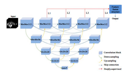
阿尔茨海默症(Alzheimer's Disease,AD)是一种神经退行性疾病,高效准确的早期诊断对其治疗至关重要.本文提出了一种融合多语义级别的深度卷积神经网络结构,基于磁共振图像,用于区分AD患者与正常受试者的方法.首先,在传统UNet++网络的基础上改进了深度监督整合算法;然后,构建了一种新的特征融合结构,进一步细化了不同语义级别的特征;最后,基于不同组织区域(白质、灰质和脑脊液)的磁共振图像,使用本文所提出的方法区分AD患者和正常受试者,并探究了从不同组织获得的信息对分类准确率的影响.实验结果表明,使用本文提出的方法区分两类人群的最高准确率为98.74%,平均准确率为98.47%,高于目前文献报道的其他方法.
Alzheimer's disease (AD) is one of the most common forms of dementia and a degenerative mental disorder that seriously affects people's daily lives. Rapid and effective diagnosis is essential for the treatment of patients with Alzheimer's disease. To solve this problem, this paper proposes a deep convolutional neural network structure with multiple semantic levels to classify AD patients and healthy controls from magnetic resonance imaging (MRI) data. Firstly, the deep supervision integration algorithm and the classification model of Alzheimer's disease based on the traditional UNet++ network were improved. Then, a new feature fusion structure was constructed, which further refined the different semantic levels. Lastly, the proposed protocol was applied to different tissue regions (e.g., white matter, gray matter and cerebrospinal fluid), and the effects of different tissue information combinations on the classification outcome were explored. The method proposed was applied to the Alzheimer's Disease Neuroimaging Initiative (ADNI) dataset to classify the AD patients. The results demonstrated that the highest accuracy of 98.74%, and an average accuracy of 98.47%.
[1] QUERFURTH H W, LAFERLA F M. Alzheimer's mechanisms of disease[J]. New Engl J Med, 2010, 362(4):329-344.
[2] ALZHEIMER'S ASSOCIATION. 2011 Alzheimer's disease facts and figures[R]. Alzheimer's & Dementia, 2011, 7(2):208.
[3] KIVIPELTO M, HELKALA E L, LAAKSO M P, et al. Midlife vascular risk factors and Alzheimer's disease in later life:longitudinal, population based study[J]. Brit Med J, 2001, 322(7300):1447-1451.
[4] JACK JR C R, BERNSTEIN M A, FOX N C, et al. The Alzheimer's disease neuroimaging initiative (ADNI):MRI methods[J]. J Magn Reson Imaging, 2008, 27(4):685-691.
[5] BRON E E, SMITS M, VAN DER FLIER W M, et al. Standardized evaluation of algorithms for computer-aided diagnosis of dementia based on structural MRI:the CADDementia challenge[J]. NeuroImage, 2015, 111:562-579.
[6] CHENG H T, WANG S S, KE Z W, et al. Deep recursive cascaded convolutional network for parallel MRI[J]. Chinese J Magn Reson, 2019, 36(4):437-445. 程慧涛, 王珊珊, 柯子文, 等. 基于深度递归级联卷积网络的并行磁共振成像方法[J]. 波谱学杂志, 2019, 36(4):437-445.
[7] LIU S D, CAI W D, WEN L F, et al. Neuroimaging biomarker based prediction of Alzheimer's disease severity with optimized graph construction[C]. San Francisco:2013 IEEE 10th International Symposium on Biomedical Imaging. 2013:1336-1339.
[8] TONG T, WOLZ R, GAO Q Q, et al. Multiple instance learning for classification of dementia in brain MRI[J]. Med Image Comput Comput Assist Interv, 2013, 16(Pt 2):599-606.
[9] SHANKAR K, LAKSHMANAPRABU S K, KHANNA A, et al. Alzheimer detection using group grey wolf optimization based features with convolutional classifier[J]. Comput Electr Eng, 2019, 77:230-243.
[10] BASKAR D, JAYANTHI V S, JAYANTHI A N. An efficient classification approach for detection of Alzheimer's disease from biomedical imaging modalities[J]. Multimed Tools Appl, 2019, 78(10):12883-12915.
[11] MADUSANKA N, CHOI H K, SO J H, et al. Alzheimer's disease classification based on multi-feature fusion[J]. Curr Med Imaging Rev, 2019, 15(2):161-169.
[12] CUI R X, LIU M H, THE ALZHEIMER'S DISEASE NEUROIMAGING INITIATIVE. RNN-based longitudinal analysis for diagnosis of Alzheimer's disease[J]. Comput Med Imag Grap, 2019, 73:1-10.
[13] LI F, LIU M H, ALZHEIMER'S DISEASE NEUROIMAGING INITIATIVE. Alzheimer's disease diagnosis based on multiple cluster dense convolutional networks[J]. Comput Med Imag Grap, 2018, 70:101-110.
[14] LIU S Q, LIU S D, CAI W D, et al. Multimodal neuroimaging feature learning for multiclass diagnosis of Alzheimer's disease[J]. IEEE T Bio-Med Eng, 2015, 62(4):1132-1140.
[15] IOFFE S, SZEGEDY C. Batch normalization:Accelerating deep network training by reducing internal covariate shift[J]. 2015. arXiv:1502.03167.
[16] LIU K W, LIU Z L, WANG X Y, et al. Prostate cancer diagnosis based on cascaded convolutional neural networks[J]. Chinese J Magn Reson, 2020, 37(2):152-161. 刘可文, 刘紫龙, 汪香玉, 等. 基于级联卷积神经网络的前列腺磁共振图像分类[J]. 波谱学杂志, 2020, 37(2):152-161.
[17] ZHOU Z W, SIDDIQUEE M M R, TAJBAKHSH N, et al. Unet++:A nested u-net architecture for medical image segmentation[C]. 4th Deep Learning in Medical Image Analysis (DLMIA) workshop, Computer Vision and Pattern Recognition, 2018. arXiv:1807.10165
[18] RONNEBERGER O, FISCHER P, BROX T. U-net:Convolutional networks for biomedical image segmentation[C]. MICCAI 2015, Computer Vision and Pattern Recognition, Cham, 2015. arXiv:1505.04597.
[19] HUANG G, LIU Z, VAN DER MAATEN L, et al. Densely connected convolutional networks[C]. 2017 IEEE Conference on Computer Vision and Pattern Recognition (CVPR), Honolulu, HI, 2017:2261-2269.
[20] DOU Q, CHEN H, JIN Y M, et al. 3D deeply supervised network for automatic liver segmentation from CT volumes[C]. MICCAI 2016, Computer Vision and Pattern Recognition, 2016, arXiv:1607.00582.
[21] LEE C Y, XIE S N, GALLAGHER P, et al. Deeply-supervised nets[C]//Artificial Intelligence and Statistics. 2015. arXiv:1409.5185.
[22] Srivastava N, Hinton G, Krizhevsky A, et al. Dropout:a simple way to prevent neural networks from overfitting[J]. J Mach Learn Res, 2014, 15(1):1929-1958.
[23] JENKINSON M, BECKMANN C F, BEHRENS T E J, et al. FSL[J]. Neuroimage, 2012, 62(2):782-790.

