

基于Faster-RCNN和Level-Set的桥小脑角区肿瘤自动精准分割
收稿日期: 2021-01-07
网络出版日期: 2021-03-22
基金资助
微创励志创新基金资助项目(193832381)
Automatic Precise Segmentation of Cerebellopontine Angle Tumor Based on Faster-RCNN and Level-Set Method
Received date: 2021-01-07
Online published: 2021-03-22
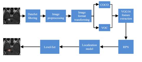
桥小脑角区(CPA)肿瘤的精准分割在手术治疗、放疗中有重要影响,本文结合更快速区域卷积神经网络(Faster-RCNN)和水平集(Level-Set)方法对CPA肿瘤的自动分割进行了研究.首先,采集317名CPA肿瘤患者的T1WI-SE序列磁共振图像,使用基于Faster-RCNN主干网络VGG16提取特征,结合区域建议网络(RPN)进行学习训练,建立带有CPA肿瘤位置信息的定位模型,再应用Level-Set对肿瘤进行精准分割.本文对比了不同CPA肿瘤区域勾画范围对分割结果产生的影响,并以精确率、召回率、均值平均精度值(mAP)和戴斯系数(Dice系数)等指标评估了模型定位和分割的性能.实验结果表明,结合Faster-RCNN和Level-Set建立的模型能更有效对CPA肿瘤进行精准分割,减轻临床医生的负担,并提升治疗效果.
刘颖 , 郭伊云 , 陈静聪 , 章浩伟 . 基于Faster-RCNN和Level-Set的桥小脑角区肿瘤自动精准分割[J]. 波谱学杂志, 2021 , 38(3) : 381 -391 . DOI: 10.11938/cjmr20212881
To meet the demands in surgical treatment and radiotherapy, this work combines the faster region convolutional neural network (Faster-RCNN) and Level-Set methods to segment cerebellopontine angle (CPA) tumors automatically and precisely. T1WI-SE magnetic resonance images from 317 CPA tumor patients were collected. Features extracted by VGG16 were combined with the region proposal network (RPN) for training. A CPA tumor localization model was then established, before the Level-Set method was applied to accurately segment the tumor. The segmentation results of different CPA tumor regions were compared in terms of precision, recall, mean average precision (mAP) and Dice coefficient. The results showed that the method proposed can effectively and precisely segment CPA tumors, thereby capable of reducing the burden on clinicians and improving the treatment effect.
| 1 | PING X X , MENG Q , TIAN X , et al. MRI findings of lesions in the cerebellopontine angle[J]. J Med Imaging, 2014, 24 (1): 12- 15. |
| 1 | 平小夏, 孟倩, 田霞, 等. 桥小脑角区病变的MRI表现[J]. 医学影像学杂志, 2014, 24 (1): 12- 15. |
| 2 | FANG F , HU S P . Comparative analysis of MRI findings and pathology in acoustic neuroma[J]. CT Theory and Applications, 2019, 28 (6): 731- 738. |
| 2 | 方芳, 胡少平. 听神经瘤MRI表现与病理对照分析[J]. CT理论与应用研究, 2019, 28 (6): 731- 738. |
| 3 | CHEN Q , LI G Q , LI J T . Study of the value MRI in the diagnosis of intracranial meningioma[J]. Chinese Jopurnal of CT and MRI, 2016, 14 (4): 23- 26. |
| 3 | 陈琪, 李国强, 李惊涛. MRI对颅内脑膜瘤的诊断价值研究[J]. 中国CT和MRI杂志, 2016, 14 (4): 23- 26. |
| 4 | ZHANG J Q , ZHANG Y , YIN Y , et al. A review of machine learning in tumor radio therapy[J]. Journal of Biomedical Engineering, 2019, 36 (5): 879- 844. |
| 4 | 张珺倩, 张远, 尹勇, 等. 机器学习在肿瘤放射治疗领域应用进展[J]. 生物医学工程学杂志, 2019, 36 (5): 879- 884. |
| 5 | LI Q , BAI K X , ZHAO L , et al. Progresss and challenges of MRI brain tumor image segmentation[J]. Journal of Image and Graphics, 2020, 25 (3): 419- 431. |
| 5 | 李锵, 白柯鑫, 赵柳, 等. MRI脑肿瘤图像分割研究进展及挑战[J]. 中国图象图形学报, 2020, 25 (3): 419- 431. |
| 6 | WANG C F , WANG B J , WANG Z , et al. The target profile of multimodal magnetic resonance imaging in the radiotherapy of craniocerebral malignant tumor[J]. Chinese Journal of Practical Nervous Diseases, 2018, 21 (19): 2118- 2124. |
| 6 | 王长福, 王斌杰, 王智, 等. 多模态磁共振成像及融合技术在颅脑恶性肿瘤放疗靶区勾画中的应用[J]. 中国实用神经疾病杂志, 2018, 21 (19): 2118- 2124. |
| 7 | ZIKIC D, IOANNOU Y, BROWN M, et al. Segmentation of brain tumor tissues with convolutional neural networks[C]. Proceedings MICCAI-BRATS, 2014: 36-39. |
| 8 | JIA Y, SHELHAMER E, DONAHUE J, et al. Caffe: Convolutional architecture for fast feature embedding[C]//Proceedings of the 22nd ACM international conference on Multimedia. 2014: 675-678. |
| 9 | IQBAL S , GHANI M U , SABA T , et al. Brain tumor segmentation in multi-spectral MRI using convolutional neural networks (CNN)[J]. Microsc Res Techniq, 2018, 81 (4): 419- 427. |
| 10 | EZHILARASI R, VARALAKSHMI P. Tumor detection in the brain using faster R-CNN[C]//20182nd International Conference on I-SMAC (IoT in Social, Mobile, Analytics and Cloud). IEEE, 2018: 388-392. |
| 11 | YANG X H , ZHANG Y . Brain tumor segmentation based on MRI multimodal information and 3D-CNNs feature extraction[J]. Chinese Journal of CT and MRI, 2020, 18 (9): 4- 6, 23. |
| 11 | 杨新焕, 张勇. 结合MRI多模态信息和3D-CNNs特征提取的脑肿瘤分割研究[J]. 中国CT和MRI杂志, 2020, 18 (9): 4- 6, 23. |
| 12 | THILLAIKKARASI R , SARAVANAN S . An enhancement of deep learning algorithm for brain tumor segmentation using kernel based CNN with M-SVM[J]. J Med Syst, 2019, 43 (4): 1- 7. |
| 13 | IQBAL S , GHANI KHAN M U , SABA T , et al. Deep learning model integrating features and novel classifiers fusion for brain tumor segmentation[J]. Microsc Res Techniq, 2019, 82 (8): 1302- 1315. |
| 14 | KASS M , WITKIN A , TERZOPOULOS D . Snakes: Active contour models[J]. Int J Comput Vision, 1988, 1 (4): 321- 331. |
| 15 | CHAN T F , VESE L A . Active contours without edges[J]. IEEE T Image Process, 2001, 10 (2): 266- 277. |
| 16 | LI C M , KAO C Y , GORE J C , et al. Minimization of region-scalable fitting energy for image segmentation[J]. IEEE T Image Process, 2008, 17 (10): 1940- 1949. |
| 17 | YIN S L , LI H , LIU D S , et al. Active contour modal based on density-oriented BIRCH clustering method for medical image segmentation[J]. Multimed Tools Appl, 2020, 79 (41): 31049- 31068. |
| 18 | YUAN C A , ZHENG Y , QIN X , et al. The medical image segmentation method of improved RSF active contour model[J]. Journal of Zhengzhou University (Natural Science Edition), 2017, 49 (1): 34- 38, 44. |
| 18 | 元昌安, 郑彦, 覃晓, 等. 改进RSF主动轮廓模型的医学图像分割方法[J]. 郑州大学学报(理学版), 2017, 49 (1): 34- 38. |
| 19 | SHI Q Y , YAN F , YANG Y , et al. Image segmentation of tooth and alveolar bone with the level set model[J]. Chinese J Magn Reson, 2021, 38 (2): 182- 193. |
| 19 | 石沁祎, 闫方, 杨阳, 等. 基于水平集的牙齿牙槽骨图像分割[J]. 波谱学杂志, 2021, 38 (2): 182- 193. |
| 20 | ZHANG D, ZHU W, ZHAO H, et al. Automatic localization and segmentation of optical disk based on faster R-CNN and level set in fundus image[C]//Medical Imaging 2018: Image Processing. International Society for Optics and Photonics, 2018, 10574: 105741U. |
| 21 | ZUO J H , ZHAO C , ZHU X L , et al. High-resolution remote sensing image building extraction combined with Faster-RCNN and Level-Set[J]. Chinese Journal of Liquid Crystals and Displays, 2019, 34 (4): 439- 447. |
| 21 | 左俊皓, 赵聪, 朱晓龙, 等. Faster-RCNN和Level-Set结合的高分遥感影像建筑物提取[J]. 液晶与显示, 2019, 34 (4): 439- 447. |
| 22 | LIU Y , CHEN J C , HU X Y , et al. Classification and localization of acoustic neuroma and meningioma in the cerebellopontine angle based on Mask RCNN[J]. Chinese J Magn Reson, 2021, 38 (1): 58- 68. |
| 22 | 刘颖, 陈静聪, 胡小洋, 等. 基于Mask RCNN的桥小脑角区听神经瘤与脑膜瘤分类定位研究[J]. 波谱学杂志, 2021, 38 (1): 58- 68. |
| 23 | REN S Q, HE K M, GIRSHICK R, et al. Faster R-CNN: Towards real-time object detection with region proposal networks[C]//Advances in neural information processing systems. 2015: 91-99. |
| 24 | OSHER S , SETHIAN J A . Fronts propagating with curvature-dependent speed: algorithms based on Hamilton-Jacobi formulations[J]. J Comput Phys, 1988, 79 (1): 12- 49. |
| 25 | YAO C W , HUANG D B , YE M Q . MR brain tumor image segmentation based on rough set adaptive granularity[J]. Computer and Modernization, 2021, (1): 34- 37. |
| 25 | 姚传文, 黄道斌, 叶明全. 基于粗糙集自适应粒度的MR脑肿瘤图像分割[J]. 计算机与现代化, 2021, (1): 34- 37. |
| 26 | THAHA M M , KUMAR K P M , MURUGAN B S , et al. Brain tumor segmentation using convolutional neural networks in MRI images[J]. J Med Syst, 2019, 43 (9): 1- 10. |
| 27 | GE T , ZHAN T M , MU S X . Brain tumor segmentation algorithm based on multi-kernel collaborative representation classification[J]. Journal of Nanjing University of Science and Technology, 2019, 43 (5): 578- 585. |
| 27 | 葛婷, 詹天明, 牟善祥. 基于多核协同表示分类的脑肿瘤分割算法[J]. 南京理工大学学报, 2019, 43 (5): 578- 585. |
| 28 | RONNEBERGER O, FISCHER P, BROX T. U-net: Convolutional networks for biomedical image segmentation[C]//International Conference on Medical image computing and computer-assisted intervention. Springer, Cham, 2015: 234-241. |
| 29 | XU Z W, WU Z Y, FENG J J. CFUN: combining faster R-CNN and U-net network for efficient whole heart segmentation[J]. arXiv preprint arXiv: 1812.04914, 2018. |
/
| 〈 |
|
〉 |