

基于全卷积网络的乳腺肿瘤动态增强磁共振图像分割
收稿日期: 2021-05-21
网络出版日期: 2021-09-16
基金资助
国家自然科学基金资助项目(81830052);上海市科技创新行动计划资助项目(18441900500);上海市自然科学基金资助项目(20ZR1438300);山东省自然科学基金资助项目(ZR2021MH160)
Segmentation of Breast Tumors Based on Fully Convolutional Network and Dynamic Contrast Enhanced Magnetic Resonance Image
Received date: 2021-05-21
Online published: 2021-09-16
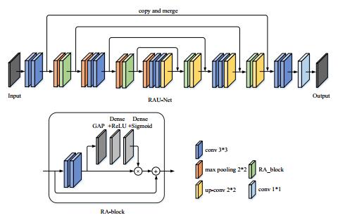
准确可靠的乳腺肿瘤分割是乳腺癌诊断、治疗、预后评估的关键.针对现有的基于动态增强磁共振成像(DCE-MRI)的乳腺肿瘤分割方法易遗漏小目标肿瘤等不足,本文提出了一种基于全卷积网络的可靠高效的乳腺肿瘤DCE-MRI图像分割方法.首先,对乳腺DCE-MRI数据进行预处理后,截取128*128大小的图像块,并以肿瘤区域像素数为依据将数据分为两个子数据集;其次,利用数据集训练CBP5-Net得到分类模型;然后,利用两个子数据集分别训练RAU-Net得到两个分割模型;最后,将测试集数据送到网络输入端,并对网络输出结果进行后处理,得到最终的乳腺肿瘤分割结果.利用本文提出的方法得到的Dice系数、敏感性、特异性和交并比(IoU)分别达到了0.938 8、0.952 3、0.998 5和0.876 8,说明利用本文方法能够有效、精确地分割乳腺肿瘤DCE-MRI图像.
关键词: 全卷积网络(FCN); 图像分割; 乳腺肿瘤; 动态对比增强磁共振成像(DCE-MRI)
邱玥 , 聂生东 , 魏珑 . 基于全卷积网络的乳腺肿瘤动态增强磁共振图像分割[J]. 波谱学杂志, 2022 , 39(2) : 196 -207 . DOI: 10.11938/cjmr20212921
Accurate and reliable breast tumor segmentation is essential for the diagnosis, treatment and prognosis of breast cancer. To address the shortcomings of existing dynamic contrast enhanced-magnetic resonance imaging (DCE-MRI)-based breast tumor segmentation methods, which tend to miss small tumors, we proposed a more reliable and efficient segmentation method for breast tumors in DCE-MRI based on a fully convolutional network (FCN). Firstly, the breast DCE-MRI data was preprocessed, followed by intercepting the image blocks of 128*128, and dividing the dataset into two sub-datasets according to the number of pixels in the tumor region. Secondly, the whole set was used to train CBP5-Net to obtain a classification model. Then, two sub-datasets were used to train RAU-Net to get two segmentation models. Finally, the test set was entered into the network, and the network outputs were post-processed to obtain the final segmentation results. The Dice coefficient, sensitivity, specificity and intersection over union (IoU) index of the method proposed in this paper reached 0.938 8, 0.952 3, 0.998 5 and 0.876 8, respectively. It proves that the proposed method can be used to segment DCE-MRI breast tumors effectively and accurately.
| 1 | SIEGEL R L , MILLE R K D , JEMAL A . Cancer statistics, 2018[J]. CA: A Cancer Journal for Clinicians, 2018, 68 (1): 7- 30. |
| 2 | FOROUZANFAR M H , FOREMAN K J , DELOSSANTOS A M , et al. Breast and cervical cancer in 187 countries betweem 1980 and 2010: a systematic analysis[J]. Lancet, 2011, 378 (9801): 1461- 1484. |
| 3 | WILD C P, WEIDERPASS E, STEWART B W. World cancer report: cancer research for cancer prevention[M]//Lyon: International Agency for Research on Cancer, 2020: 23-33. |
| 4 | FANG Z Y , QIU X G . Distribution and risk factors of breast cancer[J]. Chinese General Practice, 2002, 5 (6): 429- 430. |
| 4 | 方志沂, 邱晓光. 乳腺癌的分布规律及其危险因素[J]. 中国全科医学, 2002, 5 (6): 429- 430. |
| 5 | SMITH R A , COKKINDES V , BROOKS D , et al. Cancer screening in the United States, 2011[J]. CA-Cancer J Clin, 2011, 61 (1): 8- 30. |
| 6 | DIDKOWSKA J , WOJCIECHOWSKA U . WHO position paper on mammography screening[J]. Oncology in Clinical Practice, 2014, |
| 7 | HEYWANG S H , HAHN D , SCHMIDT H , et al. MR imaging of the breast using gadolinium-DTPA[J]. J Comput Assist Tomogr, 1986, 10 (2): 199- 204. |
| 8 | MORRIS E A . Breast cancer imaging with MRI[J]. Radiol Clin N Am, 2002, 40 (3): 443- 466. |
| 9 | BHOOSHAN N , GIGER M L , JANSEN S A , et al. Cancerous breast lesions on dynamic contrast-enhanced MR images: computerized characterization for image-based prognostic markers[J]. Radiology, 2010, 254 (3): 680- 690. |
| 10 | 杨骞. 基于DCE-MRI影像的乳腺癌早期诊断研究[D]. 杭州: 杭州电子科技大学, 2014. |
| 11 | EADIE L H , TAYLOR P , GIBSON A P . A systematic review of computer-assisted diagnosis in diagnostic cancer imaging[J]. Eur J Radiol, 2011, 81 (1): e70- e76. |
| 12 | REIG B , HEACOCK L , GERAS K J , et al. Machine learning in breast MRI[J]. J Magn Reson Imaging, 2020, 52 (4): 998- 1018. |
| 13 | TAN Z , HONG R R , YE S Z . A method to extract the region of interest in dynamic contrast-enhanced MR imaging of breast cancer[J]. Journal of Fuzhou University (Natural Science Edition), 2016, 44 (1): 33- 39. |
| 13 | 檀哲, 洪容容, 叶少珍. 一种提取乳腺癌DCE-MRI感兴趣区域的分割方法[J]. 福州大学学报(自然科学版), 2016, 44 (1): 33- 39. |
| 14 | QIAO M Y , SUO S T , CHENG F , et al. Three-dimensional breast tumor segmentation on DCE-MRI with a multilabel attention-guided joint-phase-learning network[J]. Comput Med Imag Grap, 2021, 90, 101909. |
| 15 | LIU T T , XU H Y , JIN W , et al. Medical image segmentation based on a hybrid region-based active contour model[J]. Comput Math Methods Med, 2014, 890725. |
| 16 | AL-FARIS A Q , NGAH U K , ISA N A M , et al. Computer-aided segmentation system for breast MRI tumour using modified automatic seeded region growing (BMRI-MASRG)[J]. J Digit Imaging, 2014, 27 (1): 133- 144. |
| 17 | CHEN X, WILLIAMS B M, VALLABHANENI S R, et al. Learning active contour models for medical image segmentation[C]//CVF Conference on Computer Vision and Pattern Recognition (CVPR). IEEE, 2019. |
| 18 | KANNAN S R , RAMATHILAGAM S , DEVI P , et al. Improved fuzzy clustering algorithms in segmentation of DC-enhanced breast MRI[J]. J Med Syst, 2012, 36 (1): 321- 333. |
| 19 | BAI P R , LIU Q Y , LEI L , et al. A novel region-based level set method initialized with mean shift clustering for automated medical image segmentation[J]. Comput Biol Med, 2013, 43 (11): 1827- 1832. |
| 20 | PEREIRA S , PINTO A , ALVES V , et al. Brain tumor segmentation using convolutional neural networks in MRI images[J]. IEEE T Med Imaging, 2016, 35 (5): 1240- 1251. |
| 21 | DOLZ J, AYED I B, DESROSIERS C. Dense multi-path U-Net for ischemic stroke lesion segmentation in multiple image modalities[C]//International MICCAI Brainlesion Workshop. Springer, Cham, 2018. |
| 22 | LEE P Q , GUIDA A , PATTERSON S , et al. Model-free prostate cancer segmentation from dynamic contrast-enhanced MRI with recurrent convolutional networks: A feasibility study[J]. Comput Med Imag Grap, 2019, 75, 14- 23. |
| 23 | AMIT G, HADAD O, ALPERT S, et al. Hybrid mass detection in breast MRI combining unsupervised saliency analysis and deep learning[C]//International Conference on Medical Image Computing and Computer-Assisted Intervention, MICCAI 2017. |
| 24 | BENJELLOUN M, ADOUI M E, LARHMAM M A, et al. Automated breast tumor segmentation in DCE-MRI using deep learning[C]//International Conference on Cloud Computing Technologies and Applications, 2018. |
| 25 | LONG J, SHELHAMER E, DARRELL T. Fully convolutional networks for semantic segmentation[C]//IEEE Conference on Computer Vision and Pattern Recognition. IEEE Computer Society, 2015: 3431-3440. |
| 26 | ZHANG J, SAHA A, ZHU Z, et al. Breast tumor segmentation in DCE-MRI using fully convolutional networks with an application in radiogenomics[C]//Computer-aided Diagnosis, 2018. |
| 27 | ZHANG J , SAHA A , ZHU Z , et al. Hierarchical convolutional neural networks for segmentation of breast tumors in MRI with application to radiogenomics[J]. IEEE T Med Imaging, 2019, 38 (2): 435- 447. |
| 28 | ADOUI M E , MAHMOUDI S A , LARHMAM M A , et al. MRI breast tumor segmentation using different encoder and decoder CNN architectures[J]. Computers, 2019, 8 (3): 52. |
| 29 | LOFFE S , SZEGEDY C . Batch normalization: accelerating deep network training by reducing internal covariate shift[J]. arXiv preprint, 2015, 1502.03167. |
| 30 | RONNEBERGER O , FISCHER P , BROX T . U-Net: convolutional networks for biomedical image segmentation[M]. Spring Internation Publishing, 2015, 234- 241. |
| 31 | JIN Q G , MENG Z P , SUN C M , et al. RA-UNet: a hybrid deep attention-aware network to extract liver and tumor in CT scans[J]. Front Bioeng Biotechnol, 2020, 8, 605132. |
| 32 | HE K M, ZHANG X Y, REN S Q, et al. Deep residual learning for image recognition[C]//Proceedings of 2016 IEEE Conference on Computer Vision and Pattern Recognition. Las Vegas: IEEE, 2016: 770-778. |
| 33 | HU J , SHEN L , SUN G , et al. Squeeze-and-excitation networks[J]. arXiv preprint, 2017, 1709.01507. |
| 34 | SRIVASTAVA N , HINTON G , KRIZHEVSKY A , et al. Dropout: a simple way to prevent neural networks from overfitting[J]. J Mach Learn Res, 2014, 15 (1): 1929- 1958. |
| 35 | BADRINARAYANAN V , KENDALL A , CIPOLLA R . SegNet: a deep convolutional encoder-decoder architecture for image segmentation[J]. IEEE T Pattern Anal, 2017, 39 (12): 2481- 2495. |
| 36 | ZHOU Z W, SIDDIQUEE M M R, TAJBAKHSH N, et al. UNet++: a nested U-Net architecture for medical image segmentation[C]//Deep Learning in Medical Image Anylysis and Multimodal Learning for Clinical Decision Support, 2018: 3-11. |
/
| 〈 |
|
〉 |