

基于级联网络的膝关节图像分割与模型构建
收稿日期: 2021-08-19
网络出版日期: 2021-11-16
基金资助
北京化工大学高精尖科技创新团队基金资助项目(buctylkjcx06)
Knee Joint Image Segmentation and Model Construction Based on Cascaded Network
Received date: 2021-08-19
Online published: 2021-11-16
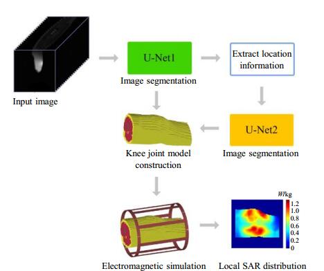
采用膝关节模型进行电磁仿真是计算膝关节局部射频功率沉积(SAR)的主要方法,为了构建膝关节模型,本文提出了一种包含两个卷积神经网络——U-Net的级联网络结构,用于膝关节磁共振图像的分割.第一个网络在整幅图上分割肌肉、脂肪等占比较大的组织,并从分割结果中预测软骨与半月板的大致位置信息,第二个网络基于该信息在一个更小的子图上分割小组织以提高分割精度.两个网络均采用焦点损失函数,它们的分割结果合并在一起构成膝关节模型.我们将该方法与其它4种方法的分割结果进行了定量指标的对比研究,并分别构建膝关节模型,计算局部SAR值.结果表明本文提出的级联网络结构可以更精确的构建用于SAR仿真的膝关节模型.
关键词: 磁共振成像(MRI); 局部射频功率沉积(SAR); 膝关节; 级联网络; 焦点损失函数
马岩 , 邢藏菊 , 肖亮 . 基于级联网络的膝关节图像分割与模型构建[J]. 波谱学杂志, 2022 , 39(2) : 184 -195 . DOI: 10.11938/cjmr20212941
Electromagnetic simulation using a knee model is the main method for calculating local specific absorption rate (SAR) values of knee joint. To construct a knee model, a cascaded network structure containing two convolutional neural networks, i.e. U-Net, was proposed for segmenting knee magnetic resonance images. The first network segmented tissues with large volume from the whole image, such as muscle and fat, and predicted the position information of cartilage and meniscus based on the segmentation results. The second network segmented tissues with small volume from a smaller sub-image based on the acquired position information to improve accuracy. Both networks adopted focal loss function and their segmentation results were merged to form the model. We evaluated the segmentation results of this method and 4 comparison methods, by quantitative metrics, and constructed separate knee joint models to calculate local SAR values. The results indicate that the cascaded network structure proposed in this paper can construct knee joint models for SAR simulation more accurately.
| 1 | PADORMO F , BEQIRI A , HAJNAL J V , et al. Parallel transmission for ultrahigh-field imaging[J]. NMR Biomed, 2016, 29 (9): 1145- 1161. |
| 2 | GRAESSLIN I , HOMANN H , BIEDERER , et al. A specific absorption rate prediction concept for parallel transmission MR[J]. Magn Reson Med, 2012, 68 (5): 1664- 1674. |
| 3 | GRAESSLIN I , VERNICKEI P , B?RNERT P , et al. Comprehensive RF safety concept for parallel transmission MR[J]. Magn Reson Med, 2015, 74 (2): 589- 598. |
| 4 | VAN DEN BERGEN B , VAN DEN BERG C A T , BARTELS L W , et al. 7 Tesla body MRI: B1 shimming with simultaneous SAR reduction[J]. Phys Med Biol, 2007, 52 (17): 5429- 5441. |
| 5 | ?ZTURKC N, ALBAYRAKS. Automatic segmentation of cartilage in high-field magnetic resonance images of the knee joint with an improved voxel-classification-driven region-growing algorithm using vicinity-correlated subsampling[J]. Comput Biol Med, 2016, 72, 90- 107. |
| 6 | TANG J S , MILLINGTON S , ACTON S T , et al. Surface extraction and thickness measurement of the articular cartilage from MR images using directional gradient vector flow snakes[J]. IEEE Trans Biomed Eng, 2006, 53 (5): 896- 907. |
| 7 | WILLIAMS T G , HOLMES A P , WATERTON J C , et al. Anatomically corresponded regional analysis of cartilage in asymptomatic and osteoarthritic knees by statistical shape modelling of the bone[J]. IEEE Trans Med Imaging, 2010, 29 (8): 1541- 1559. |
| 8 | SHAN L , ZACH C , CHARLES C , et al. Automatic atlas-based three-label cartilage segmentation from MR knee images[J]. Med Image Anal, 2014, 18 (7): 1233- 1246. |
| 9 | ZHANG K L , LU W M , MARZILIANO P . Automatic knee cartilage segmentation from multi-contrast MR images using support vector machine classification with spatial dependencies[J]. Magn Reson Imaging, 2013, 31 (10): 1731- 1743. |
| 10 | LONG J, SHELHAMER E, DARRELL T, et al. Fully convolutional networks for semantic segmentation[C]//IEEE Conference on Computer Vision and Pattern Recognition, 2015: 3431-3440. |
| 11 | CHEN J C , PAPANDREOU G , KOKKIONS I , et al. DeepLab: semantic image segmentation with deep convolutional nets, atrous convolution, and fully connected CRFs[J]. IEEE T Pattern Anal, 2018, 40 (4): 834- 848. |
| 12 | AMBELLAN F , TACK A , EHLKE M , et al. Automated segmentation of knee bone and cartilage combining statistical shape knowledge and convolutional neural networks: Data from the osteoarthritis initiative[J]. Med Image Anal, 2019, 52, 109- 118. |
| 13 | LIU F , ZHOU Z Y , JANG H , et al. Deep convolutional neural network and 3D deformable approach for tissue segmentation in musculoskeletal magnetic resonance imaging[J]. Magn Reson Med, 2018, 79 (4): 2379- 2391. |
| 14 | ZHOU Z Y , ZHAO G Y , KIJOWSKI R , et al. Deep convolutional neural network for segmentation of knee joint anatomy[J]. Magn Reson Med, 2018, 80 (6): 2759- 2770. |
| 15 | RONNEBERGER O, FISCHER P, BROX T. U-Net: Convolutional networks for biomedical image segmentation[C]//International Conference on MICCAI 2015, 2015: 234-241. |
| 16 | DONG H, YANG G, LIU F D, et al. Automatic brain tumor detection and segmentation using U-Net based fully convolutional networks[C]//MIUA 2017: Medical Image Understanding and Analysis, 2017: 506-517. |
| 17 | XIAO X, LIAN S, LUO Z M, et al. Weighted Res-UNet for high-quality retina vessel segmentation[C]//9th International Conference on Information Technology in Medicine and Education (ITME), Hang Zhou, 2018: 327-331. |
| 18 | WENG Y , ZHOU T B , LI Y J , et al. NAS-Unet neural architecture search for medical image segmentation[J]. IEEE Access, 2019, 7, 44247- 44257. |
| 19 | NORMAN B , PEDOIA V , MAJUMDAR S , et al. Use of 2D U-Net convolutional neural networks for automated cartilage and meniscus segmentation of knee MR imaging data to determine relaxometry and morphometry[J]. Radiology, 2018, 288 (2): 177- 185. |
| 20 | AMBELLAN F , TACK A , EHLKE M , et al. Automated segmentation of knee bone and cartilage combining statistical shape knowledge and convolutional neural networks[J]. Med Image Anal, 2019, 52, 109- 118. |
| 21 | BYRA M , WU M , ZHANG X D , et al. Knee menisci segmentation and relaxometry of 3D ultrashort echo time cones MR imaging using attention U-Net with transfer learning[J]. Magn Reson Med, 2020, 83 (3): 1109- 1122. |
| 22 | XIAO L , LOU Y K , ZHOU H Y . A U-Net network-based rapid construction of knee models for specific absorption rate estimation[J]. Chinese J Magn Reson, 2020, 27 (2): 144- 151. |
| 22 | 肖亮, 娄煜堃, 周航宇. 用于SAR估计的基于U-Net网络的快速膝关节模型重建[J]. 波谱学杂志, 2020, 27 (2): 144- 151. |
| 23 | LIN T Y , GOYAL P , GIRSHICK R , et al. Focal loss for dense object detection[J]. IEEE T Pattern Anal, 2020, 42 (2): 318- 327. |
| 24 | HOMANN H , B?RNERT P , EGGERS H , et al. Toward individualized SAR models and in vivo validation[J]. Magn Reson Med, 2011, 66 (6): 1767- 1776. |
| 25 | WOLF S , DIEHL D , GEBHARDT M , et al. SAR simulations for high-field MRI: how much detail, effort, and accuracy is needed?[J]. Magn Reson Med, 2013, 69 (4): 1157- 1168. |
| 26 | COLLINS C M , SMITH M B . Spatial resolution of numerical models of manand calculated specific absorption rate using the FDTD method: a study at 64 MHz in a magnetic resonance imaging coil[J]. Magn Reson Imaging, 2003, 18 (3): 383- 388. |
| 27 | PETERSON D M , CARRUTHERS C E , WOLVERTON B L , et al. Application of a birdcage coil at 3 Tesla to imaging of the human knee using MRI[J]. Magn Reson Med, 1999, 42 (2): 215- 221. |
| 28 | BYRA M , WU M , ZHANG X D , et al. Knee menisci segmentation and relaxometry of 3D ultrashort echo time cones MR imaging using attention U‐Net with transfer learning[J]. Magn Reson Med, 2019, 83 (3): 1109- 1122. |
| 29 | ZHOU Z W, SIDDIQUEE M M R, TAJBAKHSH N, et al. Unet++: A nest u-net architecture for medical image segmentation[C]//DLMIA 2018, ML-CDS 2018: Deep Learning in Medical Image Analysis and Multimodal Learning for Clinical Decision Support, 2018, 11045: 3-11. |
| 30 | LIU K W , LIU Z L , WANG X Y , et al. Prostate cancer diagnosis based on cascaded convolutional neural networks[J]. Chinese J Magn Reson, 2020, 37 (2): 152- 161. |
| 30 | 刘可文, 刘紫龙, 汪香玉, 等. 基于级联卷积神经网络的前列腺磁共振图像分类[J]. 波谱学杂志, 2020, 37 (2): 152- 161. |
/
| 〈 |
|
〉 |