

基于高斯平均的DTI脑模板构建方法
DTI Brain Template Construction Based on Gaussian Averaging
Received date: 2021-11-19
Online published: 2022-03-22
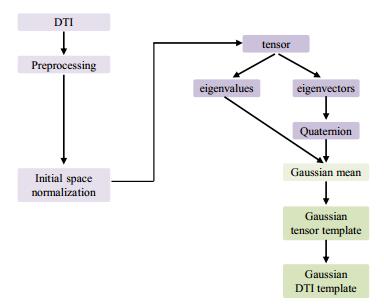
在获取被试的张量数据后通常对其进行多通道线性平均以得到张量模板.但线性平均不仅会忽略张量中的向量信息,还会使灰质和白质的交界处过于平滑,降低模板的分辨率.为了解决以上问题,本文引入了四元数及高斯加权平均来构建高斯扩散张量成像(Diffusion Tensor Imaging,DTI)脑模板.本文首先对55个健康被试的DTI数据进行预处理,使得数据伪影最小化;再通过扩散张量成像工具包(Diffusion Tensor Imaging ToolKit,DTI-TK)将预处理后的数据进行初步空间标准化;然后将张量通过特征分解得到特征向量和特征值;最后,将由特征向量转化的四元数标量和特征值分别进行高斯加权平均得到平均后的特征向量和特征值,并对其进行重建得到张量模板.实验结果表明相比于线性DTI模板,高斯DTI模板在DTED、COH、DVED、OVL、corrFA评估指标上表现更优,而IA指标较差,说明本文提出的高斯DTI模板在整体信息保留方面有所优化,但方向信息有所丢失.
邓岚 , 王远军 . 基于高斯平均的DTI脑模板构建方法[J]. 波谱学杂志, 2022 , 39(4) : 413 -427 . DOI: 10.11938/cjmr20212957
The tensor data of subjects are usually averaged linearly over multiple channels to obtain the tensor template. However, linear averaging ignores the vector information in the tensor. Additionally, it will render the interface between the gray matter and white matter too smooth, resulting in resolution reduction. To address the above problems, this paper introduced quaternion and Gaussian weighted average to construct a Gaussian diffusion tensor imaging (DTI) brain template. First, the DTI data of 55 healthy subjects were preprocessed to minimize data artifacts. The obtained data were then subjected to preliminary spatial standardization. Then, the tensor was decomposed to acquire eigenvectors and eigenvalues. Finally, the eigenvalues and quaternion converted from the eigenvectors were followed by Gaussian weighted average to gain the averaged eigenvectors and eigenvalues. The tensor template was obtained by reconstructing the averaged eigenvectors and eigenvalues. The experimental results show that compared with the linear DTI template, the Gaussian DTI template performs better on the DTED, COH, DVED, OVL, and corrFA evaluation indicators but poorer on the IA indicator. The Gaussian DTI template proposed in this paper has certain advantage on the overall information retention, but is to be further improved on the orientation information.
| 1 | BASSER P J, MATTIELLO J, LEBIHAN D Estimation of the effective self-diffusion tensor from the NMR spin echo[J]. J Magn Reson, Series B, 1994, 103 (3): 247- 254. |
| 2 | MOSELEY M E, COHEN Y, KUCHARCZYK J, et al Diffusion-weighted MR imaging of anisotropic water diffusion in cat central nervous system[J]. Radiology, 1990, 176 (2): 439- 445. |
| 3 | LE B D, MANGIN J F, POUPON C, et al Diffusion tensor imaging: concepts and applications[J]. J Magn Reson Imaging, 2010, 13 (4): 534- 546. |
| 4 | VIRTA A, BARNETT A, PIERPAOLI C Visualizing and characterizing white matter fiber structure and architecture in the human pyramidal tract using diffusion tensor MRI[J]. Magn Reson Imaging, 1999, 17 (8): 1121- 1133. |
| 5 | ERIKSSON S H Diffusion tensor imaging in patients with epilepsy and malformations of cortical development[J]. Brain, 2001, 124 (3): 617- 626. |
| 6 | SZESZKO P R, ARDEKANI B A, ASHTARI M, et al White matter abnormalities in first-episode schizophrenia or schizoaffective disorder: a diffusion tensor imaging study[J]. Am J Psychiatry, 2005, 162 (3): 602- 605. |
| 7 | BRUNO S, CERCIGNANI M, RON M A White matter abnormalities in bipolar disorder: a voxel-based diffusion tensor imaging study[J]. Bipolar Disorders, 2008, 10 (4): 460- 468. |
| 8 | OZSUNAR Y, GRANT P E, HUISMAN T A, et al Evolution of water diffusion and anisotropy in hyperacute stroke: significant correlation between fractional anisotropy and T2[J]. AJNR Am J Neuroradiol, 2004, 25 (5): 699- 705. |
| 9 | ARFANAKIS K, GUI M Z, TAMHANE A A, et al Investigating the medial temporal lobe in Alzheimer's disease and mild cognitive impairment, with Turboprop diffusion tensor imaging, MRI-volumetry, and T2-relaxometry[J]. Brain Imaging & Behavior, 2007, 1 (1): 11- 21. |
| 10 | ARFANAKIS K, HAUGHTON V M, CAREW J D, et al Diffusion tensor MR imaging in diffuse axonal injury[J]. AJNR Am J Neuroradiol, 2002, 23 (5): 794- 802. |
| 11 | FOONG J, Maier M, Clark C A, et al Neuropathological abnormalities of the corpus callosum in schizophrenia: a diffusion tensor imaging study[J]. J Neurol Neurosurg Psychiatry, 2000, 68 (2): 242- 244. |
| 12 | RUGG-GUNN F J Diffusion tensor imaging of cryptogenic and acquired partial epilepsies[J]. Brain, 2001, 124 (3): 627- 636. |
| 13 | GLAUCHE V, SACH M, KOCH M, et al Morphometry on diffusion tensor data[J]. Neuroimage, 2001, 13 (6): 128. |
| 14 | WANG Y J, JIANG F, LIU Y Reference-free brain template construction with population symmetric registration[J]. Med Biol Eng Comput, 2020, 58 (9): 2083- 2093. |
| 15 | 蒋帆, 王远军 扩散张量图像的插值方法综述[J]. 波谱学杂志, 2019, 36 (3): 393- 407. |
| 15 | JIANG F, WANG Y J A review on interpolation methods for diffusion tensor images[J]. Chinese J Magn Reson, 2019, 36 (3): 393- 407. |
| 16 | 赵尚义, 王远军 基于磁共振图像和改进的UNet++模型区分阿尔茨海默症患者和健康人群[J]. 波谱学杂志, 2020, 37 (3): 322- 331. |
| 16 | ZHAO S Y, WANG Y J Classification of Alzheimer's disease patients based on magnetic resonance images and an improved UNet++ model[J]. Chinese J Magn Reson, 2020, 37 (3): 322- 331. |
| 17 | WANG Y J, JIANG F, LIU Y Spectrum-sine interpolation framework for DTI processing[J]. Med Biol Eng Comput, 2022, 60 (1): 279- 295. |
| 18 | 蒋帆, 王远军 扩散张量成像的人脑模板构建[J]. 波谱学杂志, 2018, 35 (4): 520- 530. |
| 18 | JIANG F, WANG Y J Construction of human brain templates with diffusion tensor imaging data: a review[J]. Chinese J Magn Reson, 2018, 35 (4): 520- 530. |
| 19 | JONES D K, GRIFFIN L D, ALEXANDER D C, et al Spatial normalization and averaging of diffusion tensor MRI data sets[J]. Neuroimage, 2002, 17 (2): 592- 617. |
| 20 | PARK H J, KUBICKI M, SHENTON M E, et al Spatial normalization of diffusion tensor MRI using multiple channels[J]. Neuroimage, 2003, 20 (4): 1995- 2009. |
| 21 | ZHANG S, CAREW J D, ARFANAKIS K. Variability of diffusion tensor characteristics in human brain templates: effect of the number of subjects used for the development of the templates[C]//ISMRM Annual Meeting Proceedings, 2010: 1637. |
| 22 | MULLER H, UNRATH A A, KASSUBEK J Preservation of diffusion tensor properties during spatial normalization by use of tensor imaging and fibre tracking on a normal brain database[J]. Phys Med Biol, 2007, 52 (6): 99- 109. |
| 23 | MORI S, OISHI K, JIANG H, et al Stereotaxic white matter atlas based on diffusion tensor imaging in an ICBM template[J]. Neuroimage, 2008, 40 (2): 570- 582. |
| 24 | PENG H L, ORLICHENKO A, DAWE R J, et al Development of a human brain diffusion tensor template[J]. Neuroimage, 2009, 46 (4): 967- 980. |
| 25 | ZHANG S W, PENG H L, DAWE R J, et al Enhanced ICBM diffusion tensor template of the human brain[J]. Neuroimage, 2011, 54 (2): 974- 984. |
| 26 | VARENTSOVA A, ZHANG S, ARFANAKIS K Development of a high angular resolution diffusion imaging human brain template[J]. Neuroimage, 2014, 91, 177- 186. |
| 27 | JAHANSHAD N, KOCHUNOV P V, SPROOTEN E, et al Multi-site genetic analysis of diffusion images and voxelwise heritability analysis: A pilot project of the ENIGMA-DTI working group[J]. Neuroimage, 2013, 81 (1): 455- 469. |
| 28 | KARA?ALI B, DAVATZIKOS C Estimating topology preserving and smooth displacement fields[J]. IEEE Trans Med Imaging, 2004, 23 (7): 868- 880. |
/
| 〈 |
|
〉 |