

基于MRI影像组学的BI-RADS 3-5类乳腺病变三分类
收稿日期: 2022-01-18
网络出版日期: 2022-04-04
基金资助
上海市自然科学基金资助项目(18ZR1426900);国家自然科学基金资助项目(81971664)
Classification of BI-RADS 3-5 Breast Lesions Based on MRI Radiomics
Received date: 2022-01-18
Online published: 2022-04-04
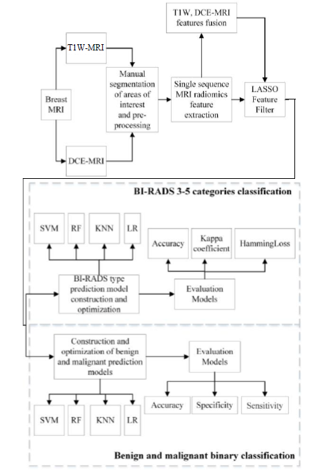
医生根据磁共振影像征象对患者的乳腺病变程度进行BI-RADS分类评估时存在一定的主观性,且 BI-RADS 3-5类病变的良恶性存在交叉,在临床诊断时极易发生因诊断类别较高而造成不必要的有创治疗.针对这些问题,本文应用影像组学技术对乳腺的T1加权(T1W)和动态对比增强(DCE)磁共振图像进行特征提取和融合,采用最小绝对收缩和选择算子(LASSO)算法筛选出各特征集的最优特征集,并分别使用支持向量机(SVM)、随机森林(RF)、K最近邻(KNN)及逻辑回归(LR)算法进行BI-RADS 3-5类乳腺病变三分类,并且在此基础上实现乳腺良恶性分类.结果显示基于特征融合的四个影像组学模型对乳腺病变BI-RADS 3-5类的分类准确率分别为81.25%、87.50%、78.38%、81.25%;对乳腺病变良恶性鉴别的准确率分别为90.91%、93.55%、92.73%、94.55%. 这表明MRI影像组学结合机器学习的算法对乳腺病变BI-RADS分类效果及良恶性鉴别效果均较好,且特征融合可进一步提高分类预测的准确率.
韩冰 , 徐晶 , 王远军 , 王中领 . 基于MRI影像组学的BI-RADS 3-5类乳腺病变三分类[J]. 波谱学杂志, 2023 , 40(1) : 52 -67 . DOI: 10.11938/cjmr20222971
The classification of the breast imaging-reporting and data system (BI-RADS) based on magnetic resonance imaging (MRI) refers to the classification of the degree of lesions according to the image signs of lesions, which is usually subjective. Moreover, the benign and malignant lesions of BI-RADS 3-5 are overlapping, which is prone to unnecessary invasive treatment due to high diagnostic categories in clinical diagnosis. To address these problems, this research applied radiomics for feature extraction and fusion of T1-weighted (T1W) and dynamic contrast-enhanced (DEC) MRI. The least absolute shrinkage and selection operator (LASSO) algorithm was used to screen out the optimal feature collection of each type of MR image. Support vector machine (SVM), random forest (RF), K-nearest neighbour (KNN) and logistic regression (LR) algorithms were applied for BI-RADS 3-5 classification, based on which the benign and malignant lesions were further classified. The results showed that the classification accuracy of breast BI-RADS 3-5 by four radiomics models based on feature fusion was 81.25%, 87.50%, 78.38%, and 81.25%, respectively. Their accuracy in distinguishing the benign and malignant breast lesions was 90.91%, 93.55%, 92.73%, and 94.55%, respectively. This indicates that the combination of radiomics and machine learning correlation algorithm has a good effect on breast MRI BI-RADS classification and benign and malignant differentiation, and feature fusion can further improve the accuracy of classification prediction.
Key words: magnetic resonance imaging; breast lesions; radiomics; BI-RADS; feature fusion; classification
| [1] | SIEGEL R L, MILLER K D, FUCHS H E, et al. Cancer statistics, 2021[J]. CA: A Cancer J Clin, 2021, 71(1): 7-33. |
| [2] | FENG R M, ZONG Y N, CAO S M, et al. Current cancer situation in China: good or bad news from the 2018 global cancer statistics?[J]. Cancer Commun, 2019, 39(1): 22-33. |
| [3] | VALDORA F, HOUSSAMI N, ROSSI F, et al. Rapid review: radiomics and breast cancer[J]. Breast Cancer Res Treat, 2018, 169(2): 217-229. |
| [4] | MANN R M, CHO N, MOY L. Breast MRI: State of the art[J]. Radiology, 2019, 292(3): 520-536. |
| [5] | WAKANA M, HYUNG W C, JOINES M M, et al. Quantitative predictors of response to neoadjuvant chemotherapy on dynamic contrast-enhanced 3T breast MRI[J]. J Breast Imaging, 2022, 4(2): 168-176. |
| [6] | SHAHAN C L, LAYNE G P. Advances in breast imaging with current screening recommendations and controversies[J]. Obstet Gynecol Clin North Am, 2022, 49(1): 1-33. |
| [7] | LIU Y, CHEN J C, HU X Y, et al. Classification and localization of meningioma and acoustic neuroma in cerebellopontine angle based on mask RCNN[J]. Chinese J Magn Reson, 2021, 38(1): 58-68. |
| [7] | 刘颖, 陈静聪, 胡小洋, 等. 基于Mask RCNN的桥小脑角区脑膜瘤与听神经瘤分类定位研究[J]. 波谱学杂志, 2021, 38(1): 58-68. |
| [8] | WEAVER O, LEUNG J W T. Biomarkers and imaging of breast cancer[J]. AJR Am J Roentgenol, 2018, 210(2): 271-278. |
| [9] | RAWASHDEH M, LEWIS S, ZAITOUN M, et al. Breast lesion shape and margin evaluation: BI-RADS based metrics understate radiologists' actual levels of agreement[J]. Comput Biol Med, 2018, 96(1): 294-298. |
| [10] | SATAKE H, ISHIGAKI S, ITO R, NAGANAWA S. Radiomics in breast MRI: current progress toward clinical application in the era of artificial intelligence[J]. Radiol Med. 2022, 127(1): 39-56. |
| [11] | LEITHNER D, HORVAT J V, OCHOA-ALBIZTEGUI R E, et al. Imaging and the completion of the omics paradigm in breast cancer[J]. Der Radiologe, 2018, 58(1): 7-13. |
| [12] | WANG N, WANG Y J, LIAN P. Prediction of preoperative T staging of rectal cancer based on radiomics[J]. Chinese J Magn Reson, 2022, 39(1): 43-55. |
| [12] | 王楠, 王远军, 廉朋. 基于影像组学的直肠癌术前T分期预测[J]. 波谱学杂志, 2022, 39(1): 43-55. |
| [13] | IMBRIACO M, CUOCOLO R. Does texture analysis of MR images of breast tumors help predict response to treatment?[J]. Radiology, 2018, 286(2): 421-423. |
| [14] | YIN X X, HADJILOUCAS S, ZHANG Y C, et al. MRI radiogenomics for intelligent diagnosis of breast tumors and accurate prediction of neoadjuvant chemotherapy responses — A review[J]. Comput Meth Prog Bio, 2021, 214: 106510. |
| [15] | NARANJO I D, GIBBS P, REINER J S, et al. Radiomics and machine learning with multiparametric breast MRI for improved diagnostic accuracy in breast cancer diagnosis[J]. Diagnostics, 2021, 11(6): 919-932. |
| [16] | PRATIKSHA Y, SURBHI C. Effectivity of combined diffusion-weighted imaging and contrast-enhanced MRI in malignant and benign breast lesions[J]. Pol J Radiol, 2018, 83: e82-e93. |
| [17] | HU Q, WHITNEY H M, GIGER M L. Radiomics methodology for breast cancer diagnosis using multiparametric magnetic resonance imaging[J]. J Med Imaging, 2020, 7(4): 044502. |
| [18] | YE D M, WANG H T, YU T. The application of radiomics in breast MRI: A review[J]. Technol Cancer Res Tr, 2020, 19: 1-16. |
| [19] | PAREKH V S, JACOBS M A. Multiparametric radiomics methods for breast cancer tissue characterization using radiological imaging[J]. Breast Cancer Res Tr, 2020, 180(2): 407-421. |
| [20] | DEMIRCIOGLU A, GRUENEISEN J, INGENWERTH M, et al. A rapid volume of interest-based approach of radiomics analysis of breast MRI for tumor decoding and phenotyping of breast cancer[J]. PLoS ONE, 2020, 15(6): e0234871. |
| [21] | TSAROUCHI M I, VLACHOPOULOS G F, KARAHALIOU A N, et al. Multi-parametric MRI lesion heterogeneity biomarkers for breast cancer diagnosis[J]. Phys Medica, 2020, 80(2): 101-110. |
| [22] | ZHANG Q, PENG Y S, LIU W, et al. Radiomics based on multimodal MRI for the differential diagnosis of benign and malignant breast lesions[J]. J Magn Reson Imaging, 2020, 52(2): 596-607. |
| [23] | HAO W, GONG J, WANG S P, et al. Application of MRI radiomics-based machine learning model to improve contralateral BI-RADS 4 lesion assessment[J]. Front Oncol, 2020. doi: 10.3389/fonc.2020.531476. |
| [24] | QIAO M Y, LI C K, SUO S T, et al. Breast DCE-MRI radiomics: a robust computer-aided system based on reproducible BI-RADS features across the influence of datasets bias and segmentation methods[J]. Int J Comput Ass Rad, 2020, 15(5): 921-930. |
| [25] | SCAPICCHIO C, GABELLONI M, BARUCCI A, et al. A deep look into radiomics[J]. La Radiologia Medica, 2021, 126: 1296-1311. |
| [26] | DOMINGUES I, ABREU P H, SANTOS J. Bi-Rads classification of breast cancer: A new pre-processing pipeline for deep models training[C]// IEEE International Conference of Image Processing (ICIP). IEEE, 2018.1378-1382. |
| [27] | SIDDEEQ S, LI J, BHATTI H M A, et al. Deep learning RN-BCNN model for breast cancer BI-RADS classification[C]. ICIGP 2021: 2021 The 4th International Conference on Image and Graphics Processing. 2021: 219-225. |
| [28] | WU P Q. Research progress of radiomics in lymph node metastasis of breast cancer[J]. Journal of Molecular Imaging, 2020, 43 (1): 31-35. |
| [28] | 吴佩琪. 影像组学在乳腺癌淋巴结转移中的研究进展[J]. 分子影像学杂志, 2020, 43(1): 31-35. |
/
| 〈 |
|
〉 |