

数据采集方案对神经扩散模型影响的评估
收稿日期: 2020-11-05
网络出版日期: 2022-05-28
基金资助
上海健康医学院师资人才百人库;上海健康医学院校级科研基金资助项目;上海高校教师产学研践习计划;国家自然科学基金资助项目(81830052);国家自然科学基金资助项目(61971257);分子影像学重点实验室建设资助项目(18DZ2260400)
Evaluation of the Influence of Data Sampling Schemes on Neural Diffusion Models
Received date: 2020-11-05
Online published: 2022-05-28
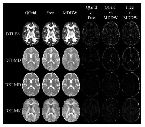
基于单次数据采集的多种扩散模型联合应用已逐渐成为临床研究的热点,本研究比较了三种采集方案对于神经扩散模型定量计算的影响,包括Q空间笛卡尔网格(QGrid)、多壳层异向(Free)和多壳层同向(MDDW)采集方案,涉及的扩散模型包含扩散张量成像(DTI);扩散峰度成像(DKI);神经突方向分散度和密度成像(NODDI);平均表观传播(MAP)模型.结果表明DTI和DKI模型对采集方案相对不敏感,而NODDI和MAP对采集方案和最大b值的设置相对较敏感,并且QGrid和Free方案一致性较高,因此在大样本和多中心研究中需要考虑采集方案的选择.此外,考虑到QGrid和Free方案分别在结合更多扩散模型和神经纤维束成像应用上更具优势,因此推荐使用.
关键词: 磁共振成像; 数据采集方案; 扩散模型; 扩散张量成像(DTI); 扩散峰度成像(DKI); 神经突方向分散度和密度成像(NODDI); 平均表观传播(MAP)
周敏雄 , 张会婷 , 王一达 , 杨光 , 姚旭峰 , 高安康 , 程敬亮 , 白洁 , 严序 . 数据采集方案对神经扩散模型影响的评估[J]. 波谱学杂志, 2022 , 39(2) : 220 -229 . DOI: 10.11938/cjmr20202870
The joint application of multiple diffusion models on single sampled dataset is becoming a hot topic in clinical research. This study investigated the influence of the three data sampling schemes on the quantification of neural diffusion models. The three sampling schemes compared were QGrid, Free and MDDW on the Siemens scanners. The diffusion models involved were diffusion tensor imaging (DTI), diffusion kurtosis imaging (DKI), neurite orientation dispersion and density imaging (NODDI) and mean apparent propagator (MAP) models. It was demonstrated that the results of NODDI and MAP were sensitive to the sampling schemes and the set of maximum b-value, while that of DTI and DKI were comparatively not sensitive to varying configurations. It was also shown that QGrid and Free schemes provided more consistent results. Thus the sampling scheme should be carefully selected in multi-center studies and studies with large sample size. QGrid and Free schemes are recommended for their advantages demonstrated in this study.
| 1 | BASSER P J , MATTIELLO J , LEBIHAN D . MR diffusion tensor spectroscopy and imaging[J]. Biophys J, 1994, 66 (1): 259- 267. |
| 2 | JIANG F , WANG Y J . A review on interpolation methods for diffusion tensor images[J]. Chinese J Magn Reson, 2019, 36 (3): 392- 407. |
| 2 | 蒋帆, 王远军. 扩散张量图像的插值方法综述[J]. 波谱学杂志, 2019, 36 (3): 392- 407. |
| 3 | XU Y H , GAO S C , HAO X F . An improved spectral quaternion interpolation method of diffusion tensor imaging[J]. J Biomed Eng, 2016, 33 (2): 362- 367. |
| 3 | 徐永红, 高上策, 郝小飞. 一种改进的扩散张量成像谱四元数插值方法[J]. 生物医学工程学杂志, 2016, 33 (2): 362- 367. |
| 4 | JENSEN J H , HELPERN J A , RAMANI A , et al. Diffusional kurtosis imaging: the quantification of non-Gaussian water diffusion by means of magnetic resonance imaging[J]. Magn Reson Med, 2005, 53 (6): 1432- 1440. |
| 5 | ZHANG H , SCHNEIDER T , WHEELER-KINGSHOTT C A , et al. NODDI: practical in vivo neurite orientation dispersion and density imaging of the human brain[J]. Neuroimage, 2012, 61 (4): 1000- 1016. |
| 6 | ?ZARSLANE, KOAYC G, SHEPHERDT M, 等. Mean apparent propagator (MAP) MRI: A novel diffusion imaging method for mapping tissue microstructure[J]. Neuroimage, 2013, 78, 16- 32. |
| 7 | FICK R, DAIANU M, PIZZOLATO M, et al. Comparison of biomarkers in transgenic alzheimer rats using multi-shell diffusion MRI[C] //MICCAI, 2017: 187-199. |
| 8 | PINES A R , CIESLAK M , LARSEN B , et al. Leveraging multi-shell diffusion for studies of brain development in youth and young adulthood[J]. Dev Cogn Neurosci, 2020, 43, 100788. |
| 9 | MA K R , ZHANG X N , ZHANG H T , et al. Mean apparent propagator-MRI: A new diffusion model which improves temporal lobe epilepsy lateralization[J]. Eur J Radiol, 2020, 126, 108914. |
| 10 | LE H B , ZENG W K , ZHANG H H , et al. Mean apparent propagator MRI is better than conventional diffusion tensor imaging for the Evaluation of Parkinson's disease: a prospective pilot study[J]. Front Aging Neurosci, 2020, 12, 563595. |
| 11 | WEDEEN V J , HAGMANN P , TSENG W Y I , et al. Mapping complex tissue architecture with diffusion spectrum magnetic resonance imaging[J]. Magn Reson Med, 2005, 54 (6): 1377- 1386. |
| 12 | CARUYER E , LENGLET C , SAPIRO G , et al. Design of multishell sampling schemes with uniform coverage in diffusion MRI[J]. Magn Reson Med, 2013, 69 (6): 1534- 1540. |
| 13 | JONES D K , HORSFIELD M A , SIMMONS A . Optimal strategies for measuring diffusion in anisotropic systems by magnetic resonance imaging[J]. Magn Reson Med, 1999, 42 (3): 515- 525. |
| 14 | PAPADAKIS N G , MURRILLS C D , HALL L D , et al. Minimal gradient encoding for robust estimation of diffusion anisotropy[J]. Magn Reson Imaging, 2000, 18 (6): 671- 679. |
| 15 | TABESH A , JENSEN J H , ARDEKANI B A , et al. Estimation of tensors and tensor-derived measures in diffusional kurtosis imaging[J]. Magn Reson Med, 2011, 65 (3): 823- 836. |
| 16 | WU Y C , ALEXANDER A L . Hybrid diffusion imaging[J]. Neuroimage, 2007, 36 (3): 617- 629. |
| 17 | HUTCHINSON E B , AVRAM A V , IRFANOGLU M O , et al. Analysis of the effects of noise, DWI sampling, and value of assumed parameters in diffusion MRI models[J]. Magn Reson Med, 2017, 78 (5): 1767- 1780. |
| 18 | ANDICA C , KAMAGATA K , HATANO T , et al. MR biomarkers of degenerative brain disorders derived from diffusion imaging[J]. J Magn Reson Imaging, 2020, 52 (6): 1620- 1636. |
| 19 | FIGINI M , RIVA M , GRAHAM M , et al. Prediction of isocitrate dehydrogenase genotype in brain gliomas with MRI: Single-shell versus multishell diffusion models[J]. Radiology, 2018, 289 (3): 788- 796. |
| 20 | SOTIROPOULOS S N , JBABDI S , XU J Q , et al. Advances in diffusion MRI acquisition and processing in the Human Connectome Project[J]. Neuroimage, 2013, 80, 125- 143. |
| 21 | JENKINSON M , BECKMANN C F , BEHRENS T E , et al. FSL[J]. Neuroimage, 2012, 62 (2): 782- 790. |
| 22 | XIE S M , CHEN L F , ZUO N M , et al. DiffusionKit: A light one-stop solution for diffusion MRI data analysis[J]. J Neurosci Methods, 2016, 273, 107- 119. |
| 23 | GARYFALLIDIS E , BRETT M , AMIRBEKIAN B , et al. Dipy, a library for the analysis of diffusion MRI data[J]. Front Neuroinform, 2014, 8, 8. |
| 24 | DADUCCI A , CANALES-RODRíGUEZ E J , ZHANG H , et al. Accelerated microstructure imaging via convex optimization (AMICO) from diffusion MRI data[J]. Neuroimage, 2015, 105, 32- 44. |
| 25 | KLEIN S , STARING M , MURPHY K , et al. elastix: a toolbox for intensity-based medical image registration[J]. IEEE Trans Med Imaging, 2010, 29 (1): 196- 205. |
| 26 | LE BIHAN D , TURNER R . Intravoxel incoherent motion imaging using spin echoes[J]. Magn Reson Med, 1991, 19 (2): 221- 227. |
| 27 | BENNETT K M , SCHMAINDA K M , BENNETT R T , et al. Characterization of continuously distributed cortical water diffusion rates with a stretched-exponential model[J]. Magn Reson Med, 2003, 50 (4): 727- 734. |
| 28 | WANG Y , WANG Q , HALDAR J P , et al. Quantification of increased cellularity during inflammatory demyelination[J]. Brain, 2011, 134 (Pt 12): 3590- 3601. |
| 29 | PARK J E , KIM H S , PARK S Y , et al. Prediction of core signaling pathway by using diffusionand perfusion-based MRI radiomics and next-generation sequencing in isocitrate dehydrogenase wild-type glioblastoma[J]. Radiology, 2020, 294 (2): 388- 397. |
/
| 〈 |
|
〉 |