

实验性脊髓损伤后大脑血管可塑性的磁共振血管尺寸成像研究
收稿日期: 2022-11-22
网络出版日期: 2023-01-29
基金资助
国家自然科学基金资助项目(U1932158);国家自然科学基金资助项目(81871085);国家自然科学基金资助项目(82271519);山东省自然科学基金肿瘤防治联合基金重点项目(ZR2019LZL018);安徽省自然科学基金杰出青年项目(2208085J10);中国科学院合肥大科学中心协同创新培育基金重要项目(2019HSC-CIP003);中国博士后科学基金项目(2019M652403);山东省博士后创新项目(202002048)
In vivo MR Vessel Size Imaging of Brain Vascular Plasticity After Experimental Spinal Cord Injury
Received date: 2022-11-22
Online published: 2023-01-29
田雨 , 周臣 , 张亚男 , 王鹏 , 张彩云 , 宋天玮 , 钱俊超 . 实验性脊髓损伤后大脑血管可塑性的磁共振血管尺寸成像研究[J]. 波谱学杂志, 2023 , 40(2) : 158 -168 . DOI: 10.11938/cjmr20223036
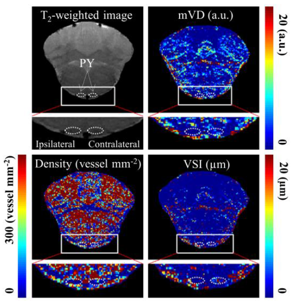
In vivo magnetic resonance vessel size imaging at 7 T high field was used to explore the changes in cerebral vascular plasticity after spinal cord hemisection injury in rats. Region of interest (ROI) analysis showed that four weeks after injury, mean vessel diameter (mVD), microvascular density (Density) and vessel size index (VSI) values were significantly increased in the contralateral side compared with the ipsilateral pyramid of the injury site, suggesting angiogenesis or vascular activation in the white matter region of the contralateral corticospinal tract (CST). Immunofluorescence results showed that the intensity of staining for platelet endothelial cell adhesion molecule 1 (CD31) and glial fibrillary acidic protein (GFAP) also increased significantly in the contralateral pyramid four weeks after injury. These results suggested that magnetic resonance vessel size imaging could provide valuable information on neovascularization in brain after spinal cord injury and may be a novel tool to diagnose brain vascular pathologies in spinal cord injury patients.
| [1] | WRIGLEY P J, GUSTIN S M, MACEY P M, et al. Anatomical changes in human motor cortex and motor pathways following complete thoracic spinal cord injury[J]. Cereb Cortex, 2009, 19(1): 224-232. |
| [2] | ILVESM?KI T, KOSKINEN E, BRANDER A, et al. Spinal cord injury induces widespread chronic changes in cerebral white matter[J]. Hum Brain Mapp, 2017, 38(7): 3637-3647. |
| [3] | HE G, QIAN J. Phase imaging of axonal integrity of cranial corticospinal tract in experimental spinal cord injury at 9.4T[J]. Microsc Res Tech, 2017, 80(9): 1009-1017. |
| [4] | COHEN-ADAD J, EL MENDILI M M, LEHéRICY S, et al. Demyelination and degeneration in the injured human spinal cord detected with diffusion and magnetization transfer MRI[J]. Neuroimage, 2011, 55(3): 1024-1033. |
| [5] | MOXON K A, OLIVIERO A, AGUILAR J, et al. Cortical reorganization after spinal cord injury: always for good?[J]. Neuroscience, 2014, 283: 78-94. |
| [6] | JURKIEWICZ M T, MIKULIS D J, MCILROY W E, et al. Sensorimotor cortical plasticity during recovery following spinal cord injury: a longitudinal fMRI study[J]. Neurorehabil Neural Repair, 2007, 21(6): 527-538. |
| [7] | FASSETT H J, TURCO C V, EL-SAYES J, et al. Alterations in Motor Cortical representation of muscles following incomplete spinal cord injury in humans[J]. Brain Sci, 2018, 8(12). |
| [8] | WANG L, CHEN N. MRI research progresses of motor imagery on brain activity and network reorganization in patients with spinal cord injury[J]. Chin J Med Imaging Technol, 2019, 35(10):1586-1589. |
| [8] | 王玲, 陈楠. 运动想象对脊髓损伤患者大脑活动和脑网络重塑MR研究进展[J]. 中国医学影像技术, 2019, 35(10): 1586-1589. |
| [9] | TSUJIOKA H, YAMASHITA T. Neural circuit repair after central nervous system injury[J]. Int Immunol, 2021, 33(6): 301-309. |
| [10] | LIU J, YANG X, JIANG L, et al. Neural plasticity after spinal cord injury[J]. Neural Regen Res, 2012, 7(5): 386-391. |
| [11] | YU S, YAO S, WEN Y, et al. Angiogenic microspheres promote neural regeneration and motor function recovery after spinal cord injury in rats[J]. Sci Rep, 2016, 6: 33428. |
| [12] | POTENTE M, GERHARDT H, CARMELIET P. Basic and therapeutic aspects of angiogenesis[J]. Cell, 2011, 146(6): 873-887. |
| [13] | LI X, LI M, TIAN L, et al. Reactive astrogliosis: implications in spinal cord injury progression and therapy[J]. Oxid Med Cell Longev, 2020, 2020: 9494352. |
| [14] | SHIMAMURA M, SATO N, SATA M, et al. Expression of hepatocyte growth factor and c-Met after spinal cord injury in rats[J]. Brain Res, 2007, 1151: 188-194. |
| [15] | NAKAMURA T, MIZUNO S. The discovery of hepatocyte growth factor (HGF) and its significance for cell biology, life sciences and clinical medicine[J]. Proc Jpn Acad Ser B Phys Biol Sci, 2010, 86(6): 588-610. |
| [16] | KITAMURA K, IWANAMI A, NAKAMURA M, et al. Hepatocyte growth factor promotes endogenous repair and functional recovery after spinal cord injury[J]. J Neurosci Res, 2007, 85(11): 2332-2342. |
| [17] | CHRISTOFORIDIS G A, YANG M, KONTZIALIS M S, et al. High resolution ultra high field magnetic resonance imaging of glioma microvascularity and hypoxia using ultra-small particles of iron oxide[J]. Invest Radiol, 2009, 44(7): 375-383. |
| [18] | XU C, SCHMIDT W U, VILLRINGER K, et al. Vessel size imaging reveals pathological changes of microvessel density and size in acute ischemia[J]. J Cereb Blood Flow Metab, 2011, 31(8): 1687-1695. |
| [19] | XU X, MENG T, WEN Q, et al. Dynamic changes in vascular size and density in transgenic mice with Alzheimer's disease[J]. Aging (Albany NY), 2020, 12(17): 17224-17234. |
| [20] | JIANG J J, ZHAO L Y, WANG C Y, et al. Initial application of magnetic resonance vessel size imaging in cerebral glioma and meningioma[J]. Radiol Practic, 2014, 29(7): 770-773. |
| [20] | 江晶晶, 赵凌云, 王承缘, 等. 磁共振血管大小成像在脑胶质瘤和脑膜瘤中的初步应用[J]. 放射学实践, 2014, 29(7): 770-773. |
| [21] | IELACQUA G D, SCHLEGEL F, FüCHTEMEIER M, et al. Magnetic resonance Q mapping reveals a decrease in microvessel density in the arcAβ mouse model of cerebral amyloidosis[J]. Front Aging Neurosci, 2015, 7: 241. |
| [22] | LEMASSON B, VALABLE S, FARION R, et al. In vivo imaging of vessel diameter, size, and density: a comparative study between MRI and histology[J]. Magn Reson Med, 2013, 69(1): 18-26. |
| [23] | HOL E M, PEKNY M. Glial fibrillary acidic protein (GFAP) and the astrocyte intermediate filament system in diseases of the central nervous system[J]. Curr Opin Cell Biol, 2015, 32: 121-130. |
| [24] | RAZAVI S M, YAHYAABADI R. Comparative study of correlation between angiogenesis markers (CD31) and Ki67 marker with behavior of aggressive and nonaggressive central giant cell granuloma with immunohistochemistry technique[J]. Asian Pac J Cancer Prev, 2018, 19(8): 2279-2283. |
| [25] | RAMU J, BOCKHORST K H, MOGATADAKALA K V, et al. Functional magnetic resonance imaging in rodents: Methodology and application to spinal cord injury[J]. J Neurosci Res, 2006, 84(6): 1235-1244. |
| [26] | SHEN Y M, ZHENG W L, CHENG Y-C N, et al. USPIO high resolution neurovascular imaging in a rat stroke model of transient middle cerebral artery occlusion[J]. Chinese J Magn Reson, 2014, 31(1): 20-31. |
| [26] | 沈伊民, 郑伟丽, CHENG Y-C N, 等. 大鼠中风模型的超小氧化铁粒子神经血管成像[J]. 波谱学杂志, 2014, 31(1): 20-31. |
| [27] | JIRJIS M B, VEDANTAM A, BUDDE M D, et al. Severity of spinal cord injury influences diffusion tensor imaging of the brain[J]. J Magn Reson Imaging, 2016, 43(1): 63-74. |
| [28] | LIDDELOW S A, BARRES B A. Reactive astrocytes: production, function, and therapeutic potential[J]. Immunity, 2017, 46(6): 957-967. |
| [29] | SOFRONIEW M V. Reactive astrocytes in neural repair and protection[J]. Neuroscientist, 2005, 11(5): 400-407. |
| [30] | OKADA S, HARA M, KOBAYAKAWA K, et al. Astrocyte reactivity and astrogliosis after spinal cord injury[J]. Neurosci Res, 2018, 126: 39-43. |
| [31] | HUSSEIN R K, MENCIO C P, KATAGIRI Y, et al. Role of chondroitin sulfation following spinal cord injury[J]. Front Cell Neurosci, 2020, 14: 208. |
| [32] | WANG Y, CHENG X, HE Q, et al. Astrocytes from the contused spinal cord inhibit oligodendrocyte differentiation of adult oligodendrocyte precursor cells by increasing the expression of bone morphogenetic proteins[J]. J Neurosci, 2011, 31(16): 6053-6058. |
| [33] | YIU G, HE Z. Glial inhibition of CNS axon regeneration[J]. Nat Rev Neurosci, 2006, 7(8): 617-627. |
| [34] | YANG T, DAI Y, CHEN G, et al. Dissecting the dual role of the glial scar and scar-forming astrocytes in spinal cord injury[J]. Front Cell Neurosci, 2020, 14: 78. |
| [35] | OKADA S, NAKAMURA M, KATOH H, et al. Conditional ablation of Stat3 or Socs3 discloses a dual role for reactive astrocytes after spinal cord injury[J]. Nat Med, 2006, 12(7): 829-834. |
| [36] | SOFRONIEW M V. Astrocyte barriers to neurotoxic inflammation[J]. Nat Rev Neurosci, 2015, 16(5): 249-263. |
| [37] | LIU D, XU G Y, PAN E, et al. Neurotoxicity of glutamate at the concentration released upon spinal cord injury[J]. Neuroscience, 1999, 93(4): 1383-1389. |
| [38] | MARAGAKIS N J, DYKES-HOBERG M, ROTHSTEIN J D. Altered expression of the glutamate transporter EAAT2b in neurological disease[J]. Ann Neurol, 2004, 55(4): 469-477. |
| [39] | CASELLA G T, MARCILLO A, BUNGE M B, et al. New vascular tissue rapidly replaces neural parenchyma and vessels destroyed by a contusion injury to the rat spinal cord[J]. Exp Neurol, 2002, 173(1): 63-76. |
| [40] | YAMANE K, MISAWA H, TAKIGAWA T, et al. Multipotent neurotrophic effects of hepatocyte growth factor in spinal cord injury[J]. Int J Mol Sci, 2019, 20(23) : 6078. |
| [41] | MENEGHINI V, PEVIANI M, LUCIANI M, et al. Delivery platforms for CRISPR/Cas9 genome editing of glial cells in the central nervous system[J]. Front Genome Ed, 2021, 3: 644319. |
| [42] | LIBERTO C M, ALBRECHT P J, HERX L M, et al. Pro-regenerative properties of cytokine-activated astrocytes[J]. J Neurochem, 2004, 89(5): 1092-1100. |
| [43] | COLLOMBET J M, FOUR E, FAUQUETTE W, et al. Soman poisoning induces delayed astrogliotic scar and angiogenesis in damaged mouse brain areas[J]. Neurotoxicology, 2007, 28(1): 38-48. |
| [44] | BARTANUSZ V, JEZOVA D, ALAJAJIAN B, et al. The blood-spinal cord barrier: morphology and clinical implications[J]. Ann Neurol, 2011, 70(2): 194-206. |
| [45] | WHETSTONE W D, HSU J Y, EISENBERG M, et al. Blood-spinal cord barrier after spinal cord injury: relation to revascularization and wound healing[J]. J Neurosci Res, 2003, 74(2): 227-239. |
| [46] | WAHIS J, HENNES M, ARCKENS L, et al. Star power: the emerging role of astrocytes as neuronal partners during cortical plasticity[J]. Curr Opin Neurobiol, 2021, 67: 174-182. |
| [47] | CHUNG W S, ALLEN N J, EROGLU C. Astrocytes control synapse formation, function, and elimination[J]. Cold Spring Harb Perspect Biol, 2015, 7(9): a020370. |
| [48] | WANG Y, FU A K Y, IP N Y. Instructive roles of astrocytes in hippocampal synaptic plasticity: neuronal activity-dependent regulatory mechanisms[J]. Febs J, 2022, 289(8): 2202-2218. |
/
| 〈 |
|
〉 |