

基于自监督网络的肝脏磁共振R2*参数图像重建
Magnetic Resonance R2* Parameter Mapping of Liver Based on Self-supervised Deep Neural Network
Received date: 2023-01-09
Online published: 2023-03-02
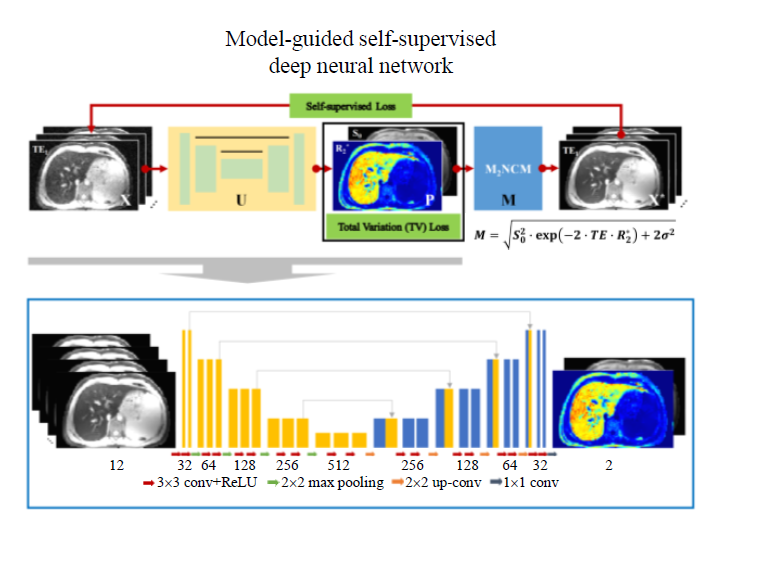
磁共振等效横向弛豫率($R_{2}^{*}$)参数量化技术已经被广泛应用于肝脏铁含量的定量测量中. 然而铁沉积肝脏$R_{2}^{*}$参数图像的重建通常会受到噪声的严重影响. 随着深度学习的兴起,深度学习网络成为磁共振参数图像重建的重要方法. 本文提出了一种模型引导的自监督深度学习网络用于铁沉积肝脏的磁共振$R_{2}^{*}$参数图像重建. 通过利用一种融合噪声校正物理模型和改进的全变分模型的损失函数来引导深度学习网络的自监督训练. 网络的训练不需要使用真实的$R_{2}^{*}$参数图像. 同时,相较于传统的参数估计算法,本文提出的方法能够快速准确地重建出铁沉积肝脏$R_{2}^{*}$参数图像,较好地抑制图像中噪声的影响, 校正噪声引起的偏差,同时保持$R_{2}^{*}$参数图像中的结构细节.
陆琪琪 , 连梓锋 , 李嘉龙 , 斯文彬 , 麦兆华 , 冯衍秋 . 基于自监督网络的肝脏磁共振R2*参数图像重建[J]. 波谱学杂志, 2023 , 40(3) : 258 -269 . DOI: 10.11938/cjmr20233050
Magnetic resonance (MR) effective transverse relaxation rate ($R_{2}^{*}$) technique has been widely applied for assessing hepatic iron concentration. However,$R_{2}^{*}$ mapping of iron-loaded liver can be severely degraded by noise. With the development of deep learning, deep neural networks have become effective tools for MR parameter mapping. In this study, a model-guided self-supervised deep neural network was designed for MR $R_{2}^{*}$ parameter mapping of iron-loaded liver. A novel loss function that integrated a noise-corrected physical model and an improved total variation model was used to train the network, which did not require reference $R_{2}^{*}$ parameter maps. Meanwhile, compared to the conventional parameter fitting methods, model-guided self-supervised deep learning method enabled accurate and efficient $R_{2}^{*}$ mapping of iron-loaded liver, suppressed the effect of noise, corrected the bias introduced by noise, and preserved the detailed structure of $R_{2}^{*}$ map.
| [1] | LABRANCHE R, GILBERT G, CERNY M, et al. Liver iron quantification with MR imaging: A primer for radiologists[J]. Radiographics, 2018, 38(2): 392-412. |
| [2] | WOOD J C, ENRIQUEZ C, GHUGRE N, et al. MRI R2 and R2* mapping accurately estimates hepatic iron concentration in transfusion-dependent thalassemia and sickle cell disease patients[J]. Blood, 2005, 106(4): 1460-1465. |
| [3] | HUANG J W, CHENG Z L, YANG Q H, et al. MRI-T2* technique in quantitative analysis of myocardium, liver and pancreas iron deposition in β-thalassemia major and the correlations with glucose metabolism[J]. Chin J Med Imaging Technol, 2021, 37(4): 557-561. |
| [3] | 黄静文, 程子亮, 杨绮华, 等. MRI-T2*技术定量分析β-重型地中海贫血心脏、肝脏、胰腺铁沉积及其与糖代谢的相关性[J]. 中国医学影像技术, 2021, 37(4): 557-561. |
| [4] | LU H M, ZHU J, WANG F, et al. Study on R2* combined with T1-mapping to evaluate iron overload in liver[J]. J Med Imaging, 2022, 32(8): 1036-1039. |
| [4] | 卢慧敏, 朱娟, 汪飞, 等. 磁共振R2*联合T1-mapping对肝脏铁过载评估的研究[J]. 医学影像学杂志, 2022, 32(8): 1036-1039. |
| [5] | MELONI A, ZMYEWSKI H, RIENHOFF H Y, et al. Fast approximation to pixelwise relaxivity maps: Validation in iron overloaded subjects[J]. Magn Reson Imaging, 2013, 31(7): 1074-1080. |
| [6] | CONSTANTINIDES C D, ATALAR E, MCVEIGH E R. Signal-to-noise measurements in magnitude images from NMR phased arrays[J]. Magn Reson Med, 1997, 38(5): 852-857. |
| [7] | FENG Y, HE T, GATEHOUSE P D, et al. Improved MRI R2* relaxometry of iron-loaded liver with noise correction[J]. Magn Reson Med, 2013, 70(6): 1765-1774. |
| [8] | WANG C, ZHANG X, LIU X, et al. Improved liver R2* mapping by pixel-wise curve fitting with adaptive neighborhood regularization[J]. Magn Reson Med, 2018, 80(2): 792-801. |
| [9] | FENG L, MA D, LIU F. Rapid MR relaxometry using deep learning: An overview of current techniques and emerging trends[J]. NMR Biomed, 2022, 35(4): e4416. |
| [10] | RONNEBERGER O, FISCHER P, BROX T. U-net: Convolutional networks for biomedical image segmentation[C]// Lect Notes Comput Sci (including Subser Lect Notes Artif. Intell Lect. Notes Bioinformatics), vol. 9351, Springer, Cham, 2015: 234-241. |
| [11] | LIU F, FENG L, KIJOWSKI R. MANTIS: Model-augmented neural network with incoherent k-space sampling for efficient MR parameter mapping[J]. Magn Reson Med, 2019, 82(1): 174-188. |
| [12] | LIU F, KIJOWSKI R, EL FAKHRI G, et al. Magnetic resonance parameter mapping using model-guided self-supervised deep learning[J]. Magn Reson Med, 2021, 85(6): 3211-3226. |
| [13] | GETREUER P. Rudin-Osher-Fatemi total variation denoising using split bregman[J]. Image Process Line, 2012, 2: 74-95. |
| [14] | SHI B L, ZHOU Y M, PANG Z F. Image denoising via anisotropic total-variation-based method[J]. J Nantong Univ, Nat Sci Ed, 2019, 18(4): 24-33. |
| [14] | 史宝丽, 周亚美, 庞志峰. 各向异性全变分图像去噪算法[J]. 南通大学学报(自然科学版), 2019, 18(4): 24-33. |
| [15] | LUSTIG M, DONOHO D, PAULY J M. Sparse MRI: The application of compressed sensing for rapid MR imaging[J]. Magn Reson Med, 2007, 58(6): 1182-1195. |
| [16] | LIU J, SUN Y, XU X, et al. Image restoration using total variation regularized deep image prior[C]// IEEE Int Conf Acoust Speech Signal Process, IEEE, 2019: 7715-7719. |
| [17] | STRONG D, CHAN T. Edge-preserving and scale-dependent properties of total variation regularization[J]. Inverse Probl, 2003, 19(6): S165-S187. |
| [18] | ZHU W. A first-order image restoration model that promotes image contrast preservation[J]. J Sci Comput, 2021, 88(2): 1-23. |
| [19] | HE K, ZHANG X, REN S, et al. Delving deep into rectifiers: Surpassing human-level performance on ImageNet classification[C]// 2015 IEEE Int Conf Comput Vis, IEEE, 2015: 1026-1034. |
| [20] | SANDINO C M, CHENG J Y, CHEN F, et al. Compressed sensing: From research to clinical practice with deep neural networks: shortening scan times for magnetic resonance imaging[J]. IEEE Signal Process Mag, 2020, 37(1): 117-127. |
| [21] | WANG Z, BOVIK AC, SHEIKH HR, et al. Image quality assessment: From error visibility to structural similarity[J]. IEEE Trans Image Process, 2004, 13(4): 600-612. |
| [22] | VAN DER WALT S, SCH?NBERGER JL, NUNEZ-IGLESIAS J, et al. scikit-image: image processing in Python[J]. Peer J, 2014, 2: e453. |
| [23] | CHENG H T, WANG S S, KE Z W, et al. A deep recursive cascaded convolutional network for parallel MRI[J]. Chinese J Magn Reson, 2019, 36(4): 437-445 |
| [23] | 程慧涛, 王珊珊, 柯子文, 等. 基于深度递归级联卷积神经网络的并行磁共振成像方法[J]. 波谱学杂志, 2019, 36(4): 437-445. |
| [24] | WANG Y S, DENG A Q, MAO J L, et al. Automatic segmentation of knee joint synovial magnetic resonance images based on 3D VNetTrans[J]. Chinese J Magn Reson, 2022, 39(3): 303-315 |
| [24] | 王颖珊, 邓奥琦, 毛瑾玲, 等. 基于3D VNetTrans的膝关节滑膜磁共振图像自动分割[J]. 波谱学杂志, 2022, 39(3): 303-315. |
| [25] | ZHANG T, PAULY J M, LEVESQUE I R. Accelerating parameter mapping with a locally low rank constraint[J]. Magn Reson Med, 2015, 73(2): 655-661. |
| [26] | ZHAO B, LU W, HITCHENS T K, et al. Accelerated MR parameter mapping with low-rank and sparsity constraints[J]. Magn Reson Med, 2015, 74(2): 489-498. |
| [27] | ROMANO Y, ELAD M, MILANFAR P. The little engine that could: Regularization by Denoising (RED)[J]. SIAM J Imaging Sci, 2017, 10(4): 1804-1844. |
| [28] | LANDMAN B A, BAZIN P L, SMITH S A, et al. Robust estimation of spatially variable noise fields[J]. Magn Reson Med, 2009, 62(2): 500-509. |
| [29] | HENNINGER B, ALUSTIZA J, GARBOWSKI M, et al. Practical guide to quantification of hepatic iron with MRI[J]. Eur Radiol, 2020, 30(1): 383-393. |
/
| 〈 |
|
〉 |