

全脑同时神经黑色素敏感与定量磁化率成像
收稿日期: 2023-03-28
网络出版日期: 2023-05-16
基金资助
华东师范大学“幸福之花”基金资助项目
Simultaneously Neuromelanin-sensitive Imaging and Quantitative Susceptibility Mapping in the Whole Brain
Received date: 2023-03-28
Online published: 2023-05-16
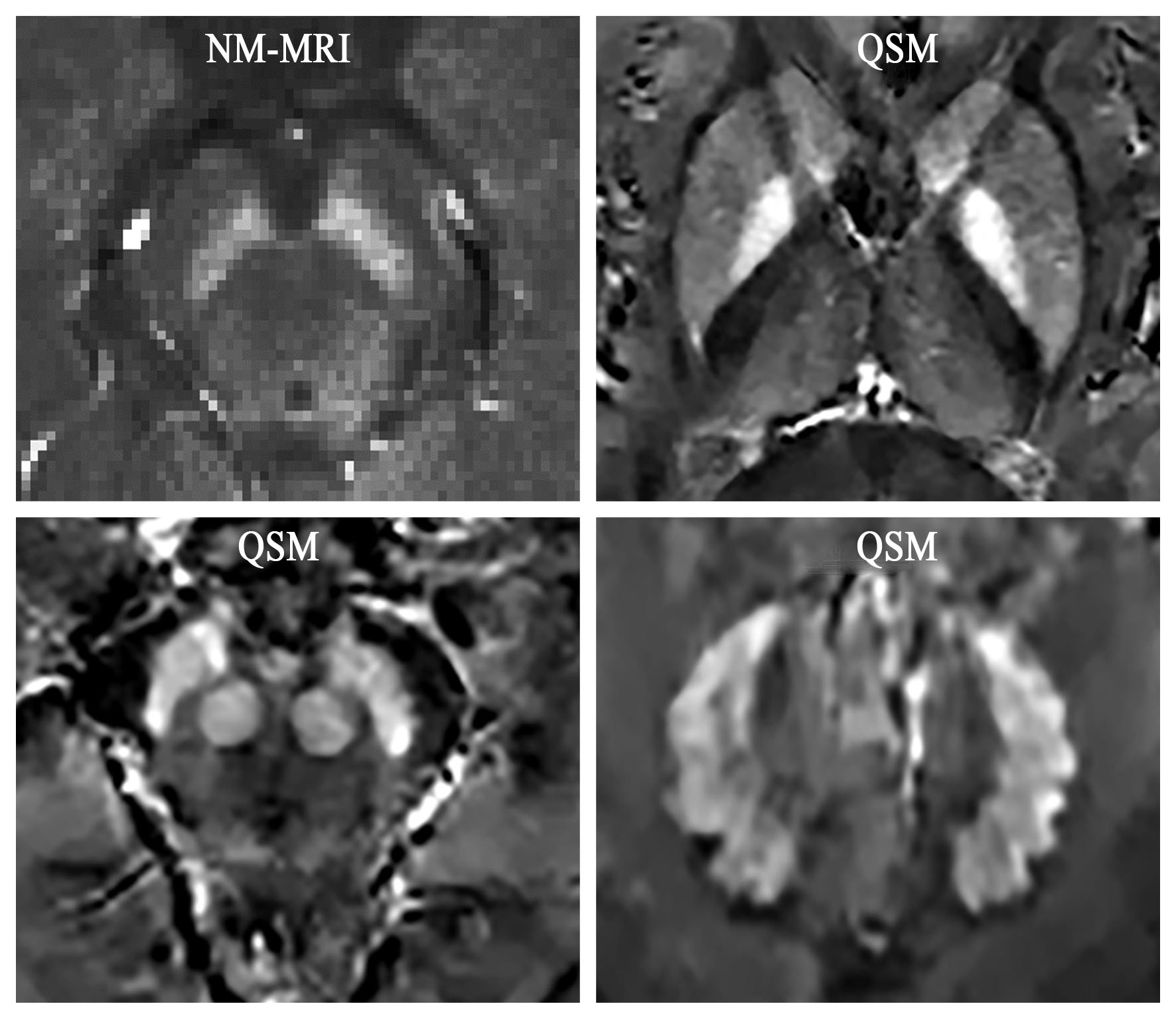
尽管具有磁化转移(Magnetization Transfer,MT)的三维梯度回波序列可以同时进行神经黑色素和定量磁化率成像,但MT预饱和脉冲耗时长,且MT对磁化率定量的影响尚不明确.本文通过开发具有可变持续时间的MT脉冲,缩短了MT脉冲持续时间,并评估了MT对磁化率值定量的影响.研究结果显示,MT脉冲持续时间为5 ms的梯度回波序列在显示神经黑色素方面不低于持续时间为8 ms、10 ms和12 ms时序列的显示效果,并且得到的大脑深部灰质核团磁化率值与未加MT的序列具有良好一致性.这表明短持续时间的MT脉冲提供了一种同时成像神经黑色素和磁化率的实用方法.
陈梦颖 , 武玉朋 , 逄奇凡 , 钟昊东 , 李改英 , 李建奇 . 全脑同时神经黑色素敏感与定量磁化率成像[J]. 波谱学杂志, 2023 , 40(4) : 385 -396 . DOI: 10.11938/cjmr20233062
Three-dimensional (3D) gradient recalled echo (GRE) sequence with magnetization transfer (MT) can simultaneously image neuromelanin and magnetic susceptibility. However, the sequence requires long duration of MT saturation pulse and the effect of MT pulse on susceptibility values remains unclear. Therefore, this paper aims to shorten the MT pulse duration and evaluate the effect of MT pulse on susceptibility value. Results showed that 3D GRE sequence with 5 ms of MT pulse provided a saturation effect no less than that of 8 ms, 10 ms and 12 ms in highlighting neuromelanin, and yielded susceptibility values in the deep gray matter nuclei similar to sequence without MT pulse. In conclusion, short MT pulse provides a practical means to simultaneously image the neuromelanin and magnetic susceptibility.
| [1] | OHTSUKA C, SASAKI M, KONNO K, et al. Changes in substantia nigra and locus coeruleus in patients with early-stage Parkinson’s disease using neuromelanin-sensitive MR imaging[J]. Neurosci Lett, 2013, 541: 93-98. |
| [2] | ZUCCA F A, SEGURA-AGUILAR J, FERRARI E, et al. Interactions of iron, dopamine and neuromelanin pathways in brain aging and Parkinson’s disease[J]. Prog Neurobiol, 2017, 155: 96-119. |
| [3] | ZECCA L, BELLEI C, COSTI P, et al. New melanic pigments in the human brain that accumulate in aging and block environmental toxic metals[J]. Proc Natl Acad Sci USA, 2008, 105(45): 17567-17572. |
| [4] | MOSHAROV E V, LARSEN K E, KANTER E, et al. Interplay between cytosolic dopamine, calcium, and alpha-synuclein causes selective death of substantia nigra neurons[J]. Neuron, 2009, 62(2): 218-229. |
| [5] | TRUJILLO P, SUMMERS P E, FERRARI E, et al. Contrast mechanisms associated with neuromelanin-MRI[J]. Magn Reson Med, 2017, 78(5): 1790-1800. |
| [6] | SASAKI M, SHIBATA E, TOHYAMA K, et al. Neuromelanin magnetic resonance imaging of locus ceruleus and substantia nigra in Parkinson’s disease[J]. Neuroreport, 2006, 17(11): 1215-1218. |
| [7] | RENEMAN L, VAN DER PLUIJM M, SCHRANTEE A, et al. Imaging of the dopamine system with focus on pharmacological MRI and neuromelanin imaging[J]. Eur J Radiol, 2021, 140: 109752. |
| [8] | LANGLEY J, HUDDLESTON D E, LIU C J, et al. Reproducibility of locus coeruleus and substantia nigra imaging with neuromelanin sensitive MRI[J]. Magn Reson Mat Phys Biol Med, 2016, 30(2): 121-125. |
| [9] | YANG J Q, YANG X F, ZHONG C Q, et al. The application value of neuromelanin sensitive magnetic resonance imaging in the diagnosis of Parkinson’s disease[J] J Clin Radiol, 2021, 40(5): 5. |
| [9] | 杨俊强, 杨晓帆, 仲崇琦, 等. 神经黑色素敏感磁共振成像对诊断帕金森病的应用价值[J]. 临床放射学杂志, 2021, 40(5): 5. |
| [10] | MATSUURA K, MAEDA M, TABEI K I, et al. A longitudinal study of neuromelanin-sensitive magnetic resonance imaging in Parkinson’s disease[J]. Neurosci Lett, 2016, 633: 112-117. |
| [11] | XIANG Y, GONG T, WU J, et al. Subtypes evaluation of motor dysfunction in Parkinson’s disease using neuromelanin-sensitive magnetic resonance imaging[J]. Neurosci Lett, 2017, 638: 145-150. |
| [12] | WANG J, HUANG Z, LI Y, et al. Neuromelanin-sensitive MRI of the substantia nigra: an imaging biomarker to differentiate essential tremor from tremor-dominant Parkinson’s disease[J]. Parkinsonism Relat Disord, 2019, 58: 3-8. |
| [13] | LEITAO R, GUERREIRO C, NUNES R G, et al. Neuromelanin magnetic resonance imaging of the substantia nigra in Huntington’s disease[J]. J Huntingtons Dis, 2020, 9(2): 143-148. |
| [14] | SHIBATA E, SASAKI M, TOHYAMA K, et al. Use of neuromelanin-sensitive MRI to distinguish schizophrenic and depressive patients and healthy individuals based on signal alterations in the substantia nigra and locus ceruleus[J]. Biol Psychiatry, 2008, 64(5): 401-406. |
| [15] | CASSIDY C M, CARPENTER K M, KONOVA A B, et al. Evidence for dopamine abnormalities in the substantia nigra in cocaine addiction revealed by neuromelanin-sensitive MRI[J]. Am J Psychiatry, 2020, 177(11): 1038-1047. |
| [16] | DE ROCHEFORT L, LIU T, KRESSLER B, et al. Quantitative susceptibility map reconstruction from MR phase data using bayesian regularization: validation and application to brain imaging[J]. Magn Reson Med, 2010, 63(1): 194-206. |
| [17] | LANGKAMMER C, SCHWESER F, KREBS N, et al. Quantitative susceptibility mapping (QSM) as a means to measure brain iron? A post mortem validation study[J]. NeuroImage, 2012, 62(3): 1593-1599. |
| [18] | LI G, WU R, TONG R, et al. Quantitative measurement of metal accumulation in brain of patients with Wilson’s disease[J]. Mov Disord, 2020, 35(10): 1787-1795. |
| [19] | BULK M, ABDELMOULA W M, GEUT H, et al. Quantitative MRI and laser ablation-inductively coupled plasma-mass spectrometry imaging of iron in the frontal cortex of healthy controls and Alzheimer’s disease patients[J]. Neuroimage, 2020, 215: 116808. |
| [20] | HUANG W, SWEENEY E M, KAUNZNER U W, et al. Quantitative susceptibility mapping versus phase imaging to identify multiple sclerosis iron rim lesions with demyelination[J]. J Neuroimaging, 2022, 32(4): 667-675. |
| [21] | WANG S, LOU M, LIU T, et al. Hematoma volume measurement in gradient echo MRI using quantitative susceptibility mapping[J]. Stroke, 2013, 44(8): 2315-2317. |
| [22] | CHEN W, ZHU W, KOVANLIKAYA I, et al. Intracranial calcifications and hemorrhages: characterization with quantitative susceptibility mapping[J]. Radiology, 2014, 270(2): 496-505. |
| [23] | AZUMA M, HIRAI T, YAMADA K, et al. Lateral asymmetry and spatial difference of iron deposition in the substantia nigra of patients with Parkinson disease measured with quantitative susceptibility mapping[J]. Am J Neuroradiol, 2016, 37(5): 782-788. |
| [24] | AN H, ZENG X, NIU T, et al. Quantifying iron deposition within the substantia nigra of Parkinson’s disease by quantitative susceptibility mapping[J]. J Neurol Sci, 2018, 386: 46-52. |
| [25] | BERGSLAND N, ZIVADINOV R, SCHWESER F, et al. Ventral posterior substantia nigra iron increases over 3 years in Parkinson’s disease[J]. Mov Disord, 2019, 34(7): 1006-1013. |
| [26] | SJOSTROM H, GRANBERG T, WESTMAN E, et al. Quantitative susceptibility mapping differentiates between parkinsonian disorders[J]. Parkinsonism Relat Disord, 2017, 44: 51-57. |
| [27] | WU M Z, LUAN J X, ZHANG C C, et al. Meta analysis of quantitative susceptibility mapping of substantia nigra in the diagnosis of Parkinson’s disease[J] Chin J Magn Reson Imaging, 2023, 14(2): 6-11. |
| [27] | 吴明振, 栾继昕, 张传臣, 等. 黑质定量磁化率成像对帕金森病诊断价值的Meta分析[J]. 磁共振成像, 2023, 14(2): 6-11. |
| [28] | TAKAHASHI H, WATANABE Y, TANAKA H, et al. Quantifying changes in nigrosomes using quantitative susceptibility mapping and neuromelanin imaging for the diagnosis of early-stage Parkinson’s disease[J]. Br J Radiol, 2018, 91(1086): 20180037. |
| [29] | TAKAHASHI H, WATANABE Y, TANAKA H, et al. Comprehensive MRI quantification of the substantia nigra pars compacta in Parkinson’s disease[J]. Eur J Radiol, 2018, 109: 48-56. |
| [30] | WANG X, ZHANG Y, ZHU C, et al. The diagnostic value of SNpc using NM-MRI in Parkinson’s disease: meta-analysis[J]. Neurol Sci, 2019, 40(12): 2479-2489. |
| [31] | HE N, GHASSABAN K, HUANG P, et al. Imaging iron and neuromelanin simultaneously using a single 3D gradient echo magnetization transfer sequence: Combining neuromelanin, iron and the nigrosome-1 sign as complementary imaging biomarkers in early stage Parkinson’s disease[J]. NeuroImage, 2021, 230: 117810. |
| [32] | JIN L, WANG J, WANG C, et al. Combined visualization of nigrosome-1 and neuromelanin in the substantia nigra using 3T MRI for the differential diagnosis of essential tremor and de novo Parkinson’s disease[J]. Front Neurol, 2019, 10: 100. |
| [33] | ARMSTRONG M J, OKUN M S. Diagnosis and treatment of Parkinson disease: a review[J]. J Am Med Assoc, 2020, 323(6): 548-560. |
| [34] | WANG J Y, ZHUANG Q Q, ZHU L B, et al. Meta-analysis of brain iron levels of Parkinson’s disease patients determined by postmortem and MRI measurements[J]. Sci Rep, 2016, 6: 36669. |
| [35] | KARSA A, PUNWANI S, SHMUELI K. The effect of low resolution and coverage on the accuracy of susceptibility mapping[J]. Magn Reson Med, 2019, 81(3): 1833-1848. |
| [36] | HENKELMAN R M, STANISZ G J, GRAHAM S J. Magnetization transfer in MRI: a review[J]. NMR Biomed, 2001, 14(2): 57-64. |
| [37] | WENGLER K, CASSIDY C, VAN DER PLUIJM M, et al. Cross-scanner harmonization of neuromelanin-sensitive MRI for multisite studies[J]. J Magn Reson Imaging, 2021, 54(4): 1189-1199. |
| [38] | BERNSTEIN M A, KING K F, ZHOU X J. Handbook of MRI pulse sequences[M]. Burlington: Academic Press, 2004: 96-124. |
| [39] | PIKE G B, GLOVER G H, HU B S, et al. Pulsed magnetization transfer spin-echo MR imaging[J]. J Magn Reson Imaging, 1993, 3(3): 531-539. |
| [40] | BIONDETTI E, KARSA A, THOMAS D L, et al. Investigating the accuracy and precision of TE-dependent versus multi-echo QSM using laplacian-based methods at 3 T[J]. Magn Reson Med, 2020, 84(6): 3040-3053. |
| [41] | SMITH S M. Fast robust automated brain extraction[J]. Hum Brain Mapp, 2002, 17(3): 143-155. |
| [42] | LIU T, WISNIEFF C, LOU M, et al. Nonlinear formulation of the magnetic field to source relationship for robust quantitative susceptibility mapping[J]. Magn Reson Med, 2013, 69(2): 467-476. |
| [43] | ZHAO X X, BO B S, LIU T, et al. Research on multi-echo phase fitting algorithm for quantitative susceptibility mapping[J]. Chinese J Magn Reson, 2016, 33(4): 609-617. |
| [43] | 赵欣欣, 薄斌仕, 刘田, 等. 定量磁化率成像多回波相位拟合算法研究[J]. 波谱学杂志, 2016, 33(4): 609-617. |
| [44] | ABDUL-RAHMAN H S, GDEISAT M A, BURTON D R, et al. Fast and robust three-dimensional best path phase unwrapping algorithm[J]. Appl Opt, 2007, 46(26): 6623-6635. |
| [45] | ZHOU D, LIU T, SPINCEMAILLE P, et al. Background field removal by solving the Laplacian boundary value problem[J]. NMR Biomed, 2014, 27(3): 312-319. |
| [46] | LIU Z, SPINCEMAILLE P, YAO Y, et al. MEDI+0: morphology enabled dipole inversion with automatic uniform cerebrospinal fluid zero reference for quantitative susceptibility mapping[J]. Magn Reson Med, 2018, 79(5): 2795-2803. |
| [47] | LIU J, LIU T, DE ROCHEFORT L, et al. Morphology enabled dipole inversion for quantitative susceptibility mapping using structural consistency between the magnitude image and the susceptibility map[J]. NeuroImage, 2012, 59(3): 2560-2568. |
| [48] | LIU T, XU W, SPINCEMAILLE P, et al. Accuracy of the morphology enabled dipole inversion (MEDI) algorithm for quantitative susceptibility mapping in MRI[J]. IEEE Trans Med Imaging, 2012, 31(3): 816-824. |
| [49] | SCHWARZ S T, RITTMAN T, GONTU V, et al. T1-weighted MRI shows stage-dependent substantia nigra signal loss in Parkinson’s disease[J]. Mov Disord, 2011, 26(9): 1633-1638. |
| [50] | OGISU K, KUDO K, SASAKI M, et al. 3D neuromelanin-sensitive magnetic resonance imaging with semi-automated volume measurement of the substantia nigra pars compacta for diagnosis of Parkinson’s disease[J]. Neuroradiology, 2013, 55(6): 719-724. |
| [51] | CHEN X, HUDDLESTON D E, LANGLEY J, et al. Simultaneous imaging of locus coeruleus and substantia nigra with a quantitative neuromelanin MRI approach[J]. Magn Reson Imaging, 2014, 32(10): 1301-1306. |
| [52] | DIMOV A V, GUPTA A, KOPELL B H, et al. High-resolution QSM for functional and structural depiction of subthalamic nuclei in DBS presurgical mapping[J]. J Neurosurg, 2018, 131(2): 360-367. |
| [53] | VITEK J L, LYONS K E, BAKAY R, et al. Standard guidelines for publication of deep brain stimulation studies in Parkinson’s disease (Guide4DBS-PD)[J]. Mov Disord, 2010, 25(11): 1530-1537. |
| [54] | ASHKAN K, BLOMSTEDT P, ZRINZO L, et al. Variability of the subthalamic nucleus: the case for direct MRI guided targeting[J]. Br J Neurosurg, 2007, 21(2): 197-200. |
| [55] | LIU T, ESKREIS-WINKLER S, SCHWEITZER A D, et al. Improved subthalamic nucleus depiction with quantitative susceptibility mapping[J]. Radiology, 2013, 269(1): 216-223. |
| [56] | ZHAO W, WANG Y, ZHOU F, et al. Automated segmentation of midbrain structures in high-resolution susceptibility maps based on convolutional neural network and transfer learning[J]. Front Neurosci, 2022, 16: 801618. |
| [57] | HUA J, HURST G C. Analysis of on- and off-resonance magnetization transfer techniques[J]. J Magn Reson Imaging, 1995, 5(1): 113-120. |
| [58] | BAOGUI Z, KUN W, TIANZI J. RF power design optimization in MRI system[J]. Magn Reson Lett, 2021, 1(1): 89-98. |
/
| 〈 |
|
〉 |