

基于生成对抗网络的膝关节模型构建与局部比吸收率估计
收稿日期: 2023-01-15
网络出版日期: 2023-06-25
基金资助
北京化工大学高精尖科技创新团队基金资助项目(buctylkjcx06)
Knee Joint Model Construction and Local Specific Absorption Rate Estimation Based on Generative Adversarial Networks
Received date: 2023-01-15
Online published: 2023-06-25
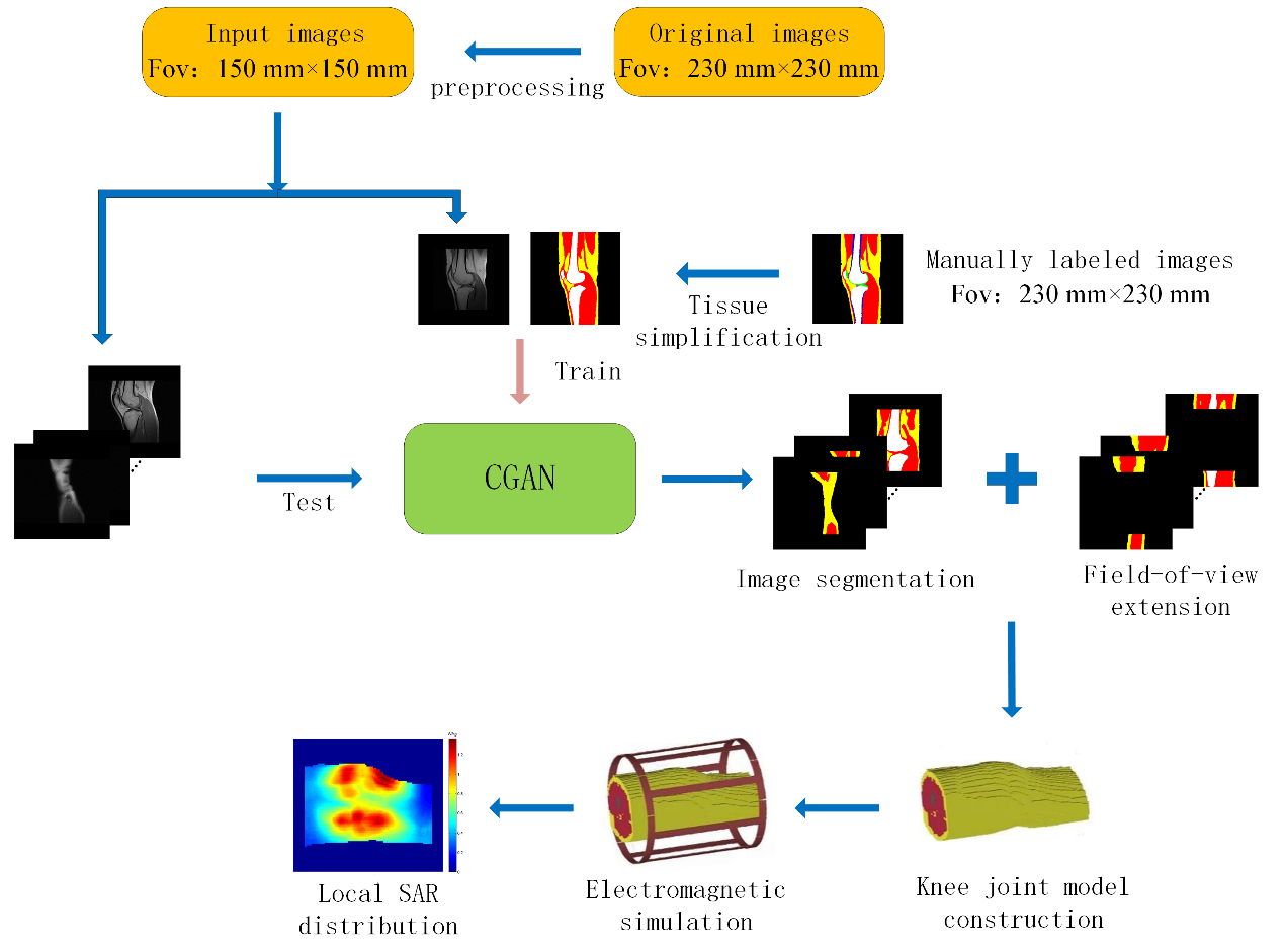
局部比吸收率(SAR)是衡量高场磁共振成像安全性的重要指标.目前主要的方法是对扫描获得的磁共振图像进行组织分割,从而构建个体特异性模型,对其进行电磁仿真以计算局部SAR.针对仿真中膝关节模型长度影响局部SAR估计准确度的问题,本文提出了一种基于条件生成对抗网络(CGAN)的膝关节磁共振图像分割与视野扩展方法,将膝关节图像简化归类为肌肉、脂肪和骨骼三种组织,通过CGAN进行像素的语义分割,采用注意力机制以提高分割的准确度,并且沿头-足方向在图像两端生成扩展区域,构建出更长的模型.实验中对所提方法以及各种对比方法得到的膝关节模型进行电磁仿真,计算它们与人工标注模型的局部SAR的相对误差,结果验证了所提方法可以获得相对精确的膝关节局部SAR估计.
任宏晋 , 马岩 , 肖亮 . 基于生成对抗网络的膝关节模型构建与局部比吸收率估计[J]. 波谱学杂志, 2023 , 40(4) : 410 -422 . DOI: 10.11938/cjmr20233053
The local specific absorption rate (SAR) is an essential metric when assessing the safety of high-field magnetic resonance imaging (MRI). Usually, the subject-specific model is created from segmented magnetic resonance (MR) images, followed by SAR estimates. However, the length of knee joint model affects the accuracy of local SAR estimation in simulation. To address the issue, this paper proposed a knee joint MR image segmentation and field-of-view extension method based on conditional generative adversarial nets (CGAN). The image of knee joint was classified into images of three tissues, namely, muscle, fat, and bone. And pixel semantic segmentation was performed using CGAN with attention mechanism to improve accuracy. The method also generated extension areas at both ends of the image along the head-foot direction to construct a longer model. The knee joint models by using the proposed method and various comparison methods were electromagnetically simulated, and the relative error of their local SAR compared to the manually annotated model was calculated. Results verified that the proposed method can achieve a relatively accurate estimation of local SAR in the knee joint.
| [1] | LIU X, XU H W, JIANG T, et al. MRI and 18F-FDG PET/CT findings of a giant cell tumor of the tendon sheath of the knee joint (pigmented villonodular synovitis): a case report and literature review[J]. Hell J Nucl Med, 2021, 24(2): 149-154. |
| [2] | YANG W H. The development of ultra-high field magnetic resonance imaging[J]. Physics, 2019, 48(4): 227-236. |
| [2] | 杨文晖. 磁共振成像发展与超高场磁共振成像技术[J]. 物理, 2019, 48(4): 227-236. |
| [3] | WANG Y S, DENG A Q, MAO J L, et al. Automatic segmentation of knee joint synovial magnetic resonance images based on 3D VNetTrans[J]. Chinese J Magn Reson, 2022, 39(3): 303-315. |
| [3] | 王颖珊, 邓奥琦, 毛瑾玲, 等. 基于3D VNetTrans的膝关节滑膜磁共振图像自动分割[J]. 波谱学杂志, 2022, 39(3): 303-315. |
| [4] | PADORMO F, BEQIRI A, HAJNAL J V, et al. Parallel transmission for ultrahigh-field imaging[J]. NMR Biomed, 2016, 29(9): 1145-1161. |
| [5] | YETISIR F, ABACI TURK E, GUERIN B, et al. Safety and imaging performance of two-channel RF shimming for fetal MRI at 3 T[J]. Magn Reson Med, 2021, 86(5): 2810-2821. |
| [6] | ZENG Q, GUO R, ZHENG J, et al. Impacts of RF shimming on local SAR caused by MRI 3 T birdcage coil near femoral plate implants[C]// IEEE International Symposium on Antennas and Propagation & USNC/URSI National Radio Science Meeting, San Diego, CA, USA: IEEE, 2017: 1005-1006. |
| [7] | GAGLIARDI V, RETICO A, BIAGI L, et al. Subject-specific knee SAR prediction using a degenerate birdcage at 7 T[C]// IEEE International Symposium on Medical Measurements and Applications (MeMeA), Rome, Italy: IEEE, 2018: 1-5. |
| [8] | LIU W, WANG H, ZHANG P, et al. Statistical evaluation of radiofrequency exposure during magnetic resonant imaging: Application of whole-body individual human model and body motion in the coil[J]. Int J Env Res Pub He, 2019, 16(6): 1069. |
| [9] | STEENSMA B R, MELIADò E F, LUIJTEN P, et al. SAR and temperature distributions in a database of realistic human models for 7 T cardiac imaging[J]. NMR Biomed, 2021, 34(7): e4525. |
| [10] | CARLUCCIO G, AKGUN C, VAUGHAN J T, et al. Temperature-based MRI safety simulations with a limited number of tissues[J]. Magn Reson Med, 2021, 86(1): 543-550. |
| [11] | VAN DEN BERGEN B, VAN DEN BERG C A, BARTELS L W, et al. 7 T body MRI: B-1 shimming with simultaneous SAR reduction[J]. Phys Med Biol, 2007, 52(17): 5429. |
| [12] | DE BUCK M H, JEZZARD P, JEONG H, et al. An investigation into the minimum number of tissue groups required for 7 T in-silico parallel transmit electromagnetic safety simulations in the human head[J]. Magn Reson Med, 2021, 85(2): 1114-1122. |
| [13] | WOLF S, DIEHL D, GEBHARDT M, et al. SAR simulations for high-field MRI: how much detail, effort, and accuracy is needed?[J] Magn Reson Med, 2013, 69(4): 1157-1168. |
| [14] | SENGAR S S, MEULENGRACHT C, BOESEN M P, et al. Multi-planar 3D knee MRI segmentation via UNet inspired architectures[J]. Int J Imag Syst Tech, 2023, 33(3): 985-998. |
| [15] | GOODFELLOW I, POUGET-ABADIE J, MIRZA M, et al. Generative adversarial networks[J]. Commun Acm, 2020, 63(11): 139-144. |
| [16] | CHANG X, CAI X, YANG G, et al. Applications of generative adversarial networks in medical image processing[J]. Chinese J Magn Reson, 2022, 39(3): 366-380. |
| [16] | 常晓, 蔡昕, 杨光, 等. 生成对抗网络在医学图像转换领域的应用[J]. 波谱学杂志, 2022, 39(3): 366-380. |
| [17] | CHEN Y, SHI F, CHRISTODOULOU A G, et al. Efficient and accurate MRI super-resolution using a generative adversarial network and 3D multi-level densely connected network[C]// Medical Image Computing and Computer Assisted Intervention-MICCAI 2018: 21st International Conference, Granada, Spain:Springer, 2018: 91-99. |
| [18] | KANG E, KOO H J, YANG D H, et al. Cycle-consistent adversarial denoising network for multiphase coronary CT angiography[J]. Med Phys, 2019, 46(2): 550-562. |
| [19] | LAHIRI A, AYUSH K, KUMAR BISWAS P, et al. Generative adversarial learning for reducing manual annotation in semantic segmentation on large scale miscroscopy images: Automated vessel segmentation in retinal fundus image as test case[C]// Proceedings of the IEEE conference on computer vision and pattern recognition workshops, Honolulu, HI, USA: IEEE, 2017: 42-48. |
| [20] | GAJ S, YANG M, NAKAMURA K, et al. Automated cartilage and meniscus segmentation of knee MRI with conditional generative adversarial networks[J]. Magn Reson Med, 2020, 84(1): 437-449. |
| [21] | ZHANG L, GOOYA A, FRANGI A F. Semi-supervised assessment of incomplete LV coverage in cardiac MRI using generative adversarial nets[C]// International Workshop on Simulation and Synthesis in Medical Imaging. Cham: Springer, 2017: 61-68. |
| [22] | MIRZA M, OSINDERO S. Conditional generative adversarial nets[J]. arXiv preprint arXiv:1411.1784, 2014. |
| [23] | MA Y, XING C J, XIAO L. Knee joint image segmentation and model construction based on cascaded network[J]. Chinese J Magn Reson, 2022, 39(2): 184-195. |
| [23] | 马岩, 邢藏菊, 肖亮. 基于级联网络的膝关节图像分割与模型构建[J]. 波谱学杂志, 2022, 39(2): 184-195. |
| [24] | SCHLEMPER J, OKTAY O, SCHAAP M, et al. Attention gated networks: Learning to leverage salient regions in medical images[J]. Med Image Anal, 2019, 53197-207. |
| [25] | LEE J, GEBHARDT M, WALD L L, et al. Local SAR in parallel transmission pulse design[J]. Magn Reson Med, 2012, 67(6): 1566-1578. |
| [26] | MILSHTEYN E, GURYEV G, TORRADO-CARVAJAL A, et al. Individualized SAR calculations using computer vision-based MR segmentation and a fast electromagnetic solver[J]. Magn Reson Med, 2021, 85(1): 429-443 |
| [27] | HARDY B M, BANIK R, YAN X Q, et al. Bench to bore ramifications of inter-subject head differences on RF shimming and specific absorption rates at 7 T[J]. Magn Reson Imaging, 2022, 92: 187-196. |
| [28] | CHRIST A, KAINZ W, HAHN E G, et al. The Virtual Family—development of surface-based anatomical models of two adults and two children for dosimetric simulations[J]. Phys Med Biol, 2009, 55(2): N23. |
/
| 〈 |
|
〉 |