

融合双解码和全局注意力上采样模块的胰腺囊性肿瘤分割网络
收稿日期: 2023-07-17
网络出版日期: 2023-09-27
Pancreatic Cystic Neoplasms Segmentation Network Combining Dual Decoding and Global Attention Upsampling Modules
Received date: 2023-07-17
Online published: 2023-09-27
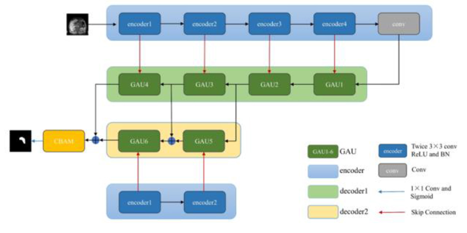
胰腺因其解剖结构复杂多变、周围环境复杂等特点,始终是医学图像分割中最具挑战性的任务之一.针对以上问题,提出一种融合双解码和全局注意力上采样模块的深度学习分割模型(Combining Dual Decoding and Global Attention Upsampling Modules Network,DGANet).模型由一个编码器和两个解码器构成,两个解码器实现了对不同深度特征信息的充分利用;模型采用全局注意力上采样模块(Global Attention Upsampling,GAU),利用高层丰富的语义信息来引导低层选择更为精准的特征信息.利用长海医院提供的数据集进行实验,结果表明平均Dice相似系数为86.28%,交并比(Intersection-over-Union,IoU)为0.77,豪斯多夫距离(Hausdorff Distance,HD)为7.7 mm,数据证实了该模型在胰腺囊性肿瘤分割中具有一定的临床意义和价值.
戴俊龙 , 何聪 , 武杰 , 边云 . 融合双解码和全局注意力上采样模块的胰腺囊性肿瘤分割网络[J]. 波谱学杂志, 2024 , 41(2) : 151 -161 . DOI: 10.11938/cjmr20233073
The pancreas has always been one of the most challenging parts in medical image segmentation due to its complex anatomical structure and complex surrounding environment. Aiming at the above problems, a deep learning segmentation model combining dual decoding and global attention upsampling module (DGANet) is proposed. The model consists of an encoder and two decoders, where the latter realizes the full utilization of different depth feature information. The model applies the global attention upsampling module and high-level rich semantic information to guide the low-level selection of more accurate feature information. The data set provided by Changhai Hospital was used for experiments. The results showed that the average Dice similarity coefficient was 86.28%, Intersection-over-Union (IoU) was 0.77, and Hausdorff distance (HD) was 7.7 mm. The data confirmed the clinical value of this model in segmenting pancreatic cystic tumors.
Key words: medical image segmentation; DGANet; deep learning; pancreas
| [1] | HUO L, HU X X, XIAO Q, et al. Automatic segmentation of breast and fibroglandular tissues in DCE-MR images based on nnU-Net[J]. Chinese J Magn Reson, 2021, 38(3): 367-380. |
| 霍璐, 胡晓欣, 肖勤, 等. 基于nnU-Net的乳腺DCE-MR图像中乳房和腺体自动分割[J]. 波谱学杂志, 2021, 38(3): 367-380. | |
| [2] | YAN S J, HAN Y S, TANG G Y. An improved level set algorithm for prostate region segmentation[J]. Chinese J Magn Reson, 2021, 38(3): 356-366. |
| 闫士举, 韩勇森, 汤光宇. 一种用于前列腺区域分割的改进水平集算法[J]. 波谱学杂志, 2021, 38(3): 356-366. | |
| [3] | WANG M, LI D. An automatic segmentation method for lung tumor based on improved region growing algorithm[J]. Diagnostics, 2022, 12(12): 2971. |
| [4] | SULTANA F, SUFIAN A, DUTTA P. Evolution of image segmentation using deep convolutional neural network: A survey[J]. Knowl Based Syst, 2020, 201: 106062. |
| [5] | JHA D, RIEGLER M A, JOHANSEN D, et al. Doubleu-net: A deep convolutional neural network for medical image segmentation[C]// 33rd International symposium on computer-based medical systems (CBMS). IEEE, 2020: 558-564. |
| [6] | DOLZ J, GOPINATH K, YUAN J, et al. HyperDense-Net: a hyper-densely connected CNN for multi-modal image segmentation[J]. IEEE T Med Imaging, 2018, 38(5): 1116-1126. |
| [7] | RONNEBERGER O, FISCHER P, BROX T. U-net: convolutional networks for biomedical image segmentation[C]// Medical Image Computing and Computer-Assisted Intervention-MICCAI 2015: 18 th International Conference, Munich, Germany, Proceedings, Part III 18. Springer International Publishing, 2015: 234-241. |
| [8] | CAI J, LU L, XING F, et al. Pancreas segmentation in CT and MRI images via domain specific network designing and recurrent neural contextual learning[J]. ArXiv: 1803.11303, 2018. |
| [9] | OKTAY O, SCHLEMPER J, FOLGOC L L, et al. Attention U-net: Learning where to look for the pancreas[J]. ArXiv: 1804.03999, 2018. |
| [10] | WANG Y, GONG G, KONG D, et al. Pancreas segmentation using a dual-input V-mesh network[J]. Med Image Anal, 2021, 69: 101958. |
| [11] | XUE J, HE K, NIE D, et al. Cascaded multitask 3-D fully convolutional networks for pancreas segmentation[J]. IEEE Trans Cybern, 2019, 51(4): 2153-2165. |
| [12] | ASATURYAN H, THOMAS E L, FITZPATRICK J, et al. Advancing pancreas segmentation in multi-protocol mri volumes using hausdorff-sine loss function[C]// Machine Learning in Medical Imaging:10th International Workshop, 2019: 27-35. |
| [13] | PROIETTO S F, BELLITTO G, IRMAKCI I, et al. Hierarchical 3D feature learning forpancreas segmentation[C]// Machine Learning in Medical Imaging: 12th International Workshop, 2021: 238-247. |
| [14] | BI X L, LU M, XIAO B, et al. Pancreas segmentation based on dual-decoding U-Net[J]. J Softw, 2022, 33(5): 1947-1958. |
| 毕秀丽, 陆猛, 肖斌, 等. 基于双解码U型卷积神经网络的胰腺分割[J]. 软件学报, 2022, 33(5): 1947-1958. | |
| [15] | LI Y, ZHANG X, CHEN D. CSRNet: dilated convolutional neural networks for understanding the highly congested scenes[C]// Proceedings of the IEEE conference on computer vision and pattern recognition. 2018: 1091-1100. |
| [16] | LI H, XIONG P, AN J, et al. Pyramid attention network for semantic segmentation[J]. ArXiv: 1805.10180, 2018. |
| [17] | WOO S, PARK J, LEE J Y, et al. CBAM: convolutional block attention module[C]// Proceedings of the European conference on computer vision (ECCV). 2018: 3-19. |
| [18] | ZHOU Z, SIDDIQUEE M M R, TAJBAKHSH N, et al. Unet++: Redesigning skip connections to exploit multiscale features in image segmentation[J]. IEEE Trans Med Imaging, 2019, 39(6): 1856-1867. |
| [19] | YU F, KOLTUN V. Multi-scale context aggregation by dilated convolutions[J]. ArXiv: 1511.07122, 2015. |
| [20] | WANG P, CHEN P, YUAN Y, et al. Understanding convolution for semantic segmentation[C]// 2018 IEEE winter conference on applications of computer vision (WACV). IEEE, 2018: 1451-1460. |
| [21] | WANG Y, ZHANG J, CUI H, et al. View adaptive learning for pancreas segmentation[J]. Biomed Signal Process Control, 2021, 66: 102347. |
| [22] | ZHANG J, XIE Y, WANG Y, et al. Inter-slice context residual learning for 3D medical image segmentation[J]. IEEE Trans Med Imaging, 2020, 40(2): 661-672. |
| [23] | ROTH H R, LU L, LAY N, et al. Spatial aggregation of holistically-nested convolutional neural networks for automated pancreas localization and segmentation[J]. Med Image Anal, 2018, 45: 94-107. |
| [24] | CAI J, LU L, XIE Y, et al. Improving deep pancreas segmentation in CT and MRI images via recurrent neural contextual learning and direct loss function[J]. ArXiv: 1707.04912, 2017. |
| [25] | YU Q, XIE L, WANG Y, et al. Recurrent saliency transformation network: Incorporating multi-stage visual cues for small organ segmentation[C]// Proceedings of the IEEE conference on computer vision and pattern recognition. 2018: 8280-8289. |
| [26] | CHEN J, LU Y, YU Q, et al. Transunet: Transformers make strong encoders for medical image segmentation[J]. ArXiv: 2102.04306, 2021. |
| [27] | WANG H Z, ZHAO D, YANG L Q, et al. An approach for training data enrichment and batch labeling in AI+MRI aided diagnosis[J]. Chinese J Magn Reson, 2018, 35(4): 447-456. |
| 汪红志, 赵地, 杨丽琴, 等. 基于AI+MRI的影像诊断的样本增广与批量标注方法[J]. 波谱学杂志, 2018, 35(4): 447-456. |
/
| 〈 |
|
〉 |