

b值范围对6种体部扩散模型在前列腺应用的影响评估
收稿日期: 2023-07-20
网络出版日期: 2023-10-18
基金资助
安徽省重点研究与开发计划项目(2022e07020065);皖南医学院教学医院科研项目(JXYY202128);上海高校教师产学研见习计划
Evaluation of the Impact of b-Value Ranges on Six Body Diffusion Models in Prostate Application
Received date: 2023-07-20
Online published: 2023-10-18
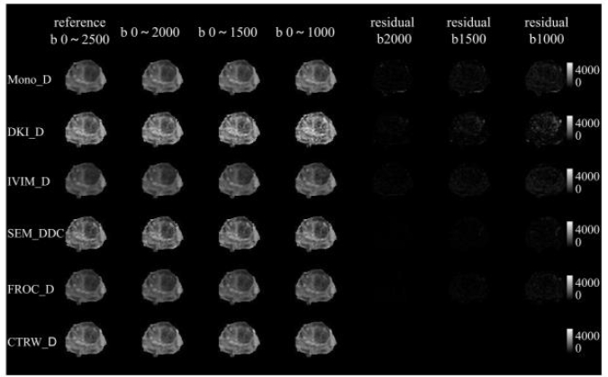
论文研究了不同b值采集范围对6种体部扩散模型定量参数计算的影响.研究涉及扩散模型包含单指数模型(Mono)、扩散峰度成像(DKI)、体素内非相干运动模型(IVIM)、扩散拉伸指数模型(SEM)、分数微积分模型(FROC)和随机游走模型(CTRW),b值范围0~2 500 s/mm2.通过扩散模型参数之间的相关性、t检验以及前列腺病灶良恶性鉴别能力三个维度,评估了不同b值采集范围对参数计算的影响.结果显示与参考采样方案相比,随着最大b值降低,所得同一扩散参数感兴趣区域(ROI)均值的差异逐渐增大,但相关性降低不明显,且前列腺病灶良恶性的鉴别能力也保持相似水平.基于实验结果,建议在临床实践中采用b值范围为0~1 500 s/mm2的采集方案.这一方案在具备较高采集效率的同时,一半以上参数与参考采样方案结果的相关性不低于0.98,且良恶性鉴别能力指标曲线下面积(AUC)值的差别小于0.01.此外,不同扩散模型对于b值方案的敏感性存在差异,其中SEM和CTRW模型的参数受b值范围的影响相对较小.
周敏雄 , 戚轩 , 杜兵 , 齐东 , 王海杰 , 杨光 , 蔡文梅 , 刘孟潇 , 张会婷 , 严序 , 聂生东 , 何永胜 . b值范围对6种体部扩散模型在前列腺应用的影响评估[J]. 波谱学杂志, 2024 , 41(1) : 9 -18 . DOI: 10.11938/cjmr20233075
This paper investigated the impact of different b-value acquisition ranges (from 0 to 2 500 s/mm2) on the quantitative parameter calculation of six body diffusion models, including mono-exponential (Mono), diffusion kurtosis imaging (DKI), intravoxel incoherent motion (IVIM), stretch exponential model (SEM), fractional-order calculus model (FROC), and continuous time random walk model (CTRW). The influence of different b-value acquisition ranges on parameter calculation was evaluated through correlation between diffusion model parameters, t-test, and the ability to differentiate benign and malignant prostate lesions. The results showed that compared with that of the reference sampling scheme (0~2 500 s/mm2), the difference of the mean value of region of interest (ROI) with the same diffusion parameters gradually increases as the maximum b-value decreases, but the correlation decreases only slightly, and the ability to differentiate between benign and malignant prostate lesions remains at a similar level. Based on the experimental results, a b-value range of 0~1 500 s/mm2 is recommended for clinical practice, because this scheme takes collection efficiency into account, the correlation of more than half of its parameters with those of the reference sampling scheme is not less than 0.98, and the difference in the values of area under the curve (AUC) between benign and malignant differentiation is less than 0.01. In addition, the sensitivity of different diffusion models to the b-value scheme varies, with the parameters of SEM and CTRW models being relatively less affected by the b-value range.
Key words: MR imaging; Data sampling; Diffusion models; FROC; CTRW
| [1] | BASSER P J, MATTIELLO J, LEBIHAN D. MR diffusion tensor spectroscopy and imaging[J]. Biophys J, 1994, 66(1): 259-267. |
| [2] | TOURNIER J D. Diffusion MRI in the brain - theory and concepts[J]. Prog Nucl Magn Reson Spectrosc, 2019, 112-113: 1-16. |
| [3] | ALEXANDER D C, DYRBY T B, NILSSON M, et al. Imaging brain microstructure with diffusion MRI: practicality and applications[J]. NMR Biomed, 2019, 32(4): e3841. |
| [4] | MALAYERI A A, EL KHOULI R H, ZAHEER A, et al. Principles and applications of diffusion-weighted imaging in cancer detection, staging, and treatment follow-up[J]. Radiographics, 2011, 31(6): 1773-91. |
| [5] | LE BIHAN D, BRETON E, LALLEMAND D, et al. Separation of diffusion and perfusion in intravoxel incoherent motion MR imaging[J]. Radiology, 1988, 168(2): 497-505. |
| [6] | JENSEN J H, HELPERN J A, Ramani A. Diffusional kurtosis imaging: the quantification of non-gaussian water diffusion by means of magnetic resonance imaging[J]. Magn Reson Med, 2005, 53(6): 1432-1440. |
| [7] | BENNETT K M, SCHMAINDA K M, Bennett R T, et al. Characterization of continuously distributed cortical water diffusion rates with a stretched-exponential model[J]. Magn Reson Med, 2003, 50(4): 727-734. |
| [8] | CHEN J, GUO Y, GUO Y, et al. Preoperative assessment of microvascular invasion of hepatocellular carcinoma using non-Gaussian diffusion-weighted imaging with a fractional order calculus model: A pilot study[J]. Magn Reson Imaging, 2023, 95: 110-117. |
| [9] | SUI Y, WANG H, LIU G, et al. Differentiation of low- and high-grade pediatric brain tumors with high b-value diffusion-weighted MR imaging and a fractional order calculus model[J]. Radiology, 2015, 277(2): 489-96. |
| [10] | METZLER R, KLAFTER J. The random walk’s guide to anomalous diffusion: a fractional dynamics approach[J]. Physics reports, 2000, 339(1): 1-77. |
| [11] | KARAMAN M M, SUI Y, WANG H, et al. Differentiating low- and high-grade pediatric brain tumors using a continuous-time random-walk diffusion model at high b-values[J]. Magn Reson Med, 2016, 76(4): 1149-57. |
| [12] | KARAMAN M M, ZHANG J, XIE K L, et al. Quartile histogram assessment of glioma malignancy using high b-value diffusion MRI with a continuous-time random-walk model[J]. NMR Biomed, 2021, 34(4): e4485. |
| [13] | QIN Y, TANG C, HU Q, et al. Assessment of prognostic factors and molecular subtypes of breast cancer with a continuous-time random-walk mr diffusion model: Using whole tumor histogram analysis[J]. J Magn Reson Imaging, 2023, 58(1): 93-105. |
| [14] | SCHMID-TANNWALD C, OTO A, REISER M F, et al. Diffusion-weighted MRI of the abdomen: current value in clinical routine[J]. J Magn Reson Imaging, 2013, 37(1): 35-47. |
| [15] | TANG L, ZHOU X J. Diffusion MRI of cancer: From low to high b-values[J]. J Magn Reson Imaging, 2019, 49(1): 23-40. |
| [16] | GAO A, ZHANG H, YAN X, et al. Whole-tumor histogram analysis of multiple diffusion metrics for glioma genotyping[J]. Radiology, 2022, 302(3): 652-661. |
| [17] | SEO N, CHUNG Y E, PARK Y N, et al. Liver fibrosis: stretched exponential model outperforms mono-exponential and bi-exponential models of diffusion-weighted MRI[J]. Eur Radiol, 2018, 28(7): 2812-2822. |
| [18] | LIU Y F, ZOU Z Y, CAI L M, et al. Characterizing sensorimotor-related area abnormalities in amyotrophic lateral sclerosis: an intravoxel incoherent motion magnetic resonance imaging study[J]. Acad Radiol, 2022, 2(3): 141-146. |
| [19] | GUO R, YANG S H, LU F, et al. Evaluation of intratumoral heterogeneity by using diffusion kurtosis imaging and stretched exponential diffusion-weighted imaging in an orthotopic hepatocellular carcinoma xenograft model[J]. Quant Imaging Med Surg, 2019, 9(9): 1566-1578. |
| [20] | SUN K, CHEN X, CHAI W, et al. Breast cancer: diffusion kurtosis MR imaging-diagnostic accuracy and correlation with clinical-pathologic factors[J]. Radiology, 2015, 277(1): 46-55. |
| [21] | WANG W T, YANG L, YANG Z X, et al. Assessment of microvascular invasion of hepatocellular carcinoma with diffusion kurtosis imaging[J]. Radiology, 2018, 286(2): 571-580. |
| [22] | MIN X, FENG Z, WANG L, et al. Multi-model analysis of diffusion-weighted imaging of normal testes at 3.0 T: preliminary findings[J]. Acad Radiol, 2018, 25(4): 445-452. |
| [23] | ZHOU M X, ZHANG H T, WANG Y D, et al. Evaluation of the influence of data acquisition schemes on multiple neural diffusion models[J]. Chinese J Magn Reson, 2022, 39(2): 220-229. |
| [23] | 周敏雄, 张会婷, 王一达, 等. 数据采集方案对神经扩散模型影响的评估[J]. 波谱学杂志, 2022, 39(2): 220-229. |
/
| 〈 |
|
〉 |