

基于心脏磁共振电影图像的压缩激励残差U形网络左心肌分割
Squeeze-and-excitation Residual U-shaped Network for Left Myocardium Segmentation Based on Cine Cardiac Magnetic Resonance Images
Received date: 2021-03-23
Online published: 2023-11-27
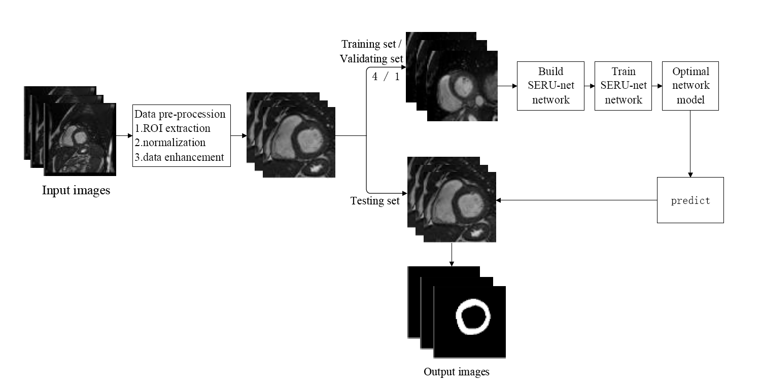
左心肌分割对心脏疾病诊疗具有重要意义.但左心肌内部毗邻乳头肌、小梁,外部与周围组织灰度相近,是分割难点.本文首先对心脏磁共振电影图像数据进行感兴趣区域提取等预处理;其次,搭建融合了压缩激励模块和残差模块的U形网络(SERU-net)分割左心肌;最后,利用75例数据训练SERU-net网络,对18例数据进行预测.基于本文方法的分割结果相对于金标准的Dice系数与豪斯多夫距离均值分别是0.902、2.697 mm;利用本文方法分割得到的舒张末期、收缩末期左心室心肌质量与金标准的相关系数和偏差均值分别是0.995、0.993和3.784 g、2.338 g.结果表明,本文方法与金标准匹配程度较高,有望辅助诊断心脏疾病.
关键词: 心脏磁共振电影图像; 左心肌分割; 压缩激励残差U形网络; 深度学习
王慧 , 王甜甜 , 王丽嘉 . 基于心脏磁共振电影图像的压缩激励残差U形网络左心肌分割[J]. 波谱学杂志, 2023 , 40(4) : 435 -447 . DOI: 10.11938/cjmr20212900
The left myocardium segmentation is significant for the diagnosis and prognosis of cardiovascular diseases. However, the internal part of the left myocardium is adjacent to the papillary muscle and trabeculae, and the external part is similar to the surrounding tissues in terms of grey level, which adds to difficulties facing segmentation. In this paper, the original datasets of cine cardiac magnetic resonance images were firstly pre-processed by extracting the region of interest. Then, squeeze-and-excitation residual U-shaped network (SERU-net), combining SE module and residual module, was built to segment the left myocardium. Finally, 75 pre-processed data were used to train SERU-net to predict the segmentation of 18 other cases. Compared with the ground truth, the average of Dice coefficient and Hausdorff distance are 0.902 and 2.697 mm. The correlation coefficient and mean deviation of end diastolic-left ventricular mass are 0.995 and 3.784 g, and that of end systolic-left ventricular mass are 0.993 and 2.338 g, respectively. The results show that SERU-net segmentation is close to the ground truth, and is prospective in assisting the diagnosis of heart diseases.
| [1] | HU S S, GAO R L, LIU L S, et al. Summary of the 2018 report on cardiovascular diseases in China[J]. Chinese Circulation Journal, 2019, 34(3): 209-220. |
| [1] | 胡盛寿, 高润霖, 刘力生, 等. 《中国心血管病报告2018》概要[J]. 中国循环杂志, 2019, 34(3): 209-220. |
| [2] | HUANG X Q, ZHAO L L, CHEN L Y, et al. Accelerated cardiac CINE imaging with CAIPIRINHA and partial parallel acquisition[J]. Chinese J Magn Reson, 2017, 34(3): 283-293. |
| [2] | 黄小倩, 赵乐乐, 陈利勇, 等. 基于同时多层激发和并行成像的心脏磁共振电影成像[J]. 波谱学杂志, 2017, 34(3): 283-293 |
| [3] | SU X Y, WANG L J, NIE S D, et al. Progress of right ventricle segmentation from short-axis images acquired with cardiac cine MRI[J]. Chinese J Magn Reson, 2019, 36(3): 377-391. |
| [3] | 苏新宇, 王丽嘉, 聂生东, 等. 基于心脏磁共振短轴电影图像的右心室分割新进展[J]. 波谱学杂志, 2019, 36(3): 377-391. |
| [4] | PETITJEAN C, DACHER J N. A review of segmentation methods in short axis cardiac MR images[J]. Med Image Anal, 2011, 15(2): 169-184. |
| [5] | SANTIAGO C, NASCIMENTO J C, MARQUES J S. Fast segmentation of the left ventricle in cardiac MRI using dynamic programming[J]. Comput Methods Programs Biomed, 2018, 154: 9-23. |
| [6] | YANG C, WU W G, SU Y Q, et al. Left ventricle segmentation via two-layer level sets with circular shape constraint[J]. Magn Reson Imaging, 2017, 38: 202-213. |
| [7] | LEE H Y, CODELLA N C, CHAM M D, et al. Automatic left ventricle segmentation using iterative thresholding and an active contour model with adaptation on short-axis cardiac MRI[J]. IEEE Trans Biomed Eng, 2010, 57(4): 905-913. |
| [8] | BERNARD O, LALANDE A, ZOTTI C, et al. Deep learning techniques for automatic MRI cardiac multi-structures segmentation and diagnosis: Is the problem solved?[J]. IEEE Trans Med Imaging, 2018, 37(11): 2514-2525. |
| [9] | AVENDI M R, KHERADVAR A, JAFARKHANI H. A combined deep-learning and deformable-model approach to fully automatic segmentation of the left ventricle in cardiac MRI[J]. Med Image Anal, 2016, 30: 108-119. |
| [10] | NGO T A, LU Z, CARNEIRO G. Combining deep learning and level set for the automated segmentation of the left ventricle of the heart from cardiac cine magnetic resonance[J]. Med Image Anal, 2017, 35: 159-171. |
| [11] | YANG H R, SUN J, LI H B, et al. Deep fusion net for multi-atlas segmentation: Application to cardiac MR images[C]// Medical Image Computing and Computer-Assisted Intervention-MICCAI 2016, Cham: Springer International Publishing, 2016: 521-528. |
| [12] | ROMAGUERA L V, ROMERO F P, FILHO C F, et al. Myocardial segmentation in cardiac magnetic resonance images using fully convolutional neural networks[J]. Biomed Signal Proces, 2018, 44: 48-57. |
| [13] | ZHAO H S, SHI J P, QI X J, et al. Pyramid scene parsing network[J]. Computer Vision and Pattern Recognition, 2017. arXiv:1612.01105. |
| [14] | BADRINARAYANAN V, KENDALL A, CIPOLLA R. SegNet: A deep convolutional encoder-decoder architecture for image segmentation[J]. IEEE T Pattern Anal, 2017, 39(12): 2481-2495. |
| [15] | RONNEBERGER O, FISCHER P, BROX T. U-Net: Convolutional networks for biomedical image segmentation[C]// International Conference on Medical Image Computing and Computer-Assisted Intervention. Springer, Cham, 2015. |
| [16] | ZHOU Z W, SIDDIQUEE M M R, TAJBAKHSH N, et al. UNet++: A nested U-net architecture for medical image segmentation[C]// Deep Learning in Medical Image Analysis and Multimodal Learning for Clinical Decision Support:4th International Workshop. Granada, Spain, 2018:11045. |
| [17] | ZOTTI C, LUO Z, HUMBERT O, et al. GridNet with automatic shape prior registration for automatic MRI cardiac segmentation[C]// Statistical Atlases and Computational Models of the Heart. ACDC and MMWHS Challenges. Cham: Springer International Publishing, 2018: 73-81. |
| [18] | KHENED M, ALEX V, KRISHNAMURTHI G. Densely connected fully convolutional network for short-axis cardiac cine MR image segmentation and heart diagnosis using random forest[C]// Statistical Atlases and Computational Models of the Heart. ACDC and MMWHS Challenges. Cham: Springer International Publishing, 2018: 140-151. |
| [19] | KHENED M, KOLLERATHU V A, KRISHNAMURTHI G. Fully convolutional multi-scale residual DenseNets for cardiac segmentation and automated cardiac diagnosis using ensemble of classifiers[J]. Med Image Anal, 2019, 51: 21-45. |
| [20] | HU J, SHEN L, ALBANIE S, et al. Squeeze-and-excitation networks[J]. IEEE T Pattern Anal, 2020, 42(8): 1-13. |
| [21] | HE K M, ZhANG X Y, REN S Q, et al. Deep residual learning for image recognition[C]// 2016 IEEE Conference on Computer Vision and Pattern Recognition. Seattle, WA: IEEE, 2016: 770-778. |
| [22] | WANG T T, WANG H, ZHU Y C, et al. Motion tracking of left myocardium in cardiac cine magnetic resonance image based on displacement flow U-Net and variational autoencoder[J]. Acta Phys Sin, 2021, 70(22): 228701. |
| [22] | 王甜甜, 王慧, 朱艳春, 等. 基于位移流U-Net和变分自动编码器的心脏电影磁共振图像左心肌运动追踪[J]. 物理学报, 2021, 70(22): 228701. |
| [23] | LONG J, SHELHAMER E, DARREL T. Fully convolutional networks for semantic segmentation[J]. IEEE T Pattern Anal, 2014, 39(4): 640-651. |
/
| 〈 |
|
〉 |