

传统方法和深度学习用于不同模态心脏医学图像的分割研究进展
收稿日期: 2023-10-19
网络出版日期: 2023-12-27
Research Progress on Cardiac Segmentation in Different Modal Medical Images by Traditional Methods and Deep Learning
Received date: 2023-10-19
Online published: 2023-12-27
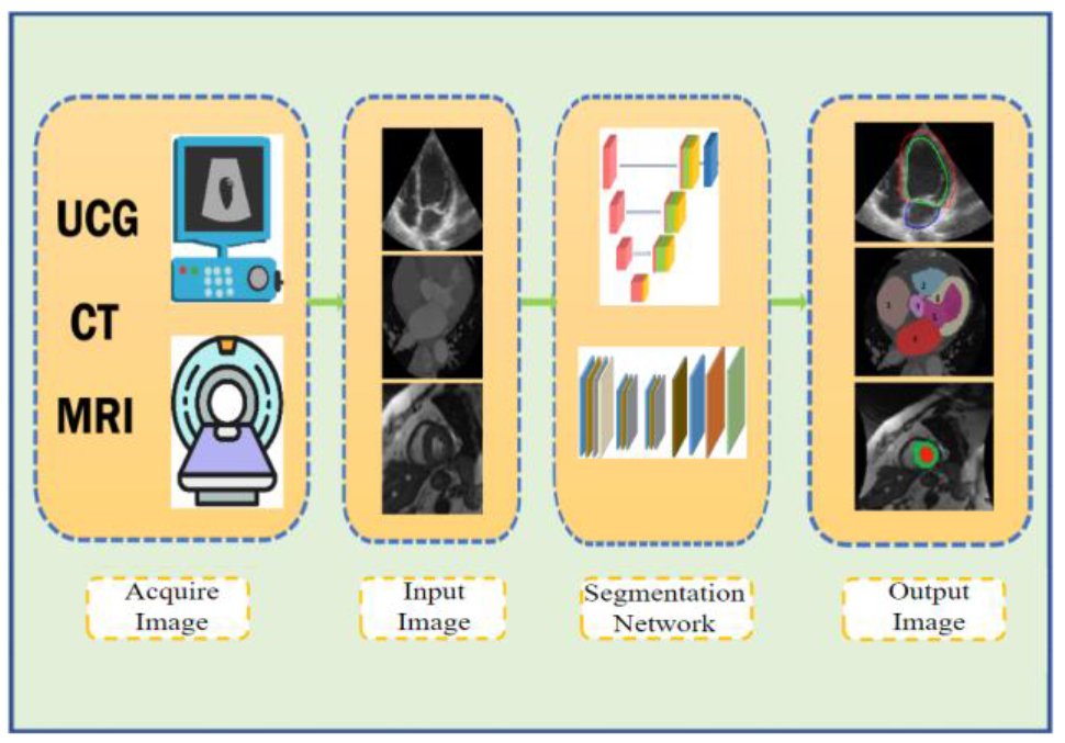
随着老龄化加剧,心血管疾病患病人数逐年增加,借助医学图像实现心脏功能的评估在诊疗过程中起着重要作用.心脏分割是评估心脏功能的前提,一直受到临床医生和科学研究者的密切关注.本文从传统方法和深度学习方法角度梳理了近十年以来关于心脏分割研究的文献.重点介绍了基于主动轮廓和图谱模型的传统分割方法,以及基于U-Net和全卷积神经网络(FCN)的深度学习算法.其中针对通过增加局部模块、优化损失函数、强化网络结构等方式改进深度学习网络以实现心脏特定区域精准分割这一主题进行了详细展开,并从心脏磁共振、X射线计算机断层扫描(CT)和超声3种成像模态对上述方法进行总结.最后总结了该领域目前的研究现状并对未来研究方向进行了展望.
常博 , 孙灏芸 , 高清宇 , 王丽嘉 . 传统方法和深度学习用于不同模态心脏医学图像的分割研究进展[J]. 波谱学杂志, 2024 , 41(2) : 224 -244 . DOI: 10.11938/cjmr20233086
As the aging population increases, the prevalence of cardiovascular disease rises annually. In this context, the evaluation of cardiac function using medical imaging techniques plays a pivotal role in the diagnosis and treatment of cardiovascular disease. Cardiac segmentation is a prerequisite for assessing cardiac function and has been closely studied by clinicians and scientific researchers. This paper provides a comprehensive review of the literature from the past decade on cardiac segmentation, categorizing the studies into traditional segmentation approaches and deep learning methodologies. Emphasis is placed on the detailed discussion of segmentation methods based on active contours and atlas models; deep learning algorithms based on U-Net and full convolution neural network (FCN) are also extensively discussed. In particular, this paper elaborates various approaches to enhance deep learning networks and achieve accurate segmentation of specific cardiac regions. These approaches include incorporating local modules, optimizing loss functions, and enhancing network architectures. A comprehensive summary of the aforementioned methods is presented, considering three imaging modalities: cardiac magnetic resonance imaging, computed tomography, and ultrasonic cardiogram. Lastly, the article concludes by summarizing the current research status and discussing research directions for further exploration.
Key words: cardiac image segmentation; deep learning; U-Net; FCN
| [1] | MA L Y, WANG Z W, FAN J, et al. Interpretation of report on cardiovascular health and diseases in China 2022[J]. Chinese General Practice, 2023, 26(32): 3975-3994. |
| 马丽媛, 王增武, 樊静, 等. 《中国心血管健康与疾病报告2022》要点解读[J]. 中国全科医学, 2023, 26(32): 3975-3994. | |
| [2] | SENTHILKUMARAN N, VAITHEGI S. Image segmentation by using thresholding techniques for medical images[J]. Computer Sci & Eng, 2016, 6(1): 1-13. |
| [3] | LEE J, KIM N, LEE H, et al. Efficient liver segmentation using a level-set method with optimal detection of the initial liver boundary from level-set speed images[J]. Comput Meth Prog Bio, 2007, 88(1): 26-38. |
| [4] | XU L, ZHU Y, ZHANG Y, et al. Liver segmentation based on region growing and level set active contour model with new signed pressure force function[J]. Optik, 2020, 202: 163705. |
| [5] | FARAG A A, ABD EL MUNIM H E, GRAHAM J H, et al. A novel approach for lung nodules segmentation in chest CT using level sets[J]. IEEE T Image Process, 2013, 22(12): 5202-5213. |
| [6] | SWIERCZYNSKI P, PAPIE? B W, SCHNABEL J A, et al. A level-set approach to joint image segmentation and registration with application to CT lung imaging[J]. Comput Med Image Grap, 2018, 65: 58-68. |
| [7] | SHRIVASTAVA N, BHARTI J. Automatic seeded region growing image segmentation for medical image segmentation: a brief review[J]. Int J Image Graph, 2020, 20(3): 2050018. |
| [8] | ZHOU S, WANG J, ZHANG S, et al. Active contour model based on local and global intensity information for medical image segmentation[J]. Neurocomputing, 2016, 186: 107-118. |
| [9] | IGLESIAS J E, SABUNCU M R. Multi-atlas segmentation of biomedical images: a survey[J]. Med Image Anal, 2015, 24(1): 205-219. |
| [10] | ZHANG Z, DUAN C, LIN T, et al. GVFOM: a novel external force for active contour based image segmentation[J]. Inform Sciences, 2020, 506: 1-18. |
| [11] | YANG C, WU W, SU Y, et al. Left ventricle segmentation via two-layer level sets with circular shape constraint[J]. Magn Reson Imaging, 2017, 38: 202-213. |
| [12] | HU H F, Liu H H, Gao Z Y, et al. Hybrid segmentation of left ventricle in cardiac MRI using gaussian-mixture model and region restricted dynamic programming[J]. Magn Reson Imaging, 2013, 31: 575-584. |
| [13] | BAI W, SHI W, LEDIG C, et al. Multi-atlas segmentation with augmented features for cardiac MR images[J]. Med Image Analysis, 2015, 19(1): 98-109. |
| [14] | NU?EZ-GARCIA M, ZHUANG X, SANROMA G, et al. Left atrial segmentation combining multi-atlas whole heart labeling and shape-based atlas selection[C]// Statistical Atlases and Computational Models of the Heart. MICCAI 2018, Granada, Spain:Springer International Publishing, 2019: 302-310. |
| [15] | CHAN T F, VESE L A. Active contours without edges[J]. IEEE T Image Process, 2001, 10(2): 266-277. |
| [16] | SAINI K, DEWAL M L, ROHIT M. A fast region-based active contour model for boundary detection of echocardiographic images[J]. J Digit Imaging, 2012, 25: 271-278. |
| [17] | LIU L X, MA Z M, Z H B, et al. A method for segmenting cardiac magnetic resonance images using active contours[J]. Chinese Journal of Computers, 2012, 35(1): 146-153. |
| 刘利雄, 马忠梅, 赵恒博, 等. 一种基于主动轮廓模型的心脏核磁共振图像分割方法[J]. 计算机学报, 2012, 35(1): 146-153. | |
| [18] | ZHAO H C, YUAN J H, ZHU E R, et al. An improved double level set algorithm for left ventricular segmentation of cardiac MRI images[J]. Comp Technol Dev, 2022, 32(6): 162-166. |
| 赵昊宸, 苑金辉, 朱恩嵘, 等. 改进的双水平集心脏MRI图像左心室分割算法[J]. 计算机技术与发展, 2022, 32(6): 162-166. | |
| [19] | QIAO M, WANG Y, VAN DER GEEST R J, et al. Fully automated left atrium cavity segmentation from 3D GE-MRI by multi-atlas selection and registration[C]// Statistical Atlases and Computational Models of the Heart. Held in Conjunction with MICCAI 2018. Granada, Spain:Springer International Publishing, 2019: 230-236. |
| [20] | WANG L J, SU X Y, LI Y, et al. Segmentation of right ventricle in cardiac cine MRI using COLLATE fusion-based multi-atlas[J]. Chinese J Magn Reson, 2018, 35(4): 407-416. |
| 王丽嘉, 苏新宇, 李亚, 等. 基于COLLATE融合多图谱的心脏电影MRI右心室分割[J]. 波谱学杂志, 2018, 35(4): 407-416. | |
| [21] | SU X Y, WANG L J, ZHU Y C. A new method of multi- atlas segmentation of right ventricle based on cardiac film magnetic resonance images[J]. Acta Phys Sin, 2019, 68(19): 50-60. |
| 苏新宇, 王丽嘉, 朱艳春. 基于心脏电影磁共振图像的一种新的右心室多图谱分割方法[J]. 物理学报, 2019, 68(19): 50-60. | |
| [22] | YANG G, SUN C, CHEN Y, et al. Automatic whole heart segmentation in CT images based on multi-atlas image registration[C]// ACDC and MMWHS Challenges:8th International Workshop, STACOM 2017. Canada: Springer International Publishing, 2018: 250-257. |
| [23] | GALISOT G, BROUARD T, RAMEL J Y. Local probabilistic atlases and a posteriori correction for the segmentation of heart images[C]// ACDC and MMWHS Challenges:8th International Workshop, STACOM 2017, Held in Conjunction with MICCAI 2017. Quebec City, Canada: Springer International Publishing, 2018: 207-214. |
| [24] | LI Z H, MEI X, GUO X Y, et al. Fuzzy level set segmentation method for cardiac CT image sequence[J]. Computer Engineering and Design, 2015, 36(11): 3030-3034+3045. |
| 李振华, 梅雪, 郭笑妍, 等. 模糊水平集心脏CT图像序列分割方法[J]. 计算机工程与设计, 2015, 36(11): 3030-3034+3045. | |
| [25] | WU X. Cardiac CT segmentation based on distance regularized level set[C]// 2021 International Conference on Big Data Analytics for Cyber-Physical System in Smart City. Springer Singapore, 2022, 2: 123-131. |
| [26] | HE C B, MA X L, YU C M. Study of left atrium segmentation in dual source CT image with random walks algorithms[J]. Electronic Measurement Technology, 2016, 39(5): 75-79. |
| 何昌保, 马秀丽, 余长明. 基于Random Walks算法的心脏双源CT左心房分割[J]. 电子测量技术, 2016, 39(5): 75-79. | |
| [27] | LEUNG K Y E, BOSCH J G. Automated border detection in three-dimensional echocardiography: principles and promises[J]. Eur J Echocardiogr, 2010, 11(2): 97-108. |
| [28] | CHALANA V, LINKER D T, HAYNOR D R, et al. A multiple active contour model for cardiac boundary detection on echocardiographic sequences[J]. IEEE T Med Imaging, 1996, 15(3): 290-298. |
| [29] | HANSEGARD J, ORDERUD F, RABBEN S I. Real-time active shape models for segmentation of 3D cardiac ultrasound[C]// International Conference on Computer Analysis of Images and Patterns. Berlin, Heidelberg: Springer Berlin Heidelberg, 2007: 157-164. |
| [30] | SMISTAD E, LINDSETH F. Real-time tracking of the left ventricle in 3D ultrasound using Kalman filter and mean value coordinates[C]// CETU2014. Boston: Springer International Publishing, 2014: 65-72. |
| [31] | HUANG X, ZHU H, WANG J. Adoption of snake variable model-based method in segmentation and quantitative calculation of cardiac ultrasound medical images[J]. J Health Eng, 2021, 2021:2425482. |
| [32] | LECUN Y, BOTTOU L, BENGIO Y, et al. Gradient-based learning applied to document recognition[J]. P IEEE, 1998, 86(11): 2278-2324. |
| [33] | KRIZHEVSKY A, SUTSKEVER I, HINTON G E. ImageNet classification with deep convolutional neural networks[J]. Commun Acm, 2017, 60(6): 84-90. |
| [34] | LONG J, SHELHAMER E, DARRELL T. Fully convolutional networks for semantic segmentation[C]// Proceedings of the IEEE conference on computer vision and pattern recognition. USA: IEEE, 2015: 3431-3440. |
| [35] | RONNEBERGER O, FISCHER P, BROX T. U-net: Convolutional networks for biomedical image segmentation[C]// Medical Image Computing and Computer-Assisted Intervention-MICCAI 2015: 18th International Conference. Munich, Germany, Proceedings, Part III 18: Springer International Publishing, 2015: 234-241. |
| [36] | PENSO M, MOCCIA S, SCAFURI S, et al. Automated left and right ventricular chamber segmentation in cardiac magnetic resonance images using dense fully convolutional neural network[J]. Comput Meth Prog Bio, 2021, 204: 106059. |
| [37] | WANG H, WANG T T, WANG L J. Squeeze-and-excitation residual U-shaped network for left myocardium segmentation based on cine cardiac magnetic resonance images[J]. Chinese J Magn Reson, 2023, 40(4): 435-447. |
| 王慧, 王甜甜, 王丽嘉. 基于心脏磁共振电影图像的压缩激励残差U形网络左心肌分割[J]. 波谱学杂志, 2023, 40(4): 435-447. | |
| [38] | SIMANTIRIS G, TZIRITAS G. Cardiac MRI segmentation with a dilated CNN incorporating domain-specific constraints[J]. IEEE J-STSP, 2020, 14(6): 1235-1243. |
| [39] | CUI H, YUWEN C, JIANG L, et al. Multiscale attention guided U-Net architecture for cardiac segmentation in short-axis MRI images[J]. Comput Meth Prog Bio, 2021, 206: 106142. |
| [40] | DONG S, PAN Z, FU Y, et al. DeU-Net 2.0: Enhanced deformable U-Net for 3D cardiac cine MRI segmentation[J]. Med Image Anal, 2022, 78: 102389. |
| [41] | DONG Z, DU X, LIU Y. Automatic segmentation of left ventricle using parallel end-end deep convolutional neural networks framework[J]. Knowl-Based Syst, 2020, 204: 106210. |
| [42] | WANG T C, XIONG J J, XU X W, et al. Msu-net: Multiscale statistical U-Net for real-time 3D cardiac MRI video segmentation[C]// Medical Image Computing and Computer Assisted Intervention-MICCAI 2019. Shenzhen, China: Springer International Publishing, 2019: 614-622. |
| [43] | LIU P, ZHONG Y M, WANG L J. Automatic segmentation of right ventricle in cine cardiac magnetic resonance image based on a dense and multi-scale U-net method[J]. Chinese J Magn Reson, 2020, 37(4): 456-468. |
| 刘鹏, 钟玉敏, 王丽嘉. 基于密集多尺度U-net网络的电影心脏磁共振图像右心室自动分割[J]. 波谱学杂志, 2020, 37(4): 456-468. | |
| [44] | WANG L, SU H, LIU P. Automatic right ventricular segmentation for cine cardiac magnetic resonance images based on a new deep atlas network[J]. Med Phys, 2023, 50(11): 7060-7070. |
| [45] | ZHANG Y, WANG F, WU H, et al. An automatic segmentation method with self-attention mechanism on left ventricle in gated PET/CT myocardial perfusion imaging[J]. Comput Meth Prog Bio, 2023, 229: 107267. |
| [46] | ABDELTAWAB H, KHALIFA F, TAHER F, et al. A deep learning-based approach for automatic segmentation and quantification of the left ventricle from cardiac cine MR images[J]. Comput Med Image Grap, 2020, 81: 101717. |
| [47] | ZHANG Y, YANG J, HOU F, et al. Semi-supervised cardiac image segmentation via label propagation and style transfer[C]// Statistical Atlases and Computational Models of the Heart, M&Ms and EMIDEC Challenges. Lima, Peru: Springer International Publishing, 2021: 219-227. |
| [48] | TRAN P V. A fully convolutional neural network for cardiac segmentation in short-axis MRI[J]. arXiv preprint, arXiv:1604.00494, 2016. |
| [49] | DA SILVA I F S, SILVA A C, DE PAIVA A C, et al. A cascade approach for automatic segmentation of cardiac structures in short-axis cine-MR images using deep neural networks[J]. Expert Syst Appl, 2022, 197: 116704. |
| [50] | WU B, FANG Y, LAI X. Left ventricle automatic segmentation in cardiac MRI using a combined CNN and U-net approach[J]. Comput Med Image Grap, 2020, 82: 101719. |
| [51] | SHAAF Z F, JAMIL M M A, AMBAR R, et al. Automatic left ventricle segmentation from short-axis cardiac MRI images based on fully convolutional neural network[J]. Diagnostics, 2022, 12(2): 414. |
| [52] | WANG Z, XIE L, QI J. Dynamic pixel-wise weighting-based fully convolutional neural networks for left ventricle segmentation in short-axis MRI[J]. Magn Reson Imaging, 2020, 66: 131-140. |
| [53] | XIONG J, PO L M, CHEUNG K W, et al. Edge-sensitive left ventricle segmentation using deep reinforcement learning[J]. Sensors, 2021, 21(7): 2375. |
| [54] | XU Z, WU Z, FENG J. CFUN: Combining faster R-CNN and U-net network for efficient whole heart segmentation[J]. arXiv preprint, arXiv:1812.04914, 2018. |
| [55] | TONG Q, NING M, SI W, et al. 3D deeply-supervised U-Net based whole heart segmentation[C]// ACDC and MMWHS Challenges, STACOM 2017, Held in Conjunction with MICCAI 2017. Canada: Springer International Publishing, 2018: 224-232. |
| [56] | WANG C, MACGILLIVRAY T, MACNAUGHT G, et al. A two-stage 3D Unet framework for multi-class segmentation on full resolution image[J]. arXiv preprint, arXiv:1804.04341, 2018. |
| [57] | Guo B J, HE X, LEI Y, et al. Automated left ventricular myocardium segmentation using 3D deeply supervised attention U-net for coronary computed tomography angiography; CT myocardium segmentation[J]. Med Physic, 2020, 47(4): 1775-1785. |
| [58] | YE C, WANG W, ZHANG S, et al. Multi-depth fusion network for whole-heart CT image segmentation[J]. IEEE Access, 2019, 7: 23421-23429. |
| [59] | WANG C, SMEDBY ?. Automatic whole heart segmentation using deep learning and shape context[C]// ACDC and MMWHS Challenges, STACOM 2017, Held in Conjunction with MICCAI 2017. Canada: Springer International Publishing, 2018: 242-249. |
| [60] | YE M, HUANG Q, YANG D, et al. PC-U net: Learning to jointly reconstruct and segment the cardiac walls in 3D from CT data[C]// Statistical Atlases and Computational Models of the Heart. M&Ms and EMIDEC Challenges, Held in Conjunction with MICCAI 2020. Lima, Peru: Springer International Publishing, 2021: 117-126. |
| [61] | HE X, GUO B J, LEI Y, et al. Automatic epicardial fat segmentation in cardiac CT imaging using 3D deep attention U-Net[C]//Medical Imaging 2020:Image Processing. SPIE, 2020, 11313: 589-595. |
| [62] | CHEN Q Y, WEI R H, SHI L Y, et al. Whole-heart CT image segmentation based on improved U-Net[J]. Mode Inf Tech, 2021, 5(13): 76-80. |
| 陈秋叶, 韦瑞华, 石璐莹, 等. 基于改进U-Net的全心脏CT图像分割[J]. 现代信息科技, 2021, 5(13): 76-80. | |
| [63] | VASWANI A, SHAZEER N, PARMAR N, et al. Attention is all you need[J]. Adv Neural Inf Process Syst, 2017, 30: 5998-6008. |
| [64] | YANG X, TIAN X. TransUnet: Using attention mechanism for whole heart segmentation[C]// 2022 IEEE 2nd International Conference on Power, ICPECA. IEEE, 2022: 553-556. |
| [65] | YANG X, BIAN C, YU L, et al. Hybrid loss guided convolutional networks for whole heart parsing[C]// ACDC and MMWHS Challenges, STACOM 2017, Held in Conjunction with MICCAI 2017. Canada: Springer International Publishing, 2018: 215-223. |
| [66] | KOO H J, LEE J G, KO J Y, et al. Automated segmentation of left ventricular myocardium on cardiac computed tomography using deep learning[J]. Korean J Radio, 2020, 21(6): 660-669. |
| [67] | DEGEL M A, NAVAB N, ALBARQOUNI S. Domain and geometry agnostic CNNs for left atrium segmentation in 3D ultrasound[C]// Medical Image Computing and Computer Assisted Intervention-MICCAI 2018. Granada, Spain:Springer International Publishing, 2018: 630-637. |
| [68] | OKTAY O, FERRANTE E, KAMNITSAS K, et al. Anatomically constrained neural networks (ACNNs): application to cardiac image enhancement and segmentation[J]. IEEE T Med Imaging, 2017, 37(2): 384-395. |
| [69] | SFAKIANAKIS C, SIMANTIRIS G, TZIRITAS G. GUDU: Geometrically-constrained ultrasound data augmentation in U-Net for echocardiography semantic segmentation[J]. Biomed Signal Proces, 2023, 82: 104557. |
| [70] | DOZEN A, KOMATSU M, SAKAI A, et al. Image segmentation of the ventricular septum in fetal cardiac ultrasound videos based on deep learning using time-series information[J]. Biomolecules, 2020, 10(11): 1526. |
| [71] | YU L, GUO Y, WANG Y, et al. Segmentation of fetal left ventricle in echocardiographic sequences based on dynamic convolutional neural networks[J]. IEEE T Bio-Med Eng, 2016, 64(8): 1886-1895. |
| [72] | PHILIP M E, FERRIEIRA A, TOMAR A, et al. A machine learning framework for fully automatic 3D fetal cardiac ultrasound evaluation[C]// 2022 ISBI: IEEE, 2022: 1-5. |
| [73] | LIU F, WANG K, LIU D, et al. Deep pyramid local attention neural network for cardiac structure segmentation in two-dimensional echocardiography[J]. Med Image Anal, 2021, 67: 101873. |
| [74] | AWASTHI N, VERMEER L, FIXSEN L S, et al. LVNet: lightweight model for left ventricle segmentation for short axis views in echocardiographic imaging[J]. IEEE Trans Ultra, 2022, 69(6): 2115-2128. |
| [75] | LECLERC S, SMISTAD E, GRENIER T, et al. Deep learning applied to multi-structure segmentation in 2D echocardiography: A preliminary investigation of the required database size[C]// IUS. Kobe, Japan: IEEE, 2018: 1-4. |
| [76] | JAFARI M H, GIRGIS H, LIAO Z, et al. A unified framework integrating recurrent fully-convolutional networks and optical flow for segmentation of the left ventricle in echocardiography data[C]// Deep Learning in Medical Image Analysis and Multimodal Learning for Clinical Decision Support: DLMIA 2018, ML-CDS 2018, Held in Conjunction with MICCAI 2018. Granada, Spain:Springer International Publishing, 2018: 29-37. |
| [77] | JAFARI M H, GIRGIS H, VAN WOUDENBERG N, et al. Automatic biplane left ventricular ejection fraction estimation with mobile point-of-care ultrasound using multi-task learning and adversarial training[J]. Int J Comput Ass Rad, 2019, 14: 1027-1037. |
| [78] | CHEN H, ZHENG Y, PARK J H, et al. Iterative multi-domain regularized deep learning for anatomical structure detection and segmentation from ultrasound images[C]// Medical Image Computing and Computer-Assisted Intervention-MICCAI 2016. Athens, Greece: Proceedings. Springer International Publishing, 2016: 487-495. |
| [79] | NGO T A, LU Z, CARNEIRO G. Combining deep learning and level set for the automated segmentation of the left ventricle of the heart from cardiac cine magnetic resonance[J]. Med Image Anal, 2017, 35: 159-171. |
| [80] | AVENDI M R, KHERADVAR A, JAFARKHANI H. A combined deep-learning and deformable-model approach to fully automatic segmentation of the left ventricle in cardiac MRI[J]. Med Image Anal, 2016, 30: 108-119. |
| [81] | DONG S, LUO G, WANG K, et al. A combined fully convolutional networks and deformable model for automatic left ventricle segmentation based on 3D echocardiography[J]. Biomed Res Int, 2018. |
| [82] | DU L Y, HU L W, ZHANG X Y, et al. Unsupervised segmentation framework with active contour models for cine cardiac MRI[C]// 2021 IEEE International Conference on Image Processing (ICIP). Anchorage, AK, USA: IEEE, 2021: 56-60. |
| [83] | POUDEL R P K, LAMATA P, MONTANA G. Recurrent fully convolutional neural networks for multi-slice MRI cardiac segmentation[C]// Reconstruction, Segmentation, and Analysis of Medical Images:RAMBO 2016 and HVSMR 2016, Held in Conjunction with MICCAI 2016. Athens, Greece: Springer International Publishing, 2017: 83-94. |
| [84] | BADRINARAYANAN V, KENDALL A, CIPOLLA R. SegNet: A deep convolutional encoder-decoder architecture for image segmentation[J]. IEEE T Pattern Anal, 2017, 39(12): 2481-2495. |
| [85] | YAN Z, SU Y, SUN H, et al. SegNet-based left ventricular MRI segmentation for the diagnosis of cardiac hypertrophy and myocardial infarction[J]. Comp Meth Prog Bio, 2022, 227: 107197. |
| [86] | AHMAD I, QAYYUM A, GUPTA B B, et al. Ensemble of 2D residual neural networks integrated with atrous spatial pyramid pooling module for myocardium segmentation of left ventricle cardiac MRI[J]. Mathematics, 2022, 10(4): 627. |
| [87] | CHEN X, WILLIAMS B M, VALLABHANENI S R, et al. Learning active contour models for medical image segmentation[C]// Proceedings of the IEEE/CVF. California: IEEE, 2019: 11632-11640. |
| [88] | BUDAI A, SUHAI F I, CSORBA K, et al. Fully automatic segmentation of right and left ventricle on short-axis cardiac MRI images[J]. Comput Med Image Grap, 2020, 85: 101786. |
| [89] | WANG X, ZHAI S, NIU Y. Left ventricle landmark localization and identification in cardiac MRI by deep metric learning-assisted CNN regression[J]. Neurocomputing, 2020, 399: 153-170. |
| [90] | YAN J R, YAO F Z, WANG L H. Left ventricular myocardium segmentation method of cardiac Cine-MRI based on optical flow and semantic feature fusion[J]. Comput Sys & App, 2022, 31(9): 368-375. |
| 闫景瑞, 姚发展, 王丽会, 基于光流场与语义特征融合的心脏Cine-MRI左心室心肌分割方法[J]. 计算机系统应用, 2022, 31(9): 368-375. | |
| [91] | LI Y J, YANG X Y, YANG X M. Magnetic resonance image reconstruction of multi-scale residual Unet fused with attention mechanism[J]. Chinese J Magn Reson, 2023, 40(3): 307-319. |
| 李奕洁, 杨馨雨, 杨晓梅. 融合注意力机制的多尺度残差Unet的磁共振图像重建[J]. 波谱学杂志, 2023, 40(3): 307-319. | |
| [92] | BIFFI C, CERROLAZA J J, TARRONI G, et al. 3D high-resolution cardiac segmentation reconstruction from 2D views using conditional variational autoencoders[C]// ISBI 2019. IEEE, 2019: 1643-1646. |
| [93] | ZREIK M, LEINER T, DE VOS B D, et al. Automatic segmentation of the left ventricle in cardiac CT angiography using convolutional neural networks[C]// ISBI 2016. IEEE, 2016: 40-43. |
| [94] | PAYER C, ?TERN D, BISCHOF H, et al. Multi-label whole heart segmentation using CNNs and anatomical label configurations[C]// ACDC and MMWHS Challenges: STACOM 2017, Held in Conjunction with MICCAI 2017. Quebec City, Canada: Springer International Publishing, 2018: 190-198. |
| [95] | XU F, LIN L, LI Z, et al. MRDFF: A deep forest based framework for CT whole heart segmentation[J]. Methods, 2022, 208: 48-58. |
| [96] | GOODFELLOW I, POUGET-ABADIE J, MIRZA M, et al. Generative adversarial networks[J]. Commun Acm, 2020, 63(11): 139-144. |
| [97] | YU WEN C, JIANG L, CUI H. Multiple GANs guided by self-attention mechanism for automatic cardiac image segmentation[C]// Thirteenth ICGIP 2021. SPIE, 2022, 12083: 509-515. |
| [98] | LE K, LOU Z, HUO W, et al. Auto whole heart segmentation from CT images using an improved Unet-Gan[C]// Journal of Physics: Conference Series. Dalian: ACM, 2021, 1769(1): 012016. |
| [99] | MORTAZI A, BURT J, BAGCI U. Multi-planar deep segmentation networks for cardiac substructures from MRI and CT[C]// ACDC and MMWHS Challenges: STACOM 2017, Held in Conjunction with MICCAI 2017. Canada: Springer International Publishing, 2018: 199-206. |
| [100] | ZHAO F, LIU J. Cardiac CT image segmentation based on convolutional neural network and image saliency[J]. Beijing Bio Eng, 2020, 39(1): 48-55. |
| 赵飞, 刘杰. 基于卷积神经网络和图像显著性的心脏CT图像分割[J]. 北京生物医学工程, 2020, 39(1): 48-55. | |
| [101] | ZHANG E, SIMA M, WANG J, et al. Weakly Supervised Whole Cardiac Segmentation via Attentional CNN[C]// International Conference on Intelligence Science. Cham: Springer International Publishing, 2022: 76-83. |
| [102] | DORMER J D, MA L, HALICEK M, et al. Heart chamber segmentation from CT using convolutional neural networks[C]// Medical Imaging 2018:Biomedical Applications in Molecular, Structural, and Functional Imaging. SPIE, 2018, 10578: 659-664. |
| [103] | YANG X, BIAN C, YU L, et al. 3D convolutional networks for fully automatic fine-grained whole heart partition[C]// ACDC and MMWHS Challenges: STACOM 2017, Held in Conjunction with MICCAI 2017. Canada: Springer International Publishing, 2018: 181-189. |
| [104] | BRUNS S, WOLTERINK J M, VAN DEN BOOGERT T P W, et al. Deep learning-based whole-heart segmentation in 4D contrast-enhanced cardiac CT[J]. Comput Biol Med, 2022, 142: 105191. |
| [105] | CARNEIRO G, NASCIMENTO J, FREITAS A. Robust left ventricle segmentation from ultrasound data using deep neural networks and efficient search methods[C]// 2010 IEEE Inter Symp Biomed Image: From Nano to Macro. IEEE, 2010: 1085-1088. |
| [106] | CARNEIRO G, NASCIMENTO J C, FREITAS A. The segmentation of the left ventricle of the heart from ultrasound data using deep learning architectures and derivative-based search methods[J]. IEEE T Image Process, 2011, 21(3): 968-982. |
| [107] | NASCIMENTO J C, CARNEIRO G. Deep learning on sparse manifolds for faster object segmentation[J]. IEEE T Image Process, 2017, 26(10): 4978-4990. |
| [108] | VENI G, MORADI M, BULU H, et al. Echocardiography segmentation based on a shape-guided deformable model driven by a fully convolutional network prior[C]// ISBI 2018. IEEE, 2018: 898-902. |
| [109] | SMISTAD E, STVIK A, SALTE I M, et al. Fully automatic real-time ejection fraction and MAPSE measurements in 2D echocardiography using deep neural networks[C]// IUS 2018. Kobe, Japan: IEEE, 2018: 1-4. |
| [110] | HSU W Y. Automatic left ventricle recognition, segmentation and tracking in cardiac ultrasound image sequences[J]. IEEE Access, 2019, 7: 140524-140533. |
| [111] | SAEED M, MUHTASEB R, YAQUB M. Contrastive pretraining for echocardiography segmentation with limited data[C]// Medical Image Understanding and Analysis:MIUA 2022. Cham: Springer International Publishing, 2022: 680-691. |
| [112] | FERREIRA D L, SALAYMANG Z, ARNAOUT R. Label-free segmentation from cardiac ultrasound using self-supervised learning[J]. arXiv preprint, arXiv:2210.04979, 2022. |
| [113] | YU C, LI S, GHISTA D, et al. Multi-level multi-type self-generated knowledge fusion for cardiac ultrasound segmentation[J]. Inform Fusion, 2023, 92: 1-12. |
| [114] | LI M, DONG S, GAO Z, et al. Unified model for interpreting multi-view echocardiographic sequences without temporal information[J]. Appl Soft Comput, 2020, 88: 106049. |
| [115] | LIN T Y, SONG L, GAO Z F, et al. Evaluation of a deep learning-based model for 2-D echocardiography segmentation on small datasets[J]. Journal of Jinan University (Natural Science & Medicine Edition), 2022, 43(2): 191-198. |
| 林天予, 宋亮, 高智凡, 等. 基于深度学习的二维心脏超声图像分割模型在小规模数据集上的性能评估[J]. 暨南大学学报(自然科学与医学版), 2022, 43(2): 191-198. | |
| [116] | JAFARI M H, GIRGIS H, ABDI A H, et al. Semi-supervised learning for cardiac left ventricle segmentation using conditional deep generative models as prior[C]// ISBI 2019. IEEE, 2019: 649-652. |
| [117] | OUYANG D, HE B, GHORBANI A, et al. Video-based AI for beat-to-beat assessment of cardiac function[J]. Nature, 2020, 580(7802): 252-256. |
| [118] | DONG S, LUO G, WANG K, et al. VoxelAtlasGAN: 3D left ventricle segmentation on echocardiography with atlas guided generation and voxel-to-voxel discrimination[C]// Medical Image Computing and Computer Assisted Intervention-MICCAI 2018. Spain:Springer International Publishing, 2018: 622-629. |
| [119] | BLVIKEN H S, VERONESI F, SAMSET E. Simultaneous segmentation of all four chambers in cardiac ultrasound images[J]. Comp M Bio Bio E-IV, 2022: 1-8. |
| [120] | DONG S, PAN Z, FU Y, et al. Partial unbalanced feature transport for cross-modality cardiac image segmentation[J]. IEEE T Med Imaging, 2023, 42(6): 1758-1773. |
| [121] | LIU J, LIU H, GONG S, et al. Automated cardiac segmentation of cross-modal medical images using unsupervised multi-domain adaptation and spatial neural attention structure[J]. Med Image Anal, 2021, 72: 102135. |
| [122] | WU F, ZHUANG X. Unsupervised domain adaptation with variational approximation for cardiac segmentation[J]. IEEE T Med Imaging, 2021, 40(12): 3555-3567. |
| [123] | ZHANG Y, WANG Y, XU L, et al. ST-GAN: A swin transformer-based generative adversarial network for unsupervised domain adaptation of cross-modality cardiac segmentation[J]. IEEE J Biomed Health, 2024, 28(2): 893-904. |
| [124] | JANG Y, HONG Y, HA S, et al. Automatic segmentation of LV and RV in cardiac MRI[C]// ACDC and MMWHS Challenges: STACOM 2017, Held in Conjunction with MICCAI 2017. Canada: Springer International Publishing, 2018: 161-169. |
| [125] | YANG X, BIAN C, YU L, et al. Class-balanced deep neural network for automatic ventricular structure segmentation[C]// ACDC and MMWHS Challenges: STACOM 2017, Held in Conjunction with MICCAI 2017. Canada: Springer International Publishing, 2018: 152-160. |
| [126] | SANDER J, DE VOS B D, WOLTERINK J M, et al. Towards increased trustworthiness of deep learning segmentation methods on cardiac MRI[C]//Medical Imaging 2019:Image Processing. SPIE, 2019, 10949: 324-330. |
| [127] | CHEN M, FANG L, LIU H. FR-NET: Focal loss constrained deep residual networks for segmentation of cardiac MRI[C]// ISBI 2019. IEEE, 2019: 764-767. |
| [128] | LIN A, WU J, YANG X. A data augmentation approach to train fully convolutional networks for left ventricle segmentation[J]. Magn Reson Imaging, 2020, 66: 152-164. |
| [129] | FENG R, DEB B, GANESAN P, et al. Automatic left atrial segmentation from cardiac CT using computer graphics imaging and deep learning[J]. Euro Heart J, 2022, 43(Supplement_2): 472-544. |
| [130] | CHARTSIAS A, JOYCE T, DHARMAKUMAR R, et al. Adversarial image synthesis for unpaired multi-modal cardiac data[C]// Simulation and Synthesis in Medical Imaging: SASHIMI 2017, Held in Conjunction with MICCAI 2017. Canada: Springer International Publishing, 2017: 3-13. |
| [131] | KIRILLOV A, MINTUN E, RAVI N, et al. Segment anything[J]. arXiv preprint, arXiv:2304.02643, 2023. |
| [132] | ZOU X, YANG J, ZHANG H, et al. Segment Everything Everywhere All at Once[J]. arXiv preprint, arXiv:2304.06718, 2023. |
| [133] | MA J, WANG B. Segment anything in medical images[J]. arXiv preprint, arXiv:2304.12306, 2023. |
/
| 〈 |
|
〉 |