

超极化129Xe MR用于COPD急性加重的肺功能研究
收稿日期: 2024-03-28
网络出版日期: 2024-04-13
基金资助
国家自然科学基金项目(81930049);国家自然科学基金项目(82127802);国家自然科学基金项目(21921004);国家自然科学基金项目(82372150);国家自然科学基金项目(82202119);国家重点研发计划(2018YFA0704000);国家重点研发计划(2022-YFC2410000);湖北省科技重大专项(2021ACA013);湖北省科技重大专项(2023BAA021);中国科学院重点项目(ZDBS-LY-JSC004);中国科学院重点项目(XDB0540000);中国科学院青年促进会专项(2020330);中国科学院青年促进会专项(2021330)
Assessment of Pulmonary Function Changes of AECOPD with Hyperpolarized 129Xe MR
Received date: 2024-03-28
Online published: 2024-04-13
李连杰 , 郑瑜 , 谢树光 , 张鸣 , 李红闯 , 刘小玲 , 赵修超 , 韩叶清 , 李海东 , 范丽 , 萧毅 , 刘士远 , 周欣 . 超极化129Xe MR用于COPD急性加重的肺功能研究[J]. 波谱学杂志, 2024 , 41(4) : 363 -372 . DOI: 10.11938/cjmr20243106
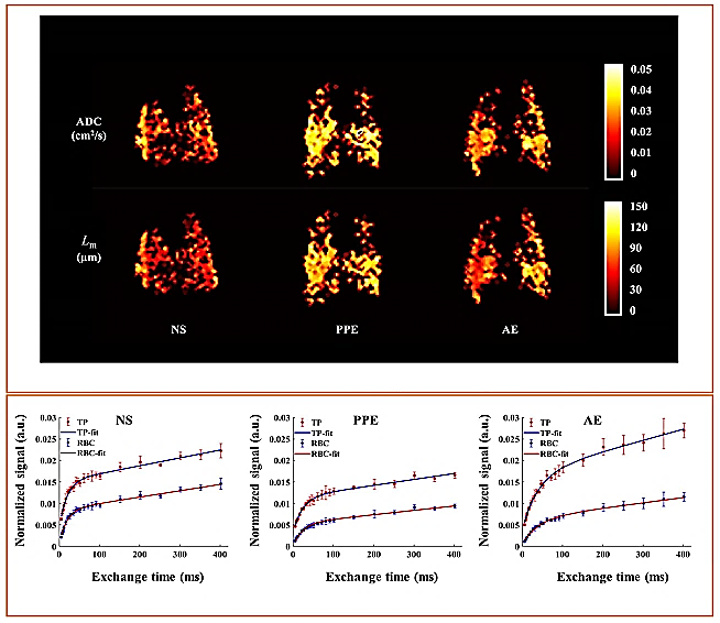
The acute exacerbation of chronic obstructive pulmonary disease (AECOPD) constitutes a significant adverse event in the trajectory of COPD. In this study, we utilized hyperpolarized 129Xe magnetic resonance (MR) to explore the alterations in alveolar microstructure and pulmonary gas exchange function in an AECOPD disease model. Our findings revealed that, in comparison to the emphysema group, the microstructure remained relatively unchanged in the acute exacerbation group, while gas exchange function exhibited a decrease. Notably, these observed changes correlated well with histological findings and pulmonary function assessments, indicating the feasibility and sensitivity of hyperpolarized 129Xe MR in detecting AECOPD.
Key words: hyperpolarized 129Xe MR; DWI; CSSR; COPD; emphysema; acute exacerbation
| [1] | SORIANO J B, ABAJOBIR A A, ABATE K H, et al. Global, regional, and national deaths, prevalence, disability-adjusted life years, and years lived with disability for chronic obstructive pulmonary disease and asthma, 1990-2015: a systematic analysis for the global burden of disease study 2015[J]. Lancet Respir Med, 2017, 5(9): 691-706. |
| [2] | DIAZ-GUZMAN E, MANNINO D M. Epidemiology and prevalence of chronic obstructive pulmonary disease[J]. Clin Chest Med, 2014, 35(1): 7-16. |
| [3] | MARCHIONI A, CASTANIERE I, TONELLI R, et al. Ultrasound-assessed diaphragmatic impairment is a predictor of outcomes in patients with acute exacerbation of chronic obstructive pulmonary disease undergoing noninvasive ventilation[J]. Crit Care, 2018, 22(1): 109. |
| [4] | RANGELOV B A, YOUNG A L, JACOB J, et al. Thoracic imaging at exacerbation of chronic obstructive pulmonary disease: a systematic review[J]. Int J Chron Obstruct Pulmon Dis, 2020, 15: 1751-1787. |
| [5] | WEI S S, LU R, ZHANG Z P, et al. MRI-assessed diaphragmatic function can predict frequent acute exacerbation of COPD: a prospective observational study based on telehealth-based monitoring system[J]. BMC Pulm Med, 2022, 22(1): 438. |
| [6] | LI H D, ZHANG Z Y, HAN Y Q, et al. Hyperpolarized gas magnetic resonance imaging of the lung[J]. Chinese J Magn Reson, 2014, 31(3): 307-320. |
| 李海东, 张智颖, 韩叶清, 等. 超极化气体肺部磁共振成像[J]. 波谱学杂志, 2014, 31(3): 307-320. | |
| [7] | ZHANG M, LI H D, LI H C, et al. Quantitative evaluation of lung injury caused by PM2.5 using hyperpolarized gas magnetic resonance[J]. Magn Reson Med, 2020, 84(2): 569-578. |
| [8] | LI H D, ZHANG Z Y, ZHAO X C, et al. Quantitative evaluation of pulmonary gas-exchange function using hyperpolarized 129Xe CEST MRS and MRI[J]. NMR Biomed, 2018, 31(9): e3961. |
| [9] | GUAN S, TUSTISON N, QING K, et al. 3D Single-breath chemical shift imaging hyperpolarized 129Xe MRI of healthy, CF, IPF, and COPD subjects[J]. Tomography, 2022, 8(5): 2574-2587. |
| [10] | RANKINE L J, LU J L, WANG Z Y, et al. Quantifying regional radiation-induced lung injury in patients using hyperpolarized 129Xe gas exchange magnetic resonance imaging[J]. Int J Radiat Oncol Biol Phys, 2024, 120(1): 216-228. |
| [11] | BOUDREAU M, XU X J, SANTYR G E. Measurement of 129Xe gas apparent diffusion coefficient anisotropy in an elastase-instilled rat model of emphysema[J]. Magn Reson Med, 2013, 69(1): 211-220. |
| [12] | MATA J F, ALTES T A, CAI J, et al. Evaluation of emphysema severity and progression in a rabbit model: comparison of hyperpolarized 3He and 129Xe diffusion MRI with lung morphometry[J]. J Appl Physiol (1985), 2007, 102(3): 1273-1280. |
| [13] | RUAN W W, ZHONG J P, WANG K, et al. Detection of the mild emphysema by quantification of lung respiratory airways with hyperpolarized xenon diffusion MRI[J]. J Magn Reson Imaging, 2017, 45(3): 879-888. |
| [14] | STEWART N J, CHAN H F, HUGHES P J C, et al. Comparison of 3He and 129Xe MRI for evaluation of lung microstructure and ventilation at 1.5 T[J]. J Magn Reson Imaging, 2018, 48(3): 632-642. |
| [15] | KAUSHIK S S, CLEVELAND Z I, COFER G P, et al. Diffusion-weighted hyperpolarized 129Xe MRI in healthy volunteers and subjects with chronic obstructive pulmonary disease[J]. Magn Reson Med, 2011, 65(4): 1154-1165. |
| [16] | SAKAI K, BILEK A M, OTEIZA E, et al. Temporal dynamics of hyperpolarized 129Xe resonances in living rats[J]. J Magn Reson B, 1996, 111(3): 300-304. |
| [17] | CHANG Y V. MOXE: A model of gas exchange for hyperpolarized 129Xe magnetic resonance of the lung[J]. Magn Reson Med, 2013, 69(3): 884-890. |
| [18] | KERN A L, GUTBERLET M, QING K, et al. Regional investigation of lung function and microstructure parameters by localized 129Xe chemical shift saturation recovery and dissolved-phase imaging: A reproducibility study[J]. Magn Reson Med, 2019, 81(1): 13-24. |
| [19] | MUMMY D G, COLEMAN E M, WANG Z Y, et al. Regional gas exchange measured by 129Xe magnetic resonance imaging before and after combination bronchodilators treatment in chronic obstructive pulmonary disease[J]. J Magn Reson Imaging, 2021, 54(3): 964-974. |
| [20] | KIRBY M, EDDY R L, PIKE D, et al. MRI ventilation abnormalities predict quality-of-life and lung function changes in mild-to-moderate COPD: longitudinal TINCan study[J]. Thorax, 2017, 72(5): 475-477. |
| [21] | TAFTI S, GARRISON W J, MUGLER J P 3rd, et al. Emphysema index based on hyperpolarized 3He or 129Xe diffusion MRI: performance and comparison with quantitative CT and pulmonary function tests[J]. Radiology, 2020, 297(1): 201-210. |
| [22] | QING K, MUGLER J P 3rd, ALTES T A, et al. Assessment of lung function in asthma and COPD using hyperpolarized 129Xe chemical shift saturation recovery spectroscopy and dissolved-phase MRI[J]. NMR Biomed, 2014, 27(12): 1490-1501. |
| [23] | STEWART N J, HORN F C, NORQUAY G, et al. Reproducibility of quantitative indices of lung function and microstructure from 129Xe chemical shift saturation recovery (CSSR) MR spectroscopy[J]. Magn Reson Med, 2017, 77(6): 2107-2113. |
| [24] | CHEN X M, ZHAO X C, SUN X P, et al. Study on the automatic accumulation-thawing device of hyperpolarized 129Xe[J]. Chinese J Magn Reson, 2022, 39(3): 316-326. |
| 陈小明, 赵修超, 孙献平, 等. 超极化129Xe自动收集-升华装置研究[J]. 波谱学杂志, 2022, 39(3): 316-326. | |
| [25] | WOODS J C, CHOONG C K, YABLONSKIY D A, et al. Hyperpolarized 3He diffusion MRI and histology in pulmonary emphysema[J]. Magn Reson Med, 2006, 56(6): 1293-1300. |
| [26] | SOHRABI F, DIANAT M, BADAVI M, et al. Gallic acid suppresses inflammation and oxidative stress through modulating Nrf2-HO-1-NF-κB signaling pathways in elastase-induced emphysema in rats[J]. Environ Sci Pollut Res Int, 2021, 28(40): 56822-56834. |
| [27] | DE OLIVEIRA M V, ROCHA N N, SANTOS R S, et al. Endotoxin-induced emphysema exacerbation: a novel model of chronic obstructive pulmonary disease exacerbations causing cardiopulmonary impairment and diaphragm dysfunction[J]. Front Physiol, 2019, 10: 664. |
| [28] | KOBAYASHI S, FUJINAWA R, OTA F, et al. A single dose of lipopolysaccharide into mice with emphysema mimics human chronic obstructive pulmonary disease exacerbation as assessed by micro-computed tomography[J]. Am J Respir Cell Mol Biol, 2013, 49(6): 971-977. |
| [29] | ZHANG M, LI H D, LI H C, et al. Dynamic evaluation of acute lung injury using hyperpolarized 129Xe magnetic resonance[J]. NMR Biomed, 2024, 37(4): e5078. |
| [30] | TOLNAI J, SZABARI M V, ALBU G, et al. Functional and morphological assessment of early impairment of airway function in a rat model of emphysema[J]. J Appl Physiol (1985), 2012, 112(11): 1932-1939. |
| [31] | KIRBY M, PIKE D, COXSON H O, et al. Hyperpolarized 3He ventilation defects used to predict pulmonary exacerbations in mild to moderate chronic obstructive pulmonary disease[J]. Radiology, 2014, 273(3): 887-896. |
| [32] | ZINELLU A, MANGONI A A. The emerging clinical significance of the red cell distribution width as a biomarker in chronic obstructive pulmonary disease: a systematic review[J]. J Clin Med, 2022, 11(19): 5642. |
/
| 〈 |
|
〉 |