

改进约束球面反卷积模型的脑灰质微结构成像
收稿日期: 2024-05-14
网络出版日期: 2024-07-10
基金资助
上海市自然科学基金(18ZR1426900)
Improved Constrained Spherical Deconvolution for Microstructural Imaging of Brain Gray Matter
Received date: 2024-05-14
Online published: 2024-07-10
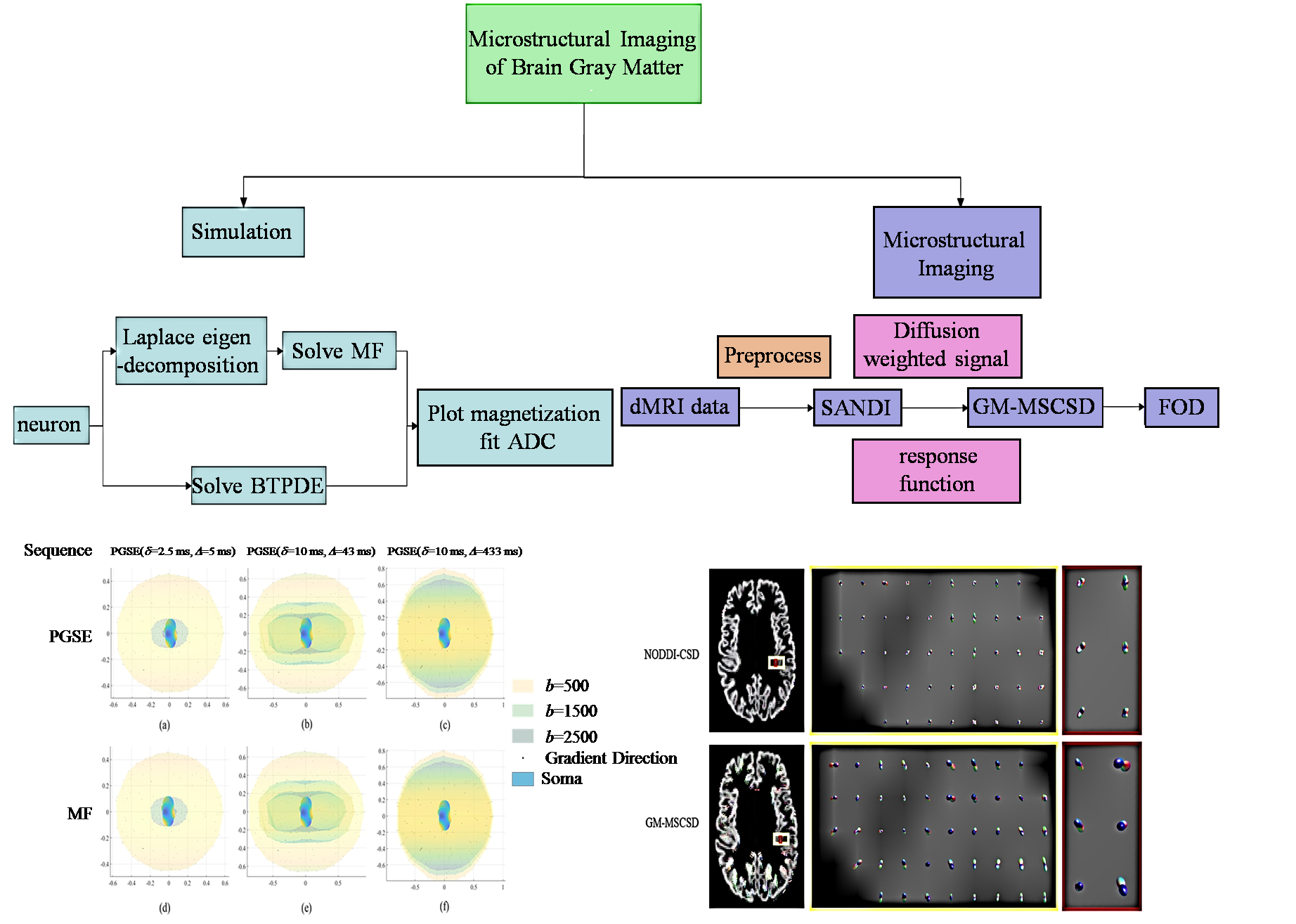
在扩散磁共振成像中,传统多壳约束球面反卷积方法通常将白质微结构建模为各向异性,而灰质微结构建模为各向同性.然而,组织学和高分辨率扩散磁共振成像的研究表明,灰质中水分子的扩散过程具有明显的各向异性特征,因此传统约束球面反卷积方法必不能准确地描述灰质微结构.针对这一问题,本文提出了一种基于体细胞和神经突密度成像(Soma And Neurite Density Imaging,SANDI)模型的灰质多壳约束球面反卷积方法,旨在更准确地揭示灰质微结构特性.利用神经元数据对胞体部分信号进行仿真,模拟灰质特有的信号模式,使用SANDI模型生成了灰质的信号以及相应的响应函数,并将其应用于真实大脑数据的灰质进行微结构重建.结果表明,该方法在提取灰质区域各向异性特征方面表现出色,提高了纤维方向估计的准确度.
杨佳铖 , 王远军 . 改进约束球面反卷积模型的脑灰质微结构成像[J]. 波谱学杂志, 2025 , 42(1) : 67 -79 . DOI: 10.11938/cjmr20243117
In diffusion magnetic resonance imaging (dMRI), conventional multi-shell constrained spherical deconvolution (CSD) methods usually model white matter microstructure as anisotropic and gray matter microstructure as isotropic. Nonetheless, investigations using high-resolution dMRI and histology have demonstrated that the diffusion process in gray matter exhibits clear anisotropic features. To better disclose the microstructural aspects of gray matter, this research suggests a gray matter multi-shell restricted spherical deconvolution method based on the SANDI (Soma And Neurite Density Imaging) model. We first simulate the signal of grey matter using the SpinDoctor with real neuronal data and then reconstruct the fibers in the grey matter portion of real brain data. Experimental results demonstrate that our algorithm effectively captures the anisotropic features of grey matter regions and estimates grey matter fiber orientations with greater accuracy, closely reflecting real-world scenarios.
| [1] | YUE Q, WANG Y J. A fiber tracking algorithm based on non-local constrained spherical deconvolution[J]. Chinese J Magn Reson, 2020, 37(4): 422-433. |
| 岳晴, 王远军. 基于非局部约束球面反卷积模型的纤维追踪算法[J]. 波谱学杂志, 2020, 37(4): 422-433. | |
| [2] | DELL'ACQUA F, RIZZO G, SCIFO P, et al. A model-based deconvolution approach to solve fiber crossing in diffusion-weighted MR imaging[J]. IEEE Trans Biomed Eng, 2007, 54(3): 462-472. |
| [3] | JIANG F, WANG Y J. Construction of human brain templates with diffusion tensor imaging data: a review[J]. Chinese J Magn Reson, 2018, 35(4): 520-530. |
| 蒋帆, 王远军. 扩散张量成像的人脑模板构建[J]. 波谱学杂志, 2018, 35(4): 520-530. | |
| [4] | CACCIOLA A, MILARDI D, CALAMUNERI A, et al. Constrained spherical deconvolution tractography reveals cerebello-mammillary connections in humans[J]. The Cerebellum, 2017, 16: 483-495. |
| [5] | GUO F, LEEMANS A, VIERGEVER M A, et al. Generalized Richardson-Lucy (GRL) for analyzing multi-shell diffusion MRI data[J]. NeuroImage, 2020, 218: 116948. |
| [6] | PIETSCH M, CHRISTIAENS D, HUTTER J, et al. A framework for multi-component analysis of diffusion MRI data over the neonatal period[J]. NeuroImage, 2019, 186: 321-337. |
| [7] | JHA R R, JASWAL G, BHAVSAR A, et al. Single-shell to multi-shell dMRI transformation using spatial and volumetric multilevel hierarchical reconstruction framework[J]. Magn Reson Imaging, 2022(87): 133-156. |
| [8] | JEURISSEN B, TOURNIER J D, DHOLLANDER T, et al. Multi-tissue constrained spherical deconvolution for improved analysis of multi-shell diffusion MRI data[J]. NeuroImage, 2014, 103: 411-426. |
| [9] | JEURISSEN B, SZCZEPANKIEWICZ F. Multi-tissue spherical deconvolution of tensor-valued diffusion MRI[J]. NeuroImage, 2021, 245: 118717. |
| [10] | KROENKE C D. Using diffusion anisotropy to study cerebral cortical gray matter development[J]. J Magn Reson, 2018, 292: 106-116. |
| [11] | ASSAF Y. Imaging laminar structures in the gray matter with diffusion MRI[J]. NeuroImage, 2019, 197: 677-688. |
| [12] | ASSAF Y, BASSER P J. Composite hindered and restricted model of diffusion (CHARMED) MR imaging of the human brain[J]. NeuroImage, 2005, 27(1): 48-58. |
| [13] | ZHANG H, SCHNEIDER T, WHEELER-KINGSHOTT C A, et al. NODDI: Practical in vivo neurite orientation dispersion and density imaging of the human brain[J]. NeuroImage, 2012, 61(4): 1000-1016. |
| [14] | PALOMBO M, IANUS A, GUERRERI M, et al. SANDI: a compartment-based model for non-invasive apparent soma and neurite imaging by diffusion MRI[J]. NeuroImage, 2020, 215: 116835. |
| [15] | ZHU Y M, WANG Y J. Review of brain microstructural imaging with diffusion magnetic resonance imaging[J]. Journal of Chinese Computer Systems, 2023, 44(8): 1763-1770. |
| 朱悦敏, 王远军. 扩散磁共振图像的大脑微结构成像研究综述[J]. 小型微型计算机系统, 2023, 44(8): 1763-1770. | |
| [16] | JESPERSEN S N, KROENKE C D, ?STERGAARD L, et al. Modeling dendrite density from magnetic resonance diffusion measurements[J]. NeuroImage, 2007, 34(4): 1473-1486. |
| [17] | FIEREMANS E, JENSEN J H, HELPERN J A. White matter characterization with diffusional kurtosis imaging[J]. NeuroImage, 2011, 58(1): 177-188. |
| [18] | KADEN E, KRUGGEL F, ALEXANDER D C. Quantitative mapping of the per-axon diffusion coefficients in brain white matter[J]. Magn Reson Med, 2016, 75(4):1752-1763. |
| [19] | YANG D M, HUETTNER J E, BRETTHORST G L, et al. Intracellular water preexchange lifetime in neurons and astrocytes[J]. Magn Reson Med, 2018(3): 1616-1627. |
| [20] | DELL" ACQUA F, SCIFO P, RIZZO G, et al. A modified damped Richardson-Lucy algorithm to reduce isotropic background effects in spherical deconvolution[J]. NeuroImage, 2010, 49(2): 1446-1458. |
| [21] | DELUCA A, GUO F H, FROELING M, et al. Spherical deconvolution with tissue-specific response functions and multi-shell diffusion MRI to estimate multiple fiber orientation distributions (mFODs)[J]. NeuroImage, 2020, (222): 117206. |
| [22] | TOURNIER J D, CALAMANTE F, CONNELLY A. Robust determination of the fibre orientation distribution in diffusion MRI: non-negativity constrained super-resolved spherical deconvolution.[J]. NeuroImage, 2007, 35(4): 1459-1472. |
| [23] | ANDERSON A W. Measurement of fiber orientation distributions using high angular resolution diffusion imaging[J]. Magn Reson Med, 2005, 54(5): 1194-1206. |
| [24] | LI J R, NGUYEN V D, TRAN T N, et al. SpinDoctor: a Matlab toolbox for diffusion MRI simulation[J]. NeuroImage, 2019, 202: 116120. |
| [25] | FANG C, NGUYEN V D, WASSERMANN D, et al. Diffusion MRI simulation of realistic neurons with SpinDoctor and the Neuron Module[J]. NeuroImage, 2020, 222: 117198. |
| [26] | STEJSKAL E O, TANNER J E. Spin diffusion measurements: spin echoes in the presence of a time-dependent field gradient[J]. J Chem Phys, 1965, 42(1): 288-292. |
| [27] | DADUCCI A, CANALES-RODRíGUEZ E J, ZHANG H, et al. Accelerated microstructure imaging via convex optimization (AMICO) from diffusion MRI data[J]. NeuroImage, 2015, 105: 32-44. |
| [28] | 潘映钰. 基于扩散磁共振成像的脑部神经纤维方向估计方法研究[D]. 上海: 上海理工大学, 2023. |
| [29] | 徐田田. 部分容积效应下的神经纤维方向估计模型与算法[D]. 杭州: 浙江工业大学, 2017. |
/
| 〈 |
|
〉 |