

基于扩散磁共振的大脑浅表白质纤维束研究进展
收稿日期: 2024-08-06
网络出版日期: 2024-10-21
基金资助
上海市自然科学基金资助项目(18ZR1426900)
Research Progress on Tractography of Superficial White Matter Based on Diffusion Magnetic Resonance Imaging
Received date: 2024-08-06
Online published: 2024-10-21
孟靖欣 , 王远军 . 基于扩散磁共振的大脑浅表白质纤维束研究进展[J]. 波谱学杂志, 2025 , 42(2) : 205 -220 . DOI: 10.11938/cjmr20243126
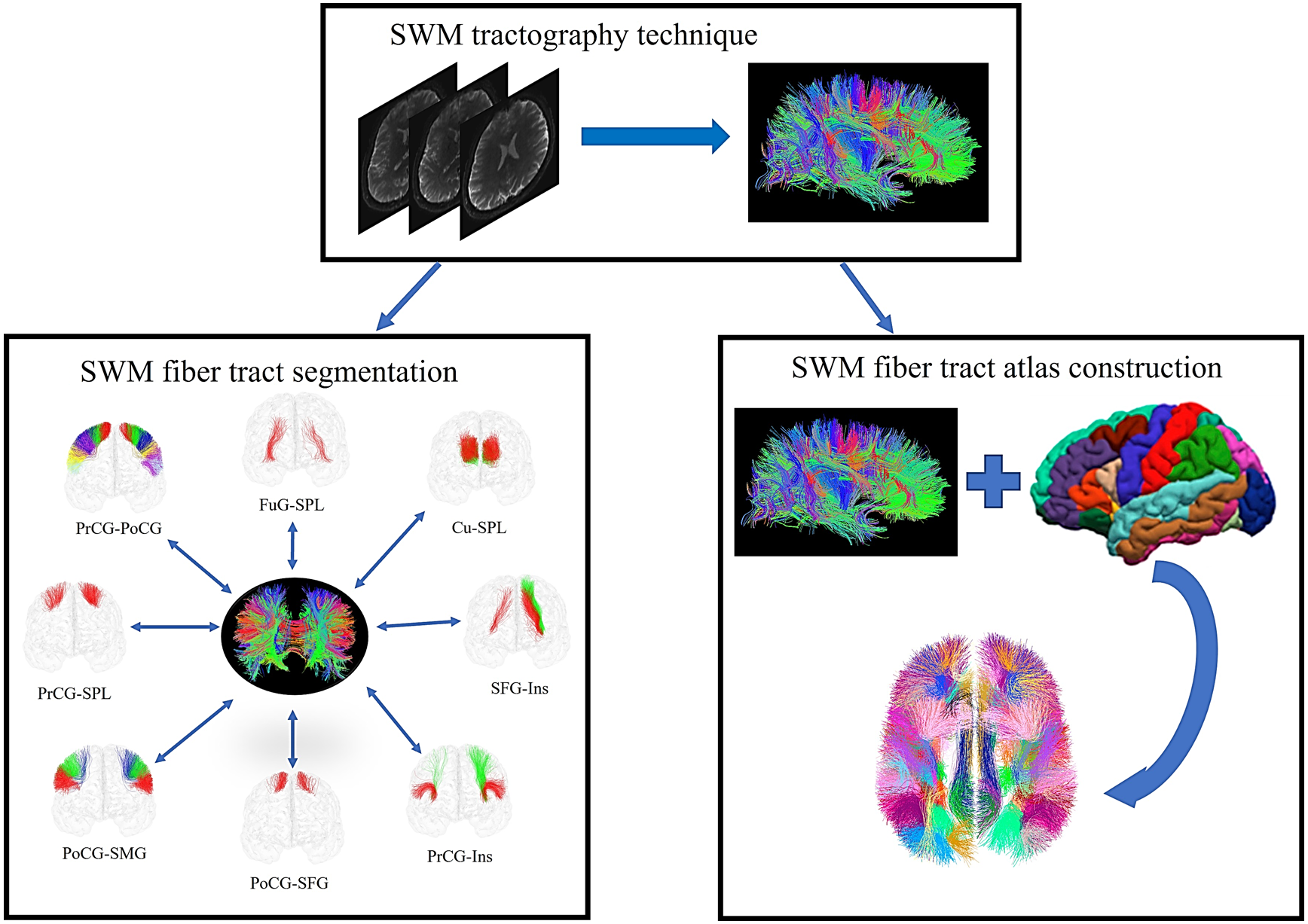
In recent years, significant progress has been made in tractography of superficial white matter in the brain based on diffusion magnetic resonance imaging. Superficial white matter fiber tracts serve as critical pathways connecting cortical and subcortical structures, playing a vital role in the construction of complete human connectome and neuropathological studies. This paper first summarizes the development of superficial white matter tractography techniques. Subsequently, it evaluates the strengths and weaknesses of different methods employed in superficial white matter fiber tract segmentation. Afterwards, the construction process of superficial white matter fiber tract atlas is discussed. Finally, this review concludes with an outlook on future research direction of superficial white matter tractography technology and segmentation methods.
| [1] | VERGANI F, MAHMOOD S, MORRIS C M, et al. Intralobar fibres of the occipital lobe: a post mortem dissection study[J]. Cortex, 2014, 56: 145-156. |
| [2] | REVELEY C, SETH A K, PIERPAOLI C, et al. Superficial white matter fiber systems impede detection of long-range cortical connections in diffusion MR tractography[J]. Proc Natl Acad Sci, 2015, 112(21): E2820-E2828. |
| [3] | VAN DYKEN P C, KHAN A R, PALANIYAPPAN L. Imaging of the superficial white matter in health and disease[J]. Imaging Neurosci, 2024, 2: 1-35. |
| [4] | KIRILINA E, HELBLING S, MORAWSKI M, et al. Superficial white matter imaging: Contrast mechanisms and whole-brain in vivo mapping[J]. Sci Adv, 2020, 6(41): eaaz9281. |
| [5] | ZHU Y, WANG Y. Brain fiber structure estimation based on principal component analysis and RINLM filter[J]. Med Biol Eng Comput, 2024, 62(3): 751-771. |
| [6] | CONTURO T E, LORI N F, CULL T S, et al. Tracking neuronal fiber pathways in the living human brain[J]. Proc Natl Acad Sci, 1999, 96(18): 10422-10427. |
| [7] | CATANI M, JONES D K, DONATO R, et al. Occipito-temporal connections in the human brain[J]. Brain, 2003, 126(9): 2093-2107. |
| [8] | GUEVARA P, DUCLAP D, POUPON C, et al. Segmentation of short association bundles in massive tractography datasets using a multi-subject bundle atlas[C]// Iberoamerican Congress on Pattern Recognition, Berlin, Heidelberg: Springer Berlin Heidelberg, 2011: 701-708. |
| [9] | XUE T, ZHANG F, ZHANG C, et al. Supwma: consistent and efficient tractography parcellation of superficial white matter with deep learning[C]// International Symposium on Biomedical Imaging, ITC Royal Bengal, Kolkata, India: IEEE, 2022: 1-5. |
| [10] | FENG Y J, WANG Z J, ZHANG G J, et al. Global white matter tractography using swarm optimization[J]. J Image Graph, 2012, 17(10): 1312-1318. |
| 冯远静, 王哲进, 张贵军, 等. 全局脑白质纤维群智能跟踪算法[J]. 中国图象图形学报, 2012, 17(10): 1312-1318. | |
| [11] | 张军. 基于邻域字典基模型的脑纤维流线微分方程跟踪算法[D]. 杭州: 浙江工业大学, 2017. |
| [12] | YUE Q, WANG Y J. A fiber tracking algorithm based on non-local constrained spherical deconvolution[J]. Chinese J Magn Reson, 2020, 37(4): 422-433. |
| 岳晴, 王远军. 基于非局部约束球面反卷积模型的纤维追踪算法[J]. 波谱学杂志, 2020, 37(4): 422-433. | |
| [13] | ZHANG T, CHEN H, GUO L, et al. Characterization of U-shape streamline fibers: methods and applications[J]. Med Image Anal, 2014, 18(5): 795-807. |
| [14] | WU Y, SUN D, WANG Y, et al. Tracing short connections of the temporo-parieto-occipital region in the human brain using diffusion spectrum imaging and fiber dissection[J]. Brain Res, 2016, 1646: 152-159. |
| [15] | ZHANG D, ZONG F, ZHANG Q, et al. Anat-SFSeg: Anatomically-guided superficial fiber segmentation with point-cloud deep learning[J]. Med Image Anal, 2024, 95: 103165. |
| [16] | LI M, HE J Z, FENG Y J. Research progress of neural fiber tracking[J]. J Image Graph, 2020, 25(8): 1513-1528. |
| 李茂, 何建忠, 冯远静. 神经纤维跟踪算法研究进展[J]. 中国图象图形学报, 2020, 25(8): 1513-1528. | |
| [17] | NIE X, RUAN J, OTADUY M C G, et al. Surface-based probabilistic fiber tracking in superficial white matter[J]. IEEE Trans Med Imaging, 2024, 43(3): 1113-1124. |
| [18] | GUEVARA M, GUEVARA P, ROMáN C, et al. Superficial white matter: A review on the dMRI analysis methods and applications[J]. NeuroImage, 2020, 212: 116673. |
| [19] | U?URBIL K, XU J, AUERBACH E J, et al. Pushing spatial and temporal resolution for functional and diffusion MRI in the Human Connectome Project[J]. NeuroImage, 2013, 80: 80-104. |
| [20] | YE W H, WANG Y J. Review of neural fiber tracking with diffusion magnetic resonance imaging[J]. J Chinese Comput Sys, 2022, 43(7): 1458-1463. |
| 叶伟红, 王远军. 扩散磁共振图像的神经纤维追踪算法研究综述[J]. 小型微型计算机系统, 2022, 43(7): 1458-1463. | |
| [21] | TOURNIER J D, CALAMANTE F, KING M D, et al. Limitations and requirements of diffusion tensor fiber tracking: an assessment using simulations[J]. Magn Reson Med, 2002, 47(4): 701-708. |
| [22] | AYDOGAN D B, SHI Y. Parallel transport tractography[J]. IEEE Trans Med Imaging, 2020, 40(2): 635-647. |
| [23] | GAHM J K, SHI Y. Surface-based tracking of U-fibers in the superficial white matter[C]// Medical Image Computing and Computer-assisted Intervention, Shenzhen, China: Springer International Publishing, 2019: 538-546. |
| [24] | SCHILLING K, GAO Y, JANVE V, et al. Confirmation of a gyral bias in diffusion MRI fiber tractography[J]. Hum Brain Mapp, 2018, 39(3): 1449-1466. |
| [25] | SOTIROPOULOS S N, HERNáNDEZ-FERNáNDEZ M, VU A T, et al. Fusion in diffusion MRI for improved fibre orientation estimation: An application to the 3T and 7T data of the Human Connectome Project[J]. NeuroImage, 2016, 134: 396-409. |
| [26] | SONG A W, CHANG H C, PETTY C, et al. Improved delineation of short cortical association fibers and gray/white matter boundary using whole-brain three-dimensional diffusion tensor imaging at submillimeter spatial resolution[J]. Brain Connect, 2014, 4(9): 636-640. |
| [27] | ST-ONGE E, DADUCCI A, GIRARD G, et al. Surface-enhanced tractography (SET)[J]. NeuroImage, 2018, 169: 524-539. |
| [28] | BASTIANI M, COTTAAR M, DIKRANIAN K, et al. Improved tractography using asymmetric fibre orientation distributions[J]. NeuroImage, 2017, 158: 205-218. |
| [29] | WU Y, HONG Y, FENG Y, et al. Mitigating gyral bias in cortical tractography via asymmetric fiber orientation distributions[J]. Med Image Anal, 2020, 59: 101543. |
| [30] | SHASTIN D, GENC S, PARKER G D, et al. Surface-based tracking for short association fibre tractography[J]. NeuroImage, 2022, 260: 119423. |
| [31] | COTTAAR M, BASTIANI M, BODDU N, et al. Modelling white matter in gyral blades as a continuous vector field[J]. NeuroImage, 2021, 227: 117693. |
| [32] | RHEAULT F, DE BENEDICTIS A, DADUCCI A, et al. Tractostorm: The what, why, and how of tractography dissection reproducibility[J]. Hum Brain Mapp, 2020, 41(7): 1859-1874. |
| [33] | WAKANA S, JIANG H, NAGAE-POETSCHER L M, et al. Fiber tract-based atlas of human white matter anatomy[J]. Radiology, 2004, 230(1): 77-87. |
| [34] | CATANI M, DELL’ACQUA F, VERGANI F, et al. Short frontal lobe connections of the human brain[J]. Cortex, 2012, 48(2): 273-291. |
| [35] | ROJKOVA K, VOLLE E, URBANSKI M, et al. Atlasing the frontal lobe connections and their variability due to age and education: a spherical deconvolution tractography study[J]. Brain Struct Funct, 2016, 221: 1751-1766. |
| [36] | BURKS J D, BOETTCHER L B, CONNER A K, et al. White matter connections of the inferior parietal lobule: a study of surgical anatomy[J]. Brain Behav, 2017, 7(4): e00640. |
| [37] | CATANI M, ROBERTSSON N, BEYH A, et al. Short parietal lobe connections of the human and monkey brain[J]. Cortex, 2017, 97: 339-357. |
| [38] | SHINOHARA H, LIU X, NAKAJIMA R, et al. Pyramid-shape crossings and intercrossing fibers are key elements for construction of the neural network in the superficial white matter of the human cerebrum[J]. Cereb Cortex, 2020, 30(10): 5218-5228. |
| [39] | OISHI K, ZILLES K, AMUNTS K, et al. Human brain white matter atlas: identification and assignment of common anatomical structures in superficial white matter[J]. NeuroImage, 2008, 43(3): 447-457. |
| [40] | ZHANG Y, ZHANG J, OISHI K, et al. Atlas-guided tract reconstruction for automated and comprehensive examination of the white matter anatomy[J]. NeuroImage, 2010, 52(4): 1289-1301. |
| [41] | PARDO E, GUEVARA P, DUCLAP D, et al. Study of the variability of short association bundles on a HARDI database[C]// Engineering in Medicine and Biology Society, Osaka, Japan: IEEE, 2013: 77-80. |
| [42] | OUYANG M, JEON T, MISHRA V, et al. Global and regional cortical connectivity maturation index (CCMI) of developmental human brain with quantification of short-range association tracts[C]// International Society for Optical Engineering, San Diego, CA, United States: SPIE, 2016, 9788: 328-334. |
| [43] | MOVAHEDIAN ATTAR F, KIRILINA E, HAENELT D, et al. Mapping short association fibers in the early cortical visual processing stream using in vivo diffusion tractography[J]. Cereb Cortex, 2020, 30(8): 4496-4514. |
| [44] | VERGANI F, LACERDA L, MARTINO J, et al. White matter connections of the supplementary motor area in humans[J]. J Neurol Neurosurg Psychiatry, 2014, 85(12): 1377-1385. |
| [45] | MAGRO E, MOREAU T, SEIZEUR R, et al. Characterization of short white matter fiber bundles in the central area from diffusion tensor MRI[J]. Neuroradiology, 2012, 54: 1275-1285. |
| [46] | YANG Z, LI X, ZHOU J, et al. Functional clustering of whole brain white matter fibers[J]. J Neurosci Meth, 2020, 335: 108626. |
| [47] | GUEVARA M, SUN Z Y, GUEVARA P, et al. Disentangling the variability of the superficial white matter organization using regional-tractogram-based population stratification[J]. NeuroImage, 2022, 255: 119197. |
| [48] | VINDAS N, AVILA N L, ZHANG F, et al. GeoLab: geometry-based tractography parcellation of superficial white matter[C]// International Symposium on Biomedical Imaging, Cartagena de Indias, Colombia: IEEE, 2023: 1-5. |
| [49] | GUEVARA P, DUCLAP D, POUPON C, et al. Automatic fiber bundle segmentation in massive tractography datasets using a multi-subject bundle atlas[J]. NeuroImage, 2012, 61(4): 1083-1099. |
| [50] | ROMáN C, GUEVARA M, VALENZUELA R, et al. Clustering of whole-brain white matter short association bundles using HARDI data[J]. Front Neuroinform, 2017, 11: 73. |
| [51] | DESIKAN R S, SéGONNE F, FISCHL B, et al. An automated labeling system for subdividing the human cerebral cortex on MRI scans into gyral based regions of interest[J]. NeuroImage, 2006, 31(3): 968-980. |
| [52] | ZHANG F, WU Y, NORTON I, et al. An anatomically curated fiber clustering white matter atlas for consistent white matter tract parcellation across the lifespan[J]. NeuroImage, 2018, 179: 429-447. |
| [53] | PRON A, BRUN L, DERUELLE C, et al. Dense and structured representations of U-shape fiber connectivity in the central sulcus[C]// International Symposium on Biomedical Imaging, Washington, DC, USA: IEEE, 2018: 700-703. |
| [54] | PRON A, DERUELLE C, COULON O. U-shape short-range extrinsic connectivity organisation around the human central sulcus[J]. Brain Struct Funct, 2021, 226(1): 179-193. |
| [55] | XUE T, ZHANG F, ZHANG C, et al. Superficial white matter analysis: An efficient point-cloud-based deep learning framework with supervised contrastive learning for consistent tractography parcellation across populations and dMRI acquisitions[J]. Med Image Anal, 2023, 85: 102759. |
| [56] | GUEVARA M, ROMáN C, HOUENOU J, et al. Reproducibility of superficial white matter tracts using diffusion-weighted imaging tractography[J]. NeuroImage, 2017, 147: 703-725. |
| [57] | ROMáN C, HERNáNDEZ C, FIGUEROA M, et al. Superficial white matter bundle atlas based on hierarchical fiber clustering over probabilistic tractography data[J]. NeuroImage, 2022, 262: 119550. |
| [58] | JIANG F, WANG Y J. Construction of human brain templates with diffusion tensor imaging data: A review[J]. Chinese J Magn Reson, 2018, 35(4): 520-530. |
| 蒋帆, 王远军. 扩散张量成像的人脑模板构建[J]. 波谱学杂志, 2018, 35(4): 520-530. |
/
| 〈 |
|
〉 |