

基于T2WI与DWI多参数MRI纹理特征融合的前列腺癌预测模型构建
Construction of Prostate Cancer Model Based on T2WI Combined with DWI Multi Parameter MRI Texture Feature Fusion
Received date: 2025-02-17
Online published: 2025-04-23
李露 , 高勇 , 周妤盼 . 基于T2WI与DWI多参数MRI纹理特征融合的前列腺癌预测模型构建[J]. 波谱学杂志, 2025 , 42(4) : 355 -363 . DOI: 10.11938/cjmr20253144
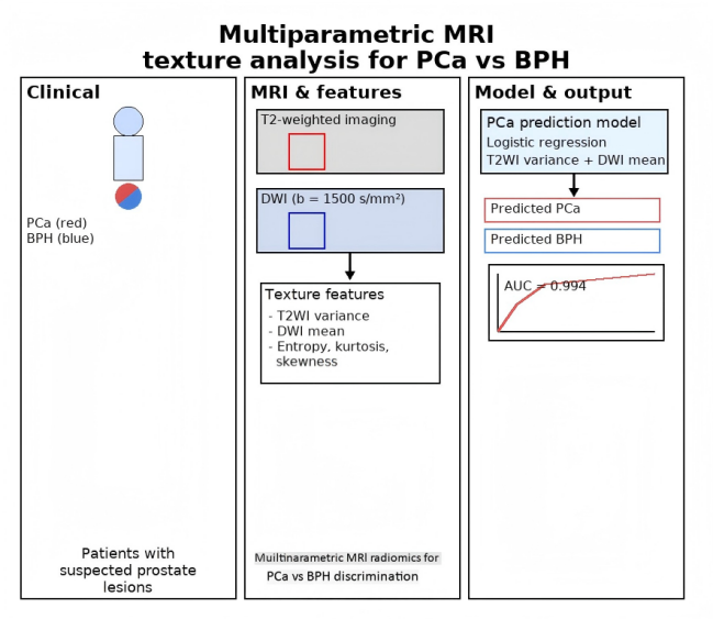
Prostate cancer (PCa) is one of the most common cancers in men, and imaging techniques have been increasingly used in its diagnosis. This study develops a prostate cancer model based on magnetic resonance T2-weighted imaging (T2WI) combined with diffusion-weighted imaging (DWI) texture analysis. Significant feature parameters were selected to construct a predictive model for prostate cancer, and the diagnostic value of texture features for disease discrimination was evaluated using the receiver operating characteristic (ROC) curve and the area under the curve (AUC). The results showed that the prostate cancer prediction model incorporating
| [1] | GUO Z, HE J, HUANG L, et al. Prevalence and risk factors of incidental prostate cancer in certain surgeries for benign prostatic hyperplasia: A systematic review and meta-analysis[J]. Int Braz J Urol, 2022, 48(6): 915-929. |
| [2] | RADEJ S, SZEWC M, MACIEJEWSKI R. Prostate infiltration by Treg and Th17 cells as an immune response to propionibacterium acnes infection in the course of benign prostatic hyperplasia and prostate cancer[J]. Int J Mol Sci, 2022, 23(16): 8849-8851. |
| [3] | SHAH A, SHAH A A, K N, et al. Mechanistic targets for BPH and prostate cancer-a review[J]. Rev Environ Health, 2020, 36(2): 261-270. |
| [4] | HOPSTAKEN J S, BOMERS J G R, SEDELAAR M J P, et al. An updated systematic review on focal therapy in localized prostate cancer: what has changed over the past 5 years?[J]. Eur Urol, 2022, 81(1): 5-33. |
| [5] | SARKAR P, MALIK S, BANERJEE A, et al. Differential microbial signature associated with benign prostatic hyperplasia and prostate cancer[J]. Front Cell Infect Microbiol, 2022, 12(8): e894777. |
| [6] | ZHOU B, LIU X, GAN H, et al. Differentiation of prostate cancer and stromal hyperplasia in the transition zone with monoexponential, stretched-exponential diffusion-weighted imaging and diffusion kurtosis imaging in a reduced number of b values: correlation with whole-mount pathology[J]. J Comput Assist Tomogr, 2022, 46(4): 545-550. |
| [7] | YAO W, LIU J, ZHENG J, et al. Study on diagnostic value of quantitative parameters of intravoxel incoherent motion diffusion-weighted imaging (IVIM-DWI) in prostate cancer[J]. Am J Transl Res, 2021, 13(4): 3696-3702. |
| [8] | INGOLE S M, MEHTA R U, KAZI Z N, et al. Multiparametric magnetic resonance imaging in evaluation of clinically significant prostate cancer[J]. Indian J Radiol Imaging, 2021, 31(1): 65-77. |
| [9] | LI P, WANG H L, WANG W L. Diagnosis of lung adenocarcinoma with brain metastases based on MRI texture features and predictive value of EGFR gene mutation in lung adenocarcinoma with brain metastases[J]. Journal of Clinical Radiology, 2023, 42(3): 498-503. |
| 李平, 王鸿礼, 王伟亮. 基于MRI纹理特征的肺腺癌脑转移瘤诊断及对肺腺癌脑转移EGFR基因突变的预测[J]. 临床放射学杂志, 2023, 42(3): 498-503. | |
| [10] | WAN T, LIU H, XU H, et al. Relationship between pre-operative MRI texture features and prognosis of patients with hepatocellular carcinoma[J]. Chinese Journal of CT and MRI, 2022, 20(10): 81-83. |
| 万涛, 刘海, 许华, 等. MRI术前肝细胞肝癌患者图像纹理参数表现与患者预后的关系[J]. 中国CT和MRI杂志, 2022, 20(10): 81-83. | |
| [11] | LIU K W, LIU Z L, WANG X Y, et al. Prostate cancer diagnosis based on cascaded convolutional neural networks[J]. Chinese J Magn Reson, 2020, 37(2): 152-161. |
| 刘可文, 刘紫龙, 汪香玉, 等. 基于级联卷积神经网络的前列腺磁共振图像分类[J]. 波谱学杂志, 2020, 37(2): 152-161. | |
| [12] | LIU J P, JIN Y B, CHENG D L, et al. Preliminary application of MR-T2WI texture analysis for detection of prostate cancer basing on FireVoxel software[J]. Journal of Clinical Radiology, 2021, 40(7): 1426-1430. |
| 刘健萍, 金亚彬, 成东亮, 等. 基于FireVoxel软件MR-T2WI纹理分析在前列腺癌诊断中的初步应用[J]. 临床放射学杂志, 2021, 40(7): 1426-1430. | |
| [13] | SUN H, DU F, LIU Y, et al. DCE-MRI and DWI can differentiate benign from malignant prostate tumors when serum PSA is ≥10 ng/mL[J]. Front Oncol, 2022, 12(8): e925186. |
| [14] | XU Q, ZHU Q Q, YE J, et al. T2-weighted based radiomics model in differentiation of prostate cancer and benign prostate hyperplasia[J]. Journal of Medical Imaging, 2021, 31(10): 1723-1726. |
| 许晴, 朱庆强, 叶靖, 等. 基于T2WI的影像组学模型对鉴别前列腺癌和前列腺增生的诊断价值[J]. 医学影像学杂志, 2021, 31(10): 1723-1726. | |
| [15] | ZHOU K P, HUANG H B, LI S Y, et al. Non-invasive MRI-based assessment of reactive stromal grade in prostate cancer using diffusion kurtosis imaging and stretched-exponential model[J]. BMC Med Imaging, 2025, 25(1): 339. |
| [16] | UEDA T, OHNO Y, YAMAMOTO K, et al. Deep learning reconstruction of diffusion-weighted MRI improves image quality for prostatic imaging[J]. Radiology, 2022, 303(2): 373-381. |
| [17] | BAI G J, WAN Y D. The value of Bp-MRI grayscale histogram in distinguishing prostate cancer from benign prostatic hyperplasia in transition zone[J]. Journal of Tianjin Medical University, 2021, 27(1): 47-51. |
| 白国杰, 万业达. Bp-MRI灰度直方图在鉴别移行带前列腺癌与良性前列腺增生中的应用价值[J]. 天津医科大学学报, 2021, 27(1): 47-51. | |
| [18] | LI Z K, DU H D, WANG Y, et al. Application of radiomics based on T2WI in diagnosis of prostate cancer[J]. Chongqing Medicine, 2021, 50(22): 3892-3895, 3899. |
| 李振凯, 杜红娣, 王莺, 等. 基于磁共振T2WI的影像组学在前列腺癌诊断中的应用研究[J]. 重庆医学, 2021, 50(22): 3892-3895, 3899. | |
| [19] | FENG J J, SHEN X F, LI Z B. The role of MRI diffusion weighted imaging in the diagnosis of benign prostatic hyperplasia and prostate cancer[J]. Systems Medicine, 2021, 6(15): 92-94. |
| 冯娟娟, 沈雪峰, 李正标. 核磁共振弥散加权成像在前列腺增生与前列腺癌诊断中的鉴别作用[J]. 系统医学, 2021, 6(15): 92-94. | |
| [20] | WANG S, MENG M, JIANG Q T, et al. Texture analysis of MR diffusion weighted imaging for discriminating high-grade from low-grade prostate cancer[J]. Chinese Journal of Medical Imaging Computing, 2021, 27(6): 540-545. |
| 王姗, 孟蒙, 姜岐涛, 等. 基于DWI的纹理分析在鉴别高、低级别前列腺癌诊断中的价值[J]. 中国医学计算机成像杂志, 2021, 27(6): 540-545. |
/
| 〈 |
|
〉 |