

磁共振成像技术在轻度创伤性脑损伤诊断与预后中的应用
收稿日期: 2025-09-15
网络出版日期: 2025-12-23
基金资助
福建省自然科学基金计划项目(2023J011722)
Advances in Magnetic Resonance Imaging for the Diagnosis and Prognosis of Mild Traumatic Brain Injury
Received date: 2025-09-15
Online published: 2025-12-23
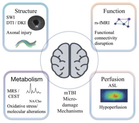
轻度创伤性脑损伤(Mild Traumatic Brain Injury,mTBI)是临床常见的神经系统损伤,其诊断和管理因症状隐匿性和病理复杂性而具有挑战性.传统影像检查技术如电子计算机断层扫描(Computed Tomography,CT)和常规磁共振成像(Magnetic Resonance Imaging,MRI)虽在急性期评估中占据主导地位,但在微小损伤检测及长期预后评估方面存在局限性.近年来,多模态MRI技术在mTBI研究中不断发展,为揭示其潜在病理机制及探索客观影像学关键指标提供了新的思路.其中磁敏感加权成像(SWI)可敏感检测微出血和脑内铁沉积,酰胺质子转移(APT)成像能够反映分子和代谢水平变化,而扩散与功能MRI技术则有助于刻画白质微结构和脑网络异常.多模态MRI的整合及影像数据库的构建,将是推动早期诊断、精准评估和人工智能辅助干预的重要方向.本文系统综述了相关MRI技术的研究进展,分析其优势与局限,并探讨其在临床转化中的前景.
傅芬芳 , 林国兵 , 李梅芳 . 磁共振成像技术在轻度创伤性脑损伤诊断与预后中的应用[J]. 波谱学杂志, 2026 , 43(2) : 214 -222 . DOI: 10.11938/cjmr20253180
Mild traumatic brain injury (mTBI) is a common neurological disorder in clinical practice, yet its diagnosis and management remain challenging due to the hidden nature of symptoms and the underlying pathological complexity. While imaging techniques such as computed tomography (CT) and conventional magnetic resonance imaging (MRI) are the mainstay for acute-phase assessment, they have limitations in detecting subtle injuries and evaluating long-term prognosis. In recent years, multimodal MRI technology has been developed in the research of mTBI, offering novel approaches for revealing its potential pathological mechanisms and exploring the key objective imaging indicators. Specifically, susceptibility weighted imaging (SWI) is sensitive for detecting microbleeds and iron deposition in the brain; amide proton transfer (APT) imaging reflects changes in molecular and metabolic levels; diffusion and functional imaging techniques help depict abnormalities in white matter microstructure and brain networks. The integration of multimodal MRI and the construction of imaging databases will be important directions for advancing early diagnosis, precise assessment, and AI-assisted intervention. This article systematically reviews research progress of related MRI techniques, analyzes their advantages and limitations, and discusses their prospects in clinical translation.
| [1] | ZENG S H, CHEN W X, DUAN Y Y, et al. Imaging detection of cerebral microhemorrhages and white matter injuries in patients with mild traumatic brain injury and their correlation with prognosis[J]. Journal of Navy Medicine, 2024, 45(8): 871-876. |
| 曾淑妍, 陈文曦, 段瑶瑶, 等. 轻度创伤性脑损伤的微出血和白质损伤的影像学检测及其与预后的相关性分析[J]. 海军医学杂志, 2024, 45(8): 871-876. | |
| [2] | RAUCHMAN S H, ALBERT J, PINKHASOV A, et al. Mild-to-moderate traumatic brain injury: A review with focus on the visual system[J]. Neurol Int, 2022, 14(2): 453-470. |
| [3] | LEFEVRE-DOGNIN C, COGNé M, PERDRIEAU V, et al. Definition and epidemiology of mild traumatic brain injury[J]. Neurochirurgie, 2021, 67(3): 218-221. |
| [4] | NI L T, QI Q, MA H C, et al. Research advances in clinical diagnosis of mild traumatic brain injury based on multimodal imaging[J]. Academic Journal of Chinese PLA Medical School, 2024, 45(7): 805-810, 817. |
| 倪立桐, 齐琦, 马珩钞, 等. 基于多模态影像学的轻度创伤性脑损伤脑功能评估的研究进展[J]. 解放军医学院学报, 2024, 45(7): 805-810, 817. | |
| [5] | LIU Y, LU L, LI F, et al. Neuropathological mechanisms of mild traumatic brain injury: A perspective from multimodal magnetic resonance imaging[J]. Front Neurosci, 2022, 16: 923662. |
| [6] | YANG X C, XIANG S Q. The value of multimodal MRI in the diagnosis of cerebral microhemorrhage caused by mild traumatic brain injury[J]. Journal of Xiangyang Polytechnic, 2024, 23(3): 102-106. |
| 杨先春, 向世强. 多模态MRI对轻型创伤性颅脑损伤性脑微出血的诊断价值[J]. 襄阳职业技术学院学报, 2024, 23(3): 102-106. | |
| [7] | RIEMANN L, MIKOLIC A, MAAS A, et al. Computed tomography lesions and their association with global outcome in young people with mild traumatic brain injury[J]. J Neurotrauma, 2023, 40(11-12): 1243-1254. |
| [8] | YUH E L, MUKHERJEE P, LINGSMA H F, et al. Magnetic resonance imaging improves 3-month outcome prediction in mild traumatic brain injury[J]. Ann Neurol, 2013, 73(2): 224-235. |
| [9] | ASHBY K, ADAMS B N, SHETTY M. Appropriate magnetic resonance imaging ordering[M/OL]. Treasure Island (FL): StatPearls Publishing, 2022. https://www.ncbi.nlm.nih.gov/books/NBK565857/. |
| [10] | HAGEMAN G, HOF J, NIHOM J. Susceptibility-Weighted MRI and microbleeds in mild traumatic brain injury: Prediction of posttraumatic complaints?[J]Eur Neurol, 2022, 85(3): 177-185. |
| [11] | LUNKOVA E, GUBERMAN G I, PTITO A, et al. Noninvasive magnetic resonance imaging techniques in mild traumatic brain injury research and diagnosis[J]. Hum Brain Mapp, 2021, 42(16): 5477-5494. |
| [12] | BELL T K, ANSARI M, JOYCE J M, et al. Quantitative susceptibility mapping in adults with persistent postconcussion symptoms after mild traumatic brain injury: an exploratory study[J]. AJNR Am J Neuroradiol, 2025, 46(2): 435-442. |
| [13] | ZHU J, SONG B. Value of multimodal head MRI in evaluation of mild traumatic brain injury[J]. Chinese Journal of CT and MRI, 2023, 21(10): 14-16. |
| 朱杰, 宋彬. 多模态头颅MRI对轻型颅脑损伤的评估价值[J]. 中国CT和MRI杂志, 2023, 21(10): 14-16. | |
| [14] | LIU W, XU K N. Research progresses of functional MRI on mild traumatic brain injury[J]. Journal of Practical Radiology, 2019, 35(4): 663-665. |
| 刘伟, 许克宁. 轻型创伤性脑损伤的功能磁共振成像研究进展[J]. 实用放射学杂志, 2019, 35(4): 663-665. | |
| [15] | BIE C, VAN ZIJL P, XU J, et al. Radiofrequency labeling strategies in chemical exchange saturation transfer MRI[J]. NMR in Biomed, 2023, 36(6): e4944. |
| [16] | BIE C, VAN ZIJL P C M, MAO D, et al. Ultrafast Z-spectroscopic imaging in vivo at 3T using through-slice spectral encoding (TS-UFZ)[J]. Magn Reson Med, 2023, 89(4): 1429-1440. |
| [17] | KIRBY A, WARD C, CALVERT N D, et al. Aldehydic load as an objective imaging biomarker of mild traumatic brain injury[J]. Npj Imaging, 2025, 3: 30. |
| [18] | ZHOU Y, BIE C, VAN ZIJL P C M, et al. The relayed nuclear Overhauser effect in magnetization transfer and chemical exchange saturation transfer MRI[J]. NMR in Biomed, 2023, 36(6): e4778. |
| [19] | MARALANI P J, CHAN R W, LAM W W, et al. Chemical exchange saturation transfer MRI: What neuro-oncology clinicians need to know[J]. Technol Cancer Res T, 2023, 22: 15330338231208613. |
| [20] | TAN X, SAJJA V, D'SOUZA M, et al. A methodology to compare biomechanical simulations with clinical brain imaging analysis utilizing two blunt impact cases[J]. Front Bioeng Biotechnol, 2021, 9: 654677. |
| [21] | CHEN B, WU F X, CAO Y, et al. Longitudinal evaluation of MR ADC for brain injury in neonatal purulent meningitis[J]. Chinese Journal of Evidence-Based Pediatrics, 2021, 16(5): 344-350. |
| 陈斌, 吴菲潇, 曹云, 等. 磁共振成像表观扩散系数变化对新生儿化脓性脑膜炎脑损伤纵向评估[J]. 中国循证儿科杂志, 2021, 16(5): 344-350. | |
| [22] | PU Z T, LI D S, JIN Z G, et al. Susceptibility-weighted imaging combined with diffusion-weighted imaging in the diagnosis of diffuse axonal injury[J]. Zhejiang Traumatic Surgery, 2021, 26(2): 343-345. |
| 浦智韬, 李殿胜, 金中高, 等. 磁敏感加权成像联合弥散加权成像诊断脑弥漫性轴索损伤的应用[J]. 浙江创伤外科, 2021, 26(2): 343-345. | |
| [23] | OEHR L E, YANG J Y, CHEN J, et al. Investigating white matter tract microstructural changes at six-twelve weeks following mild traumatic brain injury: A combined diffusion tensor imaging and neurite orientation dispersion and density imaging study[J]. J Neurotrauma, 2021, 38(16): 2255-2263. |
| [24] | JAIN B, DAS A K, AGRAWAL M, et al. Implications of DTI in mild traumatic brain injury for detecting neurological recovery and predicting long-term behavioural outcome in paediatric and young population—a systematic review[J]. Child’s Nerv Syst, 2021, 37(8): 2475-2486. |
| [25] | WEI W, LI N, DU X, et al. Rapid prediction and accurate location selection of mild traumatic brain injury (mTBI) by using multiple parameter analysis of diffusion tensor imaging (DTI): Integrating correlational and clinical approaches[J]. Biomed Res Int, 2023: 7467479. |
| [26] | PALACIOS E M, YUH E L, MAC DONALD C L, et al. Diffusion tensor imaging reveals elevated diffusivity of white matter microstructure that is independently associated with long-term outcome after mild traumatic brain injury: A TRACK-TBI study[J]. J Neurotrauma, 2022, 39(19-20): 1318-1328. |
| [27] | MCINNES K, FRIESEN C L, MACKENZIE D E, et al. Mild traumatic brain injury (mTBI) and chronic cognitive impairment: A scoping review[J]. PLoS ONE, 2017, 12(4): e0174847. |
| [28] | JENSEN J H, HELPERN J A, RAMANI A, et al. Diffusional kurtosis imaging: the quantification of non-gaussian water diffusion by means of magnetic resonance imaging[J]. Magn Reson Med, 2005, 53(6): 1432-1440. |
| [29] | STEVEN A J, ZHUO J, MELHEM E R. Diffusion kurtosis imaging: An emerging technique for evaluating the microstructural environment of the brain[J]. AJR Am J Roentgenol, 2014, 202(1): W26-W33. |
| [30] | TABESH A, JENSEN J H, ARDEKANI B A, et al. Estimation of tensors and tensor‐derived measures in diffusional kurtosis imaging[J]. Magn Reson Med, 2011, 65(3): 823-836. |
| [31] | GROSSMAN E J, GE Y, JENSEN J H, et al. Thalamus and cognitive impairment in mild traumatic brain injury: A diffusional kurtosis imaging study[J]. J Neurotrauma, 2012, 29(13): 2318-2327. |
| [32] | LINDSEY H M, HODGES C B, GREER K M, et al. Diffusion-weighted imaging in mild traumatic brain injury: A systematic review of the literature[J]. Neuropsychol Rev, 2023, 33(1): 42-121. |
| [33] | KUANG H M, GONG H H. Recent advances in magnetic resonance imaging techniques for diffuse axonal injury[J]. Journal of Practical Medicine, 2014, 30(24): 4042-4043. |
| 况红妹, 龚洪翰. 弥漫性轴索损伤的磁共振成像新技术研究进展[J]. 实用医学杂志, 2014, 30(24): 4042-4043. | |
| [34] | GIMBEL S I, WANG C C, HUNGERFORD L, et al. Associations of mTBI and post-traumatic stress to amygdala structure and functional connectivity in military service members[J]. Front Neuroimaging, 2023, 2: 1129446. |
| [35] | BIGLER E D. Volumetric MRI findings in mild traumatic brain injury (mTBI) and neuropsychological outcome[J]. Neuropsychol Rev, 2023, 33(1): 5-41. |
| [36] | XIA J, YAO J, WANG L V. Photoacoustic tomography: Principles and advances[J]. Electromagn Waves, 2014, 147: 1-22. |
| [37] | WANG Y, BARTELS H M, NELSON L D. A systematic review of ASL perfusion MRI in mild TBI[J]. Neuropsychol Rev, 2023, 33(1): 160-191. |
| [38] | DOGAHE M H, RAMEZANI S, REIHANIAN Z, et al. Role of brain metabolites during acute phase of mild traumatic brain injury in prognosis of post-concussion syndrome: A 1H-MRS study[J]. Psychiatry Res Neuroimaging, 2023, 335: 111709. |
| [39] | CHEN A M, GERHALTER T, DEHKHARGHANI S, et al. Replicability of proton MR spectroscopic imaging findings in mild traumatic brain injury: Implications for clinical applications[J]. Neuroimage: Clin, 2023, 37: 103325. |
| [40] | ALLEN J, PHAM L, BOND S T, et al. Acute effects of single and repeated mild traumatic brain injury on levels of neurometabolites, lipids, and mitochondrial function in male rats[J]. Front Mol Neurosci, 2023, 16: 1208697. |
| [41] | LY M T, SCARNEO-MILLER S E, LEPLEY A S, et al. Combining MRI and cognitive evaluation to classify concussion in university athletes[J]. Brain Imaging Behav, 2022, 16(5): 2175-2187. |
| [42] | SHI J, TENG J, DU X, LI N. Multi-modal analysis of resting-state fMRI data in mTBI patients and association with neuropsychological outcomes[J]. Front Neurol, 2021, 12: 639760. |
| [43] | RAUSA V C, SHAPIRO J, SEAL M L, et al. Neuroimaging in paediatric mild traumatic brain injury: a systematic review[J]. Neurosci Biobehav Rev, 2020, 118: 643-653. |
| [44] | LEYBA K, PAIYABHROMA N, SALVAS J P, et al. Neurovascular hypoxia after mild traumatic brain injury in juvenile mice correlates with heart-brain dysfunctions in adulthood[J]. Acta Physiologica, 2023, 238(2): e13933. |
| [45] | THORNE J, HELLEWELL S, COWEN G, et al. Neuroimaging to enhance understanding of cardiovascular autonomic changes associated with mild traumatic brain injury: a scoping review[J]. Brain Injury, 2023, 37(10): 1187-1204. |
| [46] | WHITEHOUSE D P, WILSON L, CZEITER E, et al. Association of blood-based biomarkers and 6-month patient-reported outcomes in patients with mild TBI: A CENTER-TBI analysis[J]. Neurology, 2025, 104(1): e210040. |
| [47] | LI F, ZHANG D, REN J, et al. Connectivity of the insular subdivisions differentiates posttraumatic headache-associated from nonheadache-associated mild traumatic brain injury: an arterial spin labelling study[J]. J Headache Pain, 2024, 25(1): 103. |
| [48] | MONDAL K, DEL MAR N A, GARY A A, et al. Sphingolipid changes in mouse brain and plasma after mild traumatic brain injury at the acute phases[J]. Lipids Health Dis, 2024, 23(1): 200. |
| [49] | ROBERTS C J, BARBER J, TEMKIN N R, et al. Clinical Outcomes after traumatic brain injury and exposure to extracranial surgery: A TRACK-TBI study[J]. JAMA Surg, 2024, 159(3): 248-259. |
/
| 〈 |
|
〉 |