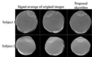
Magnetic resonance imaging (MRI) is a noninvasive intraocular tumor detection method without ionizing radiation. However, resolution limitation and motion artifacts are difficult to overcome in the imaging process. Conventional scanning methods inevitably introduce motion artifacts, or require the subjects to cooperate for accurate eye fixation, increasing the difficulty of imaging and giving the subject uncomfortable experiences. In this work, a new MRI method based on super-resolution theory is proposed, which uses a specialized orbit coil to scan a series of dynamic images of the eyeball, such that the acquisition resolution in different directions is complementary. High-resolution eyeball images with minimal motion artifacts could then be obtained after pre-processing, registration, super-resolution reconstruction and other operations. The study showed that the method proposed can be used to obtain clear eyeball images without the requirement of eye fixation.
[1]GRAAF P D,GÖRICKE S,RODJAN F,et al.Guidelines for imaging retinoblastoma:imaging principles and MRI standardization[J].Pediatr Radiol,2012,42(1):2-14. [2]BEENAKKER J W,VAN RIJN G A,LUYTEN G P,et al.High-resolution MRI of uveal melanoma using a microcoil phased array at 7 T[J].NMR Biomed,2013,26(12):1864-1869. [3]GRAESSL A,MUHLE M,SCHWERTER M,et al.Ophthalmic magnetic resonance imaging at 7 T using a 6-channel transceiver radiofrequency coil array in healthy subjects and patients with intraocular masses[J].Invest Radiol,2014,49(5):260-270. [4]BEENAKKER J W M,FERREIRA T A,SOEMARWOTO K P,et al.Clinical evaluation of ultra-high-field MRI for three-dimensional visualisation of tumour size in uveal melanoma patients,with direct relevance to treatment planning[J].MAGMA,2016,29(3):571-577. [5]HO L C,SIGAL I A,JAN N J,et al.Non-invasive MRI assessments of tissue microstructures and macromolecules in the eye upon biomechanical or biochemical modulation[J].Sci Rep,2016,6:32080. [6]HO L C,SIGAL I A,JAN N J,et al.Magic angle-enhanced MRI of fibrous microstructures in sclera and cornea with and without intraocular pressure loading[J].Invest Ophthalmol Vis Sci,2014,55(9):5662-5672. [7]HO L C,CONNER I P,DO C W,et al.In vivo assessment of aqueous humor dynamics upon chronic ocular hypertension and hypotensive drug treatment using gadolinium-enhanced MRI[J].Invest Ophthalmol Vis Sci,2014,55(6):3747-3757. [8]LEMKE A J,ALAI-OMID M,HENGST S A,et al.Eye imaging with a 3.0-T MRI using a surface coil--a study on volunteers and initial patients with uveal melanoma[J].Eur Radiol,2006,16(5):1084-1089. [9]BERKOWITZ B,MCDONALD C,ITO Y,et al.Measuring the human retinal oxygenation response to a hyperoxic challenge using MRI:Eliminating blinking artifacts and demonstrating proof of concept[J].Magn Reson Med,2001,46(2):412-416. [10]ZHANG Y,NATERAS O S E,PENG Q,et al.Lamina-specific anatomic magnetic resonance imaging of the human retina[J].Invest Ophthalmol Vis Sci,2011,52(10):7232-7237. [11]WEZEL J,GARPEBRING A,WEBB A G,et al.Automated eye blink detection and correction method for clinical MR eye imaging[J].Magn Reson Med,2017,78(1):165-171. [12]JIANG S,XUE H,GLOVER A,et al.MRI of moving subjects using multislice snapshot images with volume reconstruction (SVR):Application to fetal,neonatal,and adult brain studies[J].IEEE Trans Med Imaging,2007,26(7):967-980. [13]GREENSPAN H,PELED S,OZ G,et al.MRI inter-slice reconstruction using super-resolution[C]//International conference on medical image computing and computer-assisted intervention.Berlin Heidelberg:Springer-Verlag,2001:1204-1206. [14]GREENSPAN H,OZ G,KIRYATI N,et al.MRI inter-slice reconstruction using super-resolution[J].Magn Reson Imaging,2002,20(5):437-446. [15]SHARON P,YEHEZKEL Y.Superresolution in MRI:Application to human white matter fiber tract visualization by diffusion tensor imaging[J].Magn Reson Med,2001,45(1):29-35. [16]SCHEFFLER K.Superresolution in MRI?[J].Magn Reson Med,2002,48(2):408-409. [17]CARMI E,LIU S Y,ALON N,et al.Resolution enhancement in MRI[J].Magn Reson Imaging,2006,24(2):133-154. [18]MITCHELL R J,MAYER G S,LAUZON L M,et al.Synthetic aperture MRI:US,US7005854[P].2006. [19]MAYER G S,VRSCAY E R.Mathematical analysis of "phase ramping" for super-resolution magnetic resonance imaging[M]//CAMPILHO A,KAMEL M S.Image Analysis and Recognition.Berlin Heidelberg:Springer,2006:82-93. [20]SCHULTZ R R,STEVENSON R L.Improved definition image expansion[C].In ICASSP,IEEE international conference on a coustics,speech and signal processing-proceedings.1992,3:173-176. [21]SCHULTZ R R,STEVENSON R L.A Bayesian approach to image expansion for improved definition[J].IEEE T Image Process,1994,3(3):233-242. [22]SCHULTZ R R,STEVENSON R L.Extraction of high-resolution frames from video sequences[J].IEEE T Image Process,1996,5(6):996-1011. [23]TSAI R Y,HUANG T S.Multiframe image restoration and registration[C]//Advances in computer vision and image processing.1984. [24]IRANI M,PELEG S.Improving resolution by image registration[J].Cvgip Graphical Models&Image Processing,1991,53(3):231-239. [25]STEVENSON R L,SCHMITZ B E,DELP E J.Discontinuity preserving regularization of inverse visual problems[J].IEEE T Sys Man Cy,1994,24(3):455-469. [26]BERT R J,PATZ S,OSSIANI M,et al.High-resolution MR imaging of the human eye 20051[J].Acad Radiol,2006,13(3):368-378. [27]RICHDALE K,WASSENAAR P,TEAL B K,et al.7 Tesla MR imaging of the human eye in vivo.[J].J Magn Reson Imaging,2009,30(5):924-932. [28]DABOV K,FOI A,KATKOVNIK V,et al.Image denoising by sparse 3-D transform-domain collaborative filtering[J].IEEE T Image Process,2007,16(8):2080-2095. [29]KENNEDY J,EBERHART R.Particle swarm optimization[C]//IEEE International Conference on Neural Networks,1995,4:1942-1948. [30]EBERHART R,KENNEDY J.A new optimizer using particle swarm theory[C]//International Symposium on MICRO Machine and Human Science.IEEE,1995:39-43.