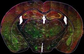
Quantitative analyses of molecular expression, cell counts and neural network connections among different brain regions are essential in brain science research. Based on Paxinos and Franklin's the Mouse Brain in Stereotaxic Coordinates (Ⅱ) and public image processing software such as Photoshop and ImageJ, we developed a convenient method for semi-automatic segmentation and cell counting on brain sections. A standard template for brain region segmentation was first obtained from the Paxinos and Franklin's mouse brain atlas. Photoshop was then used to transform the standard template semi-automatically into the space of brain sections, yielding masks of segmented brain regions. Finally, ImageJ was used to analyze the data in different brain regions. This method is useful for immunohistochemical and neuron distribution pattern analyses, as well as neural network labelling studies. The method does not require expensive commercial image analysis software, and is also easy to implement and use.
ZHU Xu-tao
,
HE Xiao-bin
,
LIU Yue
,
WEN Peng-jie
,
WANG Li
,
ZHANG Zhi-jian
,
XU Fu-qiang
. A Convenient Semi-Automatic Method for Analyzing Brain Sections: Registration, Segmentation and Cell Counting[J]. Chinese Journal of Magnetic Resonance, 2018
, 35(2)
: 133
-140
.
DOI: 10.11938/cjmr20172608
[1] ZHANG Z J, JIN S, ZHU X T, et al. Advancement in neurotropic virus-mediated trans-synaptic neural circuit tracing[J]. Chinese Bulletin of Life Sciences, 2017, 26(6):634-644. 张志建, 靳森, 朱续涛, 等. 利用嗜神经病毒跨突触追踪神经网络研究进展[J]. 生命科学, 2014, 26(6):634-644.
[2] LINDROOS O. Short Nissl staining for incubated cryostat sections of the brain[J]. Biotech Histochem, 1991, 66(4):208-209.
[3] KIERNAN J A, MACPHERSON C M, PRICE A, et al. A histochemical examination of the staining of kainate-induced neuronal degeneration by anionic dyes[J]. Biotech Histochem, 1998, 73(5):244-254.
[4] PAXINOS G, WATSON C R, EMSON P C. AChE-stained horizontal sections of the rat brain in stereotaxic coordinates[J]. J Neurosci Methods, 1980, 3(2):129-149.
[5] OSEN K, MUGNAINI E, DAHL A, et al. Histochemical localization of acetylcholinesterase in the cochlear and superior olivary nuclei. A reappraisal with emphasis on the cochlear granule cell system[J]. Arch Ital Biol, 1984, 122(3):169-212.
[6] COONS A H, CREECH H J, JONES R N. Immunological properties of an antibody containing a fluorescent group[J]. P Soc Exp Biol Med, 1941, 47(2):200-202.
[7] STERNBERGER L A, HARDY JR P H, CUCULIS J J, et al. The unlabeled antibody enzyme method of immunohistochemistry preparation and properties of soluble antigen-antibody complex (horseradish peroxidase-antihorseradish peroxidase) and its use in identification of spirochetes[J]. J Histochem Cytochem, 1970, 18(5):315-333.
[8] PAXINOS G, WATSON C. The rat brain in stereotaxic coordinates[M]. San Diego:Academic Press, 1998.
[9] FRANKLIN K B, PAXINOS G. The mouse brain in stereotaxic coordinates[M]. 3rd ed. San Diego:Academic Press, 2007.
[10] ZHOU J N, NI R J. The tree shrew (tupaia belangeri chinensis) brain in stereotaxic coordinates[M]. Singapore:Springer, 2016.
[11] BETLEY J N, STERNSON S M. Adeno-associated viral vectors for mapping, monitoring, and manipulating neural circuits[J]. Hum Gene Ther, 2011, 22(6):669-677.
[12] BEIER K T, SAUNDERS A, OLDENBURG I A, et al. Anterograde or retrograde transsynaptic labeling of CNS neurons with vesicular stomatitis virus vectors[J]. Proc Natl Acad Sci U S A, 2011, 108(37):15414-15419.
[13] WICKERSHAM I R, FINKE S, CONZELMANN K K, et al. Retrograde neuronal tracing with a deletion-mutant rabies virus[J]. Nat Methods, 2007, 4(1):47-49.
[14] TERVO D G, HWANG B Y, VISWANATHAN S, et al. A designer AAV variant permits efficient retrograde access to projection neurons[J]. Neuron, 2016, 92(2):372-382.
[15] RINAMAN L, SCHWARTZ G. Anterograde transneuronal viral tracing of central viscerosensory pathways in rats[J]. J Neurosci, 2004, 24(11):2782-2786.
[16] POMERANZ L E, REYNOLDS A E, HENGARTNER C J. Molecular biology of pseudorabies virus:impact on neurovirology and veterinary medicine[J]. Microbiol Mol Biol R, 2005, 69(3):462-500.
[17] ARENKIEL B R, EHLERS M D. Molecular genetics and imaging technologies for circuit-based neuroanatomy[J]. Nature, 2009, 461(7266):900-907.
[18] SUN N, CASSELL M, PERLMAN S. Anterograde, transneuronal transport of herpes simplex virus type 1 strain H129 in the murine visual system[J]. J Virol, 1996, 70(8):5405-5413.
[19] WU R Q, LI B, LIU Y, et al. Current state and future of optogenetic functional magnetic resonance imaging[J]. Chinese J Magn Reson, 2014, 31(2):295-305. 吴瑞琪, 李博, 刘悦, 等. 光激活磁共振脑功能成像的研究现状及前景[J]. 波谱学杂志, 2014, 31(2):295-305.
[20] YANG L Q, LIN F C, LEI H. Resting state functional connectivity in brain studied by fMRI approach[J]. Chinese J Magn Reson, 2010, 27(3):326-340. 杨丽琴, 林富春, 雷皓. 静息状态下脑功能连接的磁共振成像研究[J]. 波谱学杂志, 2010, 27(3):326-340.
[21] WEISSBOURD B, REN J, DELOACH K E, et al. Presynaptic partners of dorsal raphe serotonergic and GABAergic neurons[J]. Neuron, 2014, 83(3):645-662.
[22] POLLAK DOROCIC I, FURTH D, XUAN Y, et al. A whole-brain atlas of inputs to serotonergic neurons of the dorsal and median raphe nuclei[J]. Neuron, 2014, 83(3):663-678.
[23] OGAWA S K, COHEN J Y, HWANG D, et al. Organization of monosynaptic inputs to the serotonin and dopamine neuromodulatory systems[J]. Cell Rep, 2014, 8(4):1105-1118.
[24] PAXINOS G, FRANKLIN K B. The mouse brain in stereotaxic coordinates[M]. 2nd ed. San Diego:Academic press, 2001.
[25] JIA F, ZHU X T, XU F Q. A single adaptive point mutation in Japanese encephalitis virus capsid is sufficient to render the virus as a stable vector for gene delivery[J]. Virology, 2016, 490:109-118.

