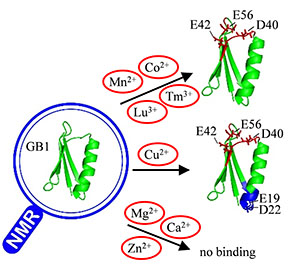
B1 domain of staphylococcal protein G (GB1) is a widely used model protein for developing in vivo and in vitro protein structural determination methods based on paramagnetic nuclear magnetic resonance (NMR) such as pseudocontact chemical shift (PCS) and paramagnetic relaxation enhancement (PRE). However, few previous studies have investigated the interactions between GB1 and metal ions, especially paramagnetic ions. In this study, the interactions between GB1 and divalent/lanthanide metal ions were studied by NMR spectroscopy. It was found that GB1 weakly bound with paramagnetic lanthanide ions and paramagnetic divalent ions, including Cu2+, Mn2+ and Co2+. In contrast, GB1 did not bind with diamagnetic divalent ions, such as Ca2+, Mg2+ and Zn2+. Furthermore, it was demonstrated that there were two binding sites for Cu2+ in GB1, but only one for lanthanide ions and divalent ions Mn2+ and Co2+. The current study demonstrated that NMR spectroscopy is a powerful tool to study weak binding between protein and metal ions. And the results indicated that care must be taken to avoid possible interference to paramagnetic NMR data when using GB1 as the model protein.
[1] GRONENBORN A M, FILPULA D R, ESSIG N Z, et al. A novel, highly stable fold of the immunoglobulin binding domain of streptococcal protein G[J]. Science, 1991, 253(5020):657-661.[2] NADAUD P S, HELMUS J J, HOFER N, et al. Long-range structural restraints in spin-labeled proteins probed by solid-state nuclear magnetic resonance spectroscopy[J]. J Am Chem Soc, 2007, 129(24):7502-7503.[3] NADAUD P S, HELMUS J J, KALL S L, et al. Paramagnetic ions enable tuning of nuclear relaxation rates and provide long-range structural restraints in solid-state NMR of proteins[J]. J Am Chem Soc, 2009, 131(23):8108-8120.[4] NADAUD P S, SENGUPTA I, HELMUS J J, et al. Evaluation of the influence of intermolecular electron-nucleus couplings and intrinsic metal binding sites on the measurement of <sup>15</sup>N longitudinal paramagnetic relaxation enhancements in proteins by solid-state NMR[J]. J Biomol NMR, 2011, 51(3):293-302.[5] SENGUPTA I, NADAUD P S, HELMUS J J, et al. Protein fold determined by paramagnetic magic-angle spinning solid-state NMR spectroscopy[J]. Nat Chem, 2012, 4(5):410-417.[6] SENGUPTA I, NADAUD P S, JARONIEC C P. Protein structure determination with paramagnetic solid-state NMR spectroscopy[J]. Acc Chem Res, 2013, 46(9):2117-2126.[7] LI J P, PILLA K B, LI Q F, et al. Magic angle spinning NMR structure determination of proteins from pseudocontact shifts[J]. J Am Chem Soc, 2013, 135(22):8294-8303.[8] SENGUPTA I, GAO M, ARACHCHIGE R J, et al. Protein structural studies by paramagnetic solid-state NMR spectroscopy aided by a compact cyclen-type Cu(Ⅱ) binding tag[J]. J Biomol NMR, 2015, 61(1):1-6.[9] TAMAKI H, EGAWA A, KIDO K, et al. Structure determination of uniformly <sup>13</sup>C, <sup>15</sup>N labeled protein using qualitative distance restraints from MAS solid-state <sup>13</sup>C-NMR observed paramagnetic relaxation enhancement[J]. J Biomol NMR, 2016, 64(1):87-101.[10] MUNTENER T, HAUSSINGER D, SELENKO P, et al. In-cell protein structures from 2D NMR experiments[J]. J Phys Chem Lett, 2016, 7(14):2821-2825.[11] PAN B B, YANG F, YE Y S, et al. 3D structure determination of a protein in living cells using paramagnetic NMR spectroscopy[J]. Chem Commun, 2016, 52(67):10237-10240.[12] SEEWALD M J, PICHUMANI K, STOWELL C, et al. The role of backbone conformational heat capacity in protein stability:temperature dependent dynamics of the B1 domain of Streptococcal protein G[J]. Protein Sci, 2000, 9(6):1177-1193.[13] BARCHI JR J J, GRASBERGER B, GRONENBORN A M, et al. Investigation of the backbone dynamics of the IgG-binding domain of streptococcal protein G by heteronuclear two-dimensional <sup>1</sup>H-<sup>15</sup>N nuclear magnetic resonance spectroscopy[J]. Protein Sci, 1994, 3(1):15-21.[14] TUNNICLIFFE R B, WABY J L, WILLIAMS R J, et al. An experimental investigation of conformational fluctuations in proteins G and L[J]. Structure, 2005, 13(11):1677-1684.[15] JEE J, BYEON I J, LOUIS J M, et al. The point mutation A34F causes dimerization of GB1[J]. Proteins, 2008, 71(3):1420-1431.[16] MCCALLISTER E L, ALM E, BAKER D. Critical role of beta-hairpin formation in protein G folding[J]. Nat Struct Biol, 2000, 7(8):669-673.[17] KARANICOLAS J, BROOKS C L. The origins of asymmetry in the folding transition states of protein L and protein G[J]. Protein Sci, 2002, 11(10):2351-2361.[18] WILTON D J, TUNNICLIFFE R B, KAMATARI Y O, et al. Pressure-induced changes in the solution structure of the GB1 domain of protein G[J]. Proteins, 2008, 71(3):1432-1440.[19] GRONENBORN A M, FILPULA D R, ESSIG N Z, et al. A novel, highly stable fold of the immunoglobulin binding domain of streptococcal protein-G[J]. Science, 1991, 253(5020):657-661.[20] THORDARSON P. Determining association constants from titration experiments in supramolecular chemistry[J]. Chem Soc Rev, 2011, 40(3):1305-1323.[21] GRZESIEK S, BAX A, CLORE G M, et al. The solution structure of HIV-1 Nef reveals an unexpected fold and permits delineation of the binding surface for the SH3 domain of Hck tyrosine protein kinase[J]. Nat Struct Biol, 1996, 3(4):340-345.[22] CHEEVER M L, SATO T K, DE BEER T, et al. Phox domain interaction with PtdIns(3)P targets the Vam7 t-SNARE to vacuole membranes[J]. Nat Cell Biol, 2001, 3(7):613-618.[23] FISHER R D, WANG B, ALAM S L, et al. Structure and ubiquitin binding of the ubiquitin-interacting motif[J]. J Biol Chem, 2003, 278(31):28976-82894.[24] OTTING G. Protein NMR using paramagnetic ions[J]. Annu Rev Biophys, 2010, 39:387-405.[25] KUSZEWSKI J, GRONENBORN A M, CLORE G M. Improving the packing and accuracy of NMR structures with a pseudopotential for the radius of gyration[J]. J Am Chem Soc, 1999, 121(10):2337-2338.[26] SCHMITZ C, STANTON-COOK M J, SU X C, et al. Numbat:an interactive software tool for fitting Delta chi-tensors to molecular coordinates using pseudocontact shifts[J]. J Biomol NMR, 2008, 41(3):179-189.[27] WILLIAMS R J P. Chemical selection of elements by cells[J]. Coordin Chem Rev, 2001, 216, 217:583-595.

