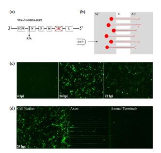
Neuron is the basic unit of nervous system. Studying the fine morphology of neurons is crucial for understanding the structure of neural network and the manner of neural information processing. At present, effective methods to visualize fine morphology of neurons with specific cell types and specific projection are still lacking. In this paper, a recombination vesicular stomatitis virus (VSV) was designed, with nucleoprotein gene mutated and glycoprotein gene deleted. The virus, VSV-△G-NR7A-EGFP, was capable of highlighted morphology of the labelled neurons in a short time. With specific transgenic animals and a helper virus rAAV-EF1α-Dio-Bfp-Tva, it was demonstrated that, a further constructed virus VSV-△G-EnvA-NR7A-EGFP could sparsely and specifically label D2R neurons with nucleus accumbens-lateral hypothalamus (NAc-LH) projection and dopamine neurons with ventral tegmental area (VTA)-NAc projection.
WEN Peng-jie
,
SU Peng
,
WANG Hao
,
HU Liang
,
LIN Kun-zhang
,
ZHU Xu-tao
,
QIU Yu-xiang
,
ZHENG Ning
,
ZHANG Zhi-jian
,
XU Fu-qiang
. Labeling Fine Morphology of Neurons with Specific Cell Types and Specific Projection Using Vesicular Stomatitis Virus[J]. Chinese Journal of Magnetic Resonance, 2018
, 35(3)
: 353
-362
.
DOI: 10.11938/cjmr20182617
[1] KOCH C, REID R C. Observatories of the mind[J]. Nature, 2012, 483:397-398.
[2] LLINAS R. The intrinsic electrophysiological properties of mammalian neurons:insights into central nervous system function[J]. Science, 1988, 242(4886):1654-1664.
[3] WU R Q, LI B, LIU Y, et al. Current state and future of optogenetic functional magnetic resonance imaging[J]. Chinese J Magn Reson, 2014, 31(2):295-305. 吴瑞琪, 李博, 刘悦, 等. 光激活磁共振脑功能成像的研究现状及前景[J]. 波谱学杂志, 2014, 31(2):295-305.
[4] ZHU X T, HE X B, LIU Y, et al. A convenient semi-automatic method for analyzing brain sections:registration, segmentation and cell counting[J]. Chinese J Magn Reson, 2018, 35(2):133-140. 朱续涛, 何晓斌, 刘悦, 等. 一种简易的脑片图像的半自动区域划分及细胞计数方法[J]. 波谱学杂志, 2018, 35(2):133-140.
[5] JEFFERIS G S, LIVET J. Sparse and combinatorial neuron labelling[J]. Curr Opin Neurobiol, 2012, 22(1):101-110.
[6] NERN A, PFEIFFER B D, RUBIN G M. Optimized tools for multicolor stochastic labeling reveal diverse stereotyped cell arrangements in the fly visual system[J]. Proc Natl Acad Sci U S A, 2015, 112(22):E2967-E2976.
[7] WU H, WILLIAMS J, NATHANS J. Morphologic diversity of cutaneous sensory afferents revealed by genetically directed sparse labeling[J]. eLife, 2012, 1:e00181.
[8] ECONOMO M N, CLACK N G, LAVIS L D, et al. A platform for brain-wide imaging and reconstruction of individual neurons[J]. eLife, 2016, 5:e10566.
[9] HOLTMAAT A, BONHOEFFER T, CHOW D K, et al. Long-term, high-resolution imaging in the mouse neocortex through a chronic cranial window[J]. Nat Protoc, 2009, 4(8):1128-1144.
[10] FENG G, MELLOR R H, BERNSTEIN M, et al. Imaging neuronal subsets in transgenic mice expressing multiple spectral variants of GFP[J]. Neuron, 2000, 28(1):41-51.
[11] LU X H, YANG X W. Genetically-directed sparse neuronal labeling in BAC transgenic mice through mononucleotide repeat frameshift[J]. Sci Rep, 2017, 7:43915.
[12] AKO R, WAKIMOTO M, EBISU H, et al. Simultaneous visualization of multiple neuronal properties with single-cell resolution in the living rodent brain[J]. Mol Cell Neurosci, 2011, 48(3):246-257.
[13] LUO W, MIZUNO H, IWATA R, et al. Supernova:A versatile vector system for single-cell labeling and gene function studies in vivo[J]. Sci Rep, 2016, 6:35747.
[14] MADISEN L, ZWINGMAN T A, SUNKIN S M, et al. A robust and high-throughput Cre reporting and characterization system for the whole mouse brain[J]. Nat Neurosci, 2010, 13(1):133-140.
[15] KUHLMAN S J, HUANG Z J. High-resolution labeling and functional manipulation of specific neuron types in mouse brain by Cre-activated viral gene expression[J]. PLoS One, 2008, 3(4):e2005.
[16] LAMMEL S, LIM B K, RAN C, et al. Input-specific control of reward and aversion in the ventral tegmental area[J]. Nature, 2012, 491(7423):212-217.
[17] KIM C K, YANG S J, PICHAMOORTHY N, et al. Simultaneous fast measurement of circuit dynamics at multiple sites across the mammalian brain[J]. Nat Methods, 2016, 13(4):325-328.
[18] LUO M. The nucleocapsid of vesicular stomatitis virus[J]. Sci China Life Sci, 2012, 55(4):291-300.
[19] HABERL M G, VIANA DA SILVA S, GUEST J M, et al. An anterograde rabies virus vector for high-resolution large-scale reconstruction of 3D neuron morphology[J]. Brain Struct Funct, 2015, 220(3):1369-1379.
[20] VAN DEN POL A N, OZDUMAN K, WOLLMANN G, et al. Viral strategies for studying the brain, including a replication-restricted self-amplifying delta-G vesicular stomatis virus that rapidly expresses transgenes in brain and can generate a multicolor golgi-like expression[J]. J Comp Neurol, 2009, 516(6):456-481.
[21] BEIER K, CEPKO C. Viral tracing of genetically defined neural circuitry[J]. J Vis Exp, 2012, 68:4253.
[22] GREEN T J, ZHANG X, WERTZ G W, et al. Structure of the vesicular stomatitis virus nucleoprotein-RNA complex[J]. Science, 2006, 313(5785):357-360.
[23] CHEN L Y, YAN Q, LU G L, et al. Several residues within the N-terminal arm of vesicular stomatitis virus nucleoprotein play a critical role in protecting viral RNA from nuclease digestion[J]. Virology, 2015, 478:9-17.
[24] STOJDL D F, LICHTY B D, TENOEVER B R, et al. VSV strains with defects in their ability to shutdown innate immunity are potent systemic anti-cancer agents[J]. Cancer Cell, 2003, 4(4):263-275.
[25] FANG X K, ZHANG S K, SUN X D, et al. Evaluation of attenuated VSVs with mutated M or/and G proteins as vaccine vectors[J]. Vaccine, 2012, 30(7):1313-1321.
[26] GENOVE G, DEMARCO U, XU H Y, et al. A new transgene reporter for in vivo magnetic resonance imaging[J]. Nat Med, 2005, 11(4):450-454.