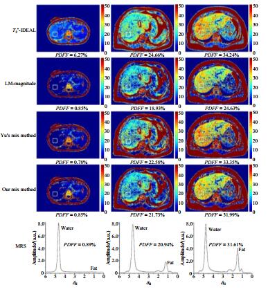
Proton density fat fraction (PDFF) is emerging as a useful quantitative magnetic resonance imaging (MRI) biomarker for the diagnosis of hepatic steatosis. In this work, a hybrid method combining complex-based and magnitude-based water-fat separation was proposed to provide accurate measurements of liver PDFF. Clinical experiments were performed to evaluate the feasibility and accuracy of the method proposed. The results demonstrated that PDFF results obtained with the proposed method had excellent correlation and agreement with those calculated from magnetic resonance spectroscopy (MRS).
ZHAI Guo-qiang
,
ZHANG Miao
,
BO Bin-shi
,
WANG Yi
,
FAN Ming-xia
,
LI Jian-qi
. Quantifying Liver Fat with Combined Complex-Based and Magnitude-Based Water-Fat Separation[J]. Chinese Journal of Magnetic Resonance, 2018
, 35(4)
: 417
-426
.
DOI: 10.11938/cjmr20182635
[1] HAUFE W M, WOLFSON T, HOOKER C A, et al. Accuracy of PDFF estimation by magnitude-based and complex-based MRI in children with MR spectroscopy as a reference[J]. J Magn Reson Imaging, 2017, 46(6):1641-1647.
[2] HERNANDO D, SHARMA S D, ALIYARI G M, et al. Multisite, multivendor validation of the accuracy and reproducibility of proton-density fat-fraction quantification at 1.5T and 3T using a fat-water phantom[J]. Magn Reson Med, 2017, 77(4):1516-1524.
[3] KINNER S, REEDER S B, YOKOO T. Quantitative Imaging Biomarkers of NAFLD[J]. Dig Dis Sci, 2016, 61(5):1337-1347.
[4] REEDER S B, HU H H, SIRLIN C B. Proton density fat-fraction:a standardized MR-based biomarker of tissue fat concentration[J]. J Magn Reson Imaging, 2012, 36(5):1011-1014.
[5] LEPORQ B, LAMBERT S A, RONOT M, et al. Quantification of the triglyceride fatty acid composition with 3.0 T MRI[J]. NMR Biomed, 2014, 27(10):1211-1221.
[6] WELLS S A. Quantification of hepatic fat and iron with magnetic resonance imaging[J]. Magn Reson Imaging Clin N Am, 2014, 22(3):397-416.
[7] WANG L L, ZHU Y, ZHONG K. Progresses in MRI and MRS studies on obesity[J]. Chinese J Magn Reson, 2016, 33(1):168-178. 王璐璐, 朱勇, 钟凯. MRI与MRS在肥胖症研究中的应用进展[J]. 波谱学杂志, 2016, 33(1):168-178.
[8] REEDER S B, WEN Z, YU H, et al. Multicoil Dixon chemical species separation with an iterative least-squares estimation method[J]. Magn Reson Med, 2004, 51(1):35-45.
[9] YU H Z, MCKENZIE C A, SHIMAKAWA A, et al. Multiecho reconstruction for simultaneous water-fat decomposition and T2* estimation[J]. J Magn Reson Imaging, 2007, 26(4):1153-1161.
[10] YU H Z, SHIMAKAWA A, HINES C D, et al. Combination of complex-based and magnitude-based multiecho water-fat separation for accurate quantification of fat-fraction[J]. Magn Reson Med, 2011, 66(1):199-206.
[11] HERNANDO D, KRAMER J H, REEDER S B. Multipeak fat-corrected complex R2* relaxometry:theory, optimization, and clinical validation[J]. Magn Reson Med, 2013, 70(5):1319-1331.
[12] REN J M, IVAN D, DEAN S A, et al. Composition of adipose tissue and marrow fat in humans by 1H NMR at 7 Tesla[J]. J Lipid Res, 2008, 49(9):2055-2062.
[13] HAMILTON G, YOKOO T, BYDDER M, et al. In vivo characterization of the liver fat 1H MR spectrum[J]. NMR Biomed, 2011, 24(7):784-790.
[14] MEISAMY S, HINES C D, HAMILTON G, et al. Quantification of hepatic steatosis with T1-independent, T2*-corrected MR imaging with spectral modeling of fat:blinded comparison with MR spectroscopy[J]. Radiology, 2011, 258(3):767-775.
[15] MACHANN J, THAMER C, SCHNOEDT B, et al. Standardized assessment of whole body adipose tissue topography by MRI[J]. J Magn Reson Imaging, 2005, 21(4):455-462.
[16] REEDER S B, SIRLIN C. Quantification of liver fat with magnetic resonance imaging[J]. Magn Reson Imaging Clin N Am, 2010, 18(3):337-357.
[17] HAACKE E M, BROWN R W, THOMPSON M R, et al. Magnetic resonance imaging:physical principles and sequence design[M]. New York:John Wiley & Sons, 1999.
[18] HU B H, YUAN H Z, GUO J X, et al. Determination of plant oils by 1H and 13C NMR[J]. Chinese J Magn Reson, 1993, 10(3):251-259. 胡邦豪, 袁汉珍, 郭建新, 等. 植物油的1H NMR和13C NMR的测定[J]. 波谱学杂志, 1993, 10(3):251-259.
[19] DONG J W, LIU T, CHEN F, et al. Simultaneous phase unwrapping and removal of chemical shift (SPURS) using graph cuts:application in quantitative susceptibility mapping[J]. IEEE T Med Imaging, 2015, 34(2):531-540.