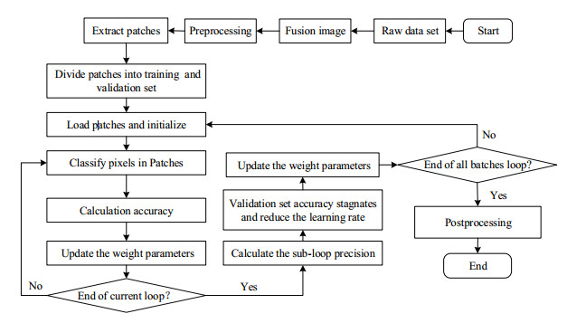
An automatic algorithm based on wavelet fusion is proposed for segmenting brain glioma with blurred boundaries and complex intratumoral structures on multimodal magnetic resonance (MR) images. Firstly, the T1, T1ce, T2 and Flair MR images of brain glioma are fused with the bias field corrected. Secondly, the image blocks to be classified are extracted, and the 3D-UNet network is trained to classify the pixels in the image blocks. Finally, the trained network model is used for segmentation, and the contour extraction method based on connected regions is used to reduce false positives. The average segmentation accuracy (DSC) of the whole, core and edema parts of the tumors was found to be 90.64%, 80.74% and 86.37%, respectively. The results indicated that the accuracy of the algorithm proposed was similar with or higher than the gold standard method. Compared with the method without multimodal image fusion, the algorithm proposed not only reduced the amount of data and redundant information in the input network, but also improved the accuracy and robustness of segmentation.
[1] MENZE B H, JAKABA, BAUER S, et al. The multimodal brain tumor image segmentation benchmark (BRATS)[J]. IEEE Trans Med Imaging, 2015, 34(10):1993-2024.
[2] DENG A, WU J, YANG S. An image fusion algorithm based on discrete wavelet transform and canny operator[M]. Berlin Heidelberg:Springer, 2011.
[3] LEVINE M D, SHAHEEN S I. A modular computer vision system for picture segmentation and interpretation[J]. IEEE Trans Pattern Anal Mach Intell, 1981, 3(5):540-556.
[4] SHIH F Y, CHENG S. Automatic seeded region growing for color image segmentation[M]. London:Butterworth-Heinemann, 2005.
[5] YAMASAKI T. GrowCut-based fast tumor segmentation for 3D magnetic resonance images[C]//Medical Imaging:Image Processing. San Diego, California, USA:International Society for Optics and Photonics, 2012.
[6] RANA R, BHADAURIA H S, SINGH A. Brain tumour extraction from MRI images using bounding-box with level set method[C]//Sixth International Conference on Contemporary Computing. Noida, India:IEEE, 2013.
[7] SELVAKUMAR J, LAKSHMI A, Arivoli T. Brain tumor segmentation and its area calculation in brain MR images using K-mean clustering and Fuzzy C-mean algorithm[C]//International Conference on Advances in Engineering. Singapore:IEEE, 2012.
[8] CLARKE L P, VELTHUIZEN R P, CAMACHO M A, et al. MRI segmentation:methods and applications[J]. Magn Reson Imaging, 1995, 13(3):343-368.
[9] LECUN Y, BENGIO Y, HINTON G. Deep learning[J]. Nature, 2015, 521(7553):436-444.
[10] ZHANG W L, LI R J, DENG H T, et al. Deep convolutional neural networks for multi-modality isointense infant brain image segmentation[J]. Proc IEEE Int Symp Biomed Imaging, 2015, 108:1342-1345.
[11] LIAO S, GAO Y Z, OTO A, et al. Representation learning:A unified deep learning framework for automatic prostate MR segmentation[J]. Med Image Comput Comput Assist Interv, 2013, 16(2):254-261.
[12] GUO Y R, GAO Y Z, SHEN D G. Deformable MR prostate segmentation via deep feature learning and sparse patch matching[J]. IEEE Trans Med Imaging, 2016, 35(4):1077-1089.
[13] WU G R, KIM M, WANG Q, et al. Unsupervised deep feature learning for deformable registration of MR brain images[J]. Med Image Comput Comput Assist Interv, 2013, 16(2):649-656.
[14] XIAO Z, HUANG R H, DING Y, et al. A deep learning-based segmentation method for brain tumor in MR images[C]//2016 IEEE 6th International Conference on Computational Advances in Bio and Medical Sciences (ICCABS). Atlanta, Georgia, USA:IEEE, 2016.
[15] WANG H Z, ZHAO D, YANG L Q, et al. An approach for training data enrichment and batch labeling in AI+MRI aided diagnosis[J]. Chinese J Magn Reson, 2018(4):447-456. 汪红志, 赵地, 杨丽琴, 等. 基于AI+MRI的影像诊断的样本增广与批量标注方法[J]. 波谱学杂志, 2018(4):447-456.
[16] HAVAEI M, DAVY A, WARDE-FARLEY D, et al. Brain tumor segmentation with deep neural networks[J]. Med Image Anal, 2015, 35:18-31.
[17] PEREIRA S, PINTO A, ALVES V, et al. Brain tumor segmentation using convolutional neural networks in MRI images[J]. IEEE Trans Med Imaging, 2016, 35(5):1240-1251.
[18] RONNEBERGER O, FISCHER P, BROX T. U-Net:Convolutional networks for biomedical image segmentation[C]//International Conference on Medical Image Computing and Computer-assisted Intervention. Munich, Germany:Springer, 2015.9351:234-241.
[19] KAMNITSAS K, LEDIG C, NEWCOMBE V F J, et al. Efficient multi-scale 3D CNN with fully connected CRF for accurate brain lesion segmentation[J]. Med Image Anal, 2017, 36:61-78.
[20] CASAMITJANA A, PUCH S, ADURIZ A, et al. 3D convolutional neural networks for brain tumor segmentation:a comparison of multi-resolution architectures[C]//International Workshop on Brainlesion:Glioma. Quebec, Canada:Springer, 2017.
[21] CHEN L L, WU Y, DSOUZA A M, et al. MRI tumor segmentation with densely connected 3D CNN[C]//Medical Imaging 2018:Image Processing. Houston, Texas, United States:International Society for Optics and Photonics, 2018, 10574:105741F.
[22] MEHTA R, ARBEL T. RS-Net:Regression-segmentation 3D CNN for synthesis of full resolution missing brain MRI in the presence of tumours[C]//International Workshop on Simulation and Synthesis in Medical Imaging. Granada, Spain:Springer, 2018.
[23] SZEGEDY C, VANHOUCKE V, IOFFE S, et al. Rethinking the inception architecture for computer vision[C]//2016 IEEE Conference on Computer Vision and Pattern Recognition (CVPR). Las Vegas, NV, USA:IEEE, 2016:2818-2826.
[24] IOFFE S, SZEGEDY C. Batch normalization:Accelerating deep network training by reducing internal covariate shift[J]. ICML, 2015.
[25] CIAMPI M. Medical image fusion for color visualization via 3D RDWT[C]//IEEE International Conference on Information Technology & Applications in Biomedicine. Corfu, Greece:IEEE, 2010.
[26] CHAI Q H, SU G Q, NIE S D. Compressive sensing low-field MRI reconstruction with dual-tree wavelet transform and wavelet tree sparsity[J]. Chinese J Magn Reson, 2018, 35(4):486-497. 柴青焕, 苏冠群, 聂生东. 双树小波变换与小波树稀疏联合的低场CS-MRI算法[J]. 波谱学杂志, 2018, 35(4):486-497.
[27] SRIVASTAVA N, HINTON G, KRIZHEVSKY A, et al. Dropout:A simple way to prevent neural networks from overfitting[J]. J Mach Learn Res, 2014, 15(1):1929-1958.
[28] BOCK S, GOPPOLD J, WEI M. An improvement of the convergence proof of the ADAM-optimizer[J]. Computer Science, 2018. arXiv:1804.10587
[29] TUSTION N J, AVANTS B B, COOK P A, et al. N4ITK:improved N3 bias correction[J]. IEEE Trans Med Imaging, 2010, 29(6):1310-1320.
[30] KAO P Y, NGO T, ZHANG A, et al. Brain tumor segmentation and tractographic feature extraction from structural MR images for overall survival prediction[C]//International MICCAI Brainlesion Workshop. Granada, Spain:Springer, 2018.
[31] WENINGER L, RIPPEL O, KOPPERS S, et al. Segmentation of brain tumors and patient survival prediction:methods for the BraTS 2018 challenge[C]//International MICCAI Brainlesion Workshop. Granada, Spain:Springer, 2018.

