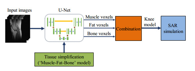
The specific absorption rate (SAR) needs to be estimated for safety concerns when performing high-field magnetic resonance imaging (MRI) on knee joint. Electromagnetic simulation can be used to calculate the local SAR on knees if a patient-specific knee model is available. In this work, a method for rapid construction of knee models from low-field magnetic resonance knee images was proposed. A convolutional neural network (CNN) was first used to segment the sagittal T1-weighted spin echo images of knees into fat, bone and muscle. All the pixels other than those from the fat and bone are classified as muscle pixels. The U-Net network, a full CNN with a convolutional layer number of four and adopting a cross entropy function, was used to perform segmentation slice-by-slice. The results from automatic segmentation were compared with those obtained with manual delineation with quantitative measures. Moreover, SAR with a 3 T quadrature birdcage coil was calculated. The simulation results validated the proposed method by showing that and a relatively accurate local SAR estimation could be obtained with the knee models constructed from low-field knee images.
XIAO Liang
,
LOU Yu-kun
,
ZHOU Hang-yu
. A U-Net Network-Based Rapid Construction of Knee Models for Specific Absorption Rate Estimation[J]. Chinese Journal of Magnetic Resonance, 2020
, 37(2)
: 144
-151
.
DOI: 10.11938/cjmr20192716
[1] PADORMO F, BEQIRI A, HAJNAL J V, et al. Parallel transmission for ultrahigh-field imaging[J]. NMR Biomed, 2016, 29(9):1145-1161.
[2] BOSS A, GRAF H, BERGER A, et al. Tissue warming and regulatory responses induced by radio frequency energy deposition on a whole-body 3-Tesla magnetic resonance imager[J]. J Magn Reson Imaging, 2007, 26(5):1334-1339.
[3] JIN J, WEBER E, DESTRUEL A, et al. An open 8-channel parallel transmission coil for static and dynamic 7T MRI of the knee and ankle joints at multiple postures[J]. Magn Reson Med, 2018, 79(3):1804-1816.
[4] GAGLIARDI V, RETICO A, BIAGI L, et al. Subject-specific knee SAR prediction using a degenerate birdcage at 7 T[C]. 2018 IEEE International Symposium on Medical Measurements and Applications (MeMeA), 2018:1-5. doi:10.1109/MeMeA.2018.8438709.
[5] 燕新强, 薛蓉, 丰宝桐, 等. 一种多通道磁共振成像设备的SAR实时监测系统及方法:中国, CN201410503941.7[P]. 2014-12-24.
[6] HUANG Q H, GAO Y, XIN X G. Study on the law of B1 field homogeneity and SAR inside human body varying with field strength at high and ultra-high field MR[J]. Chin J Biomed Eng, 2013, 32(1):21-27. 黄绮华, 高勇, 辛学刚. 高场和超高场MR下人体内B1场均匀性及SAR随场强变化规律的研究[J]. 中国生物医学工程学报, 2013, 32(1):21-27.
[7] GRAESSLIN I, HOMANN H, BIEDERER S, et al. A specific absorption rate prediction concept for parallel transmission MR[J]. Magn Reson Med, 2012, 68(5):1664-1674.
[8] GRAESSLIN I, VERNICKEL P, BÖRNERT P, et al. Comprehensive RF safety concept for parallel transmission MR[J]. Magn Reson Med, 2015, 74(2):589-598.
[9] VAN DEN BERGEN B, VAN DEN BERG CA, BARTELS LW, et al. 7 Tesla body MRI:B1 shimming with simultaneous SAR reduction[J]. Phys Med Biol, 2007, 52:5429-5441.
[10] HOMANN H, BÖRNERT P, EGGERS H, et al. Toward individualized SAR models and in vivo validation[J]. Magn Reson Med, 2011, 66(6):1767-1776.
[11] WOLF S, DIEHL D, GEBHARDT M, et al. SAR simulations for high-field MRI:how much detail, effort, and accuracy is needed?[J]. Magn Reson Med, 2013, 69(4):1157-1168.
[12] WU T N, SHAO Q, YANG L. Simplified segmented human models for whole body and localised SAR evaluation of 20 MHz to 6 GHz electromagnetic field exposures[J]. Radiat Prot Dosimetry, 2013, 153(3):266-272.
[13] PRASOON A, PETERSEN K, IGEL C, et al. Deep feature learning for knee cartilage segmentation using a triplanar convolutional neural network[C]. International conference on MICCAI 2013, 2013:246-253.
[14] LIU F, ZHOU Z Y, JANG H, et al. Deep convolutional neural network and 3D deformable approach for tissue segmentation in musculoskeletal magnetic resonance imaging[J]. Magn Reson Med, 2018, 79(4):2379-2391.
[15] ZHOU Z Y, ZHAO G Y, KIJOWSKI R, et al. Deep convolutional neural network for segmentation of knee joint anatomy[J]. Magn Reson Med, 2018, 80(6):2759-2770.
[16] RONNEBERGER O, FISCHER P, BROX T. U-net:Convolutional networks for biomedical image segmentation[C]. International Conference on MICCAI 2015, 2015:234-241.
[17] COLLINS C M, SMITH M B. Spatial resolution of numerical models of manand calculated specific absorption rate using the FDTD method:a study at 64 MHz in a magnetic resonance imaging coil[J]. J Magn Reson Imaging, 2003, 18(3):383-388.
[18] HASGALL P A, DI GENNARO F, BAUMGARTNER C, et al. IT'IS Database for thermal and electromagnetic parameters of biological tissues[R]. Version 4.0, 2018-5-15. doi:10.13099/VIP21000-04-0.itis swiss/database.
[19] HINTON G, SRIVASTAVA N, SWERSKY K. Neural networks for machine learning lecture 6a:Overview of mini-batch gradient descent[R]. Toronto University, 2012:14.
[20] YUAN K, YING B, VLASKI S, et al. Stochastic gradient descent with finite samples sizes[C]. 2016 IEEE 26th International Workshop on Machine Learning for Signal Processing (MLSP), 2016:1-6.
[21] PETERSON D M, CARRUTHERS C E, WOLVERTON B L, et al. Application of a birdcage coil at 3 Tesla to imaging of the human knee using MRI[J]. Magn Reson Med, 1999, 42(2):215-221.

