

Chinese Journal of Magnetic Resonance >
Reconstruction of Displacement Field of Left Ventricle Combined with Biomechanical Model
Received date: 2021-03-22
Online published: 2021-07-05
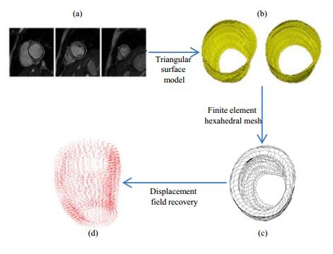
The left myocardium is the strongest cardiac muscle, representing the ability of heart to pump arterial blood throughout the body. Quantitative analysis of the contractive motion of left ventricle (LV) stands an important approach to diagnose cardiovascular diseases such as myocardial infarction. This paper utilized a biomechanical model describing the material of left myocardium to reconstruct the displacement field of LV. The mechanical model was used as an interpolation term, which was incorporated into the Bayesian estimation framework with the displacement field observed by cardiac cine magnetic resonance (CCMR) image, and the equation of displacement field was solved by finite element method. Functional parameters of LV were compared between the weak (46 cases) and normal (55 cases) ejection fraction groups of LV in the experiment. Significant differences in radial and circumferential displacement and velocity, strain and strain rate were observed between the two groups (p < 0.001), which proved that the method could effectively distinguish whether the motion of LV is normal or not. The experimental results are also highly correlated with the functional parameters of LV measured by CVI software, indicating that the method will facilitate the diagnosis of cardiovascular diseases.
Jian-sheng LIN , Li-jia WANG . Reconstruction of Displacement Field of Left Ventricle Combined with Biomechanical Model[J]. Chinese Journal of Magnetic Resonance, 2022 , 39(1) : 72 -86 . DOI: 10.11938/cjmr20212898
| 1 | DEMARINIS S . Cancer overtakes cardiovascular disease as leading cause of death in wealthy nations[J]. Explore, 2020, 16 (1): 6- 7. |
| 2 | PANG Y , LYU J , YU C , et al. Risk factors for cardiovascular disease in the Chinese population: recent progress and implications[J]. Glo Hea J, 2020, 4 (3): 65- 71. |
| 3 | FRANGI A F , NIESSEN W J , VIERGEVER M A . Three-dimensional modeling for functional analysis of cardiac images: a review[J]. IEEE Trans Med Imaging, 2001, 20 (1): 2- 5. |
| 4 | WANG H , AMINI A A . Cardiac motion and deformation recovery from MRI: A Review[J]. IEEE Trans Med Imaging, 2012, 31 (2): 487- 503. |
| 5 | ZERHOUNI E A , PARISH D M , ROGERS W J , et al. Human heart: Tagging with MR imaging-A method for noninvasive assessment of myocardial motion[J]. Radiology, 1988, 169 (1): 59- 63. |
| 6 | CHEN T , WANG X , CHUNG S , et al. Automated 3D motion tracking using Gabor filter bank, robust point matching, and deformable models[J]. IEEE Trans Med Imaging, 2009, 29 (1): 1- 11. |
| 7 | LI M , GUPTA H , LLOYD S G , et al. A graph theoretic approach for computing 3D+ time biventricular cardiac strain from tagged MRI data[J]. Med Image Anal, 2017, 35, 46- 57. |
| 8 | YU Y , ZHANG S T , LI K , et al. Deformable models with sparsity constraints for cardiac motion analysis[J]. Med Image Anal, 2014, 18 (6): 927- 937. |
| 9 | TSADOK Y , FRIEDMAN Z , HALUSKA B A , et al. Myocardial strain assessment by cine cardiac magnetic resonance imaging using non-rigid registration[J]. Magn Reson Imaging, 2016, 34 (4): 381- 390. |
| 10 | SHI P , SINUSAS A J , CONSTABLE R T , et al. Volumetric deformation analysis using mechanics-based data fusion: applications in cardiac motion recovery[J]. Int J Comput Vision, 1999, 35 (1): 87- 107. |
| 11 | REMME E W , AUGENSTEIN K F , YOUNG A A , et al. Parameter distribution models for estimation of population based left ventricular deformation using sparse fiducial markers[J]. IEEE Trans Med Imaging, 2005, 24 (3): 381- 388. |
| 12 | VERESS A I , GULLBERG G T , WEISS J A . Measurement of strain in the left ventricle during diastole with cine-MRI and deformable image registration[J]. J Biomech Eng, 2005, 127 (7): 1195- 1207. |
| 13 | BISTOQUET A , SKRINJAR O M . Left ventricular deformation recovery from cine MRI using a 4d incompressible model[J]. IEEE Trans Med Imaging, 2007, 26 (9): 1136- 1153. |
| 14 | LIU H F , SHI P C . State-space analysis of cardiac motion with biomechanical constraints[J]. IEEE T Image Process, 2007, 16 (4): 901- 917. |
| 15 | WONG K C L , WANG L W , ZHANG H Y , et al. Physiological fusion of functional and structural images for cardiac deformation recovery[J]. IEEE Trans Med Imaging, 2011, 30 (4): 990- 1000. |
| 16 | QIAO M Y , WANG Y Y , GUO Y , et al. Temporally coherent cardiac motion tracking from cine MRI: traditional registration method and modern CNN method[J]. Med Phys, 2020, 47 (9): 4189- 4198. |
| 17 | TANG J Y, GAN Z Y, YANG X. Cardiac motion tracking in short-axis MRI using siamese convolution network[C]//2019 IEEE International Conference on Bioinformatics and Biomedicine (BIBM). IEEE, 2019: 865-870. |
| 18 | ZHANG N , YANG G , GAO Z F , et al. Deep learning for diagnosis of chronic myocardial infarction on nonenhanced cardiac cine MRI[J]. Radiology, 2019, 291 (3): 606- 617. |
| 19 | ZHENG Q , DELINGETTE H , AYACHE N . Explainable cardiac pathology classification on cine MRI with motion characterization by semi-supervised learning of apparent flow[J]. Med Image Anal, 2019, 56, 80- 95. |
| 20 | MANTILLA J J , PAREDES J L , BELLANGER J J , et al. Discriminative dictionary learning for local LV wall motion classification in cardiac MRI[J]. Expert Syst Appl, 2019, 129, 286- 295. |
| 21 | SEDERBERG T W , PARRY S R . Free-form deformation of solid geometric models[J]. Siggraph, 1986, 20 (4): 151- 160. |
| 22 | COOTES T F , TAYLOR C J , COOPER D H , et al. Active shape models-their training and application[J]. Comput Vis Image Underst, 1995, 61 (1): 38- 59. |
| 23 | GONG J C , WANG Y , WANG Y J . A method for segmentation of glioma on multimodal magnetic resonance images based on wavelet fusion and deep learning[J]. Chinese J Magn Reson, 2020, 37 (2): 131- 143. |
| 23 | 宫进昌, 王宇, 王远军. 结合小波融合和深度学习的脑胶质瘤自动分割[J]. 波谱学杂志, 2020, 37 (2): 131- 143. |
| 24 | LIU P , ZHONG Y M , WANG L J . Automatic segmentation of right ventricle in cine cardiac magnetic resonance image based on a dense and multi-scale U-net method[J]. Chinese J Magn Reson, 2020, 37 (4): 456- 468. |
| 24 | 刘鹏, 钟玉敏, 王丽嘉. 基于密集多尺度U-net网络的电影心脏磁共振图像右心室自动分割[J]. 波谱学杂志, 2020, 37 (4): 456- 468. |
| 25 | GUCCIONE J M, MCCULLOCH A D. Finite element modeling of ventricular mechanics[M]. Springer New York, 1991. |
| 26 | PAPADEMETRIS X, SHI P C, DIONE D P, et al. Recovery of soft tissue object deformation from 3D image sequences using biomechanical models[C]//Proceedings of the 16th International Conference on Information Processing in Medical Imaging. Berlin, Heidelberg: Springer, 2000. |
| 27 | SHI P C , SINUSAS A J . Point-tracked quantitative analysis of left ventricular surface motion from 3-D image sequences[J]. IEEE Trans Med Imaging, 2000, 19 (1): 36- 50. |
| 28 | PAPADEMETRIS , XENOPHON , SINUSAS , et al. Estimation of 3-D left ventricular deformation from medical images using biomechanical models[J]. IEEE Trans Med Imaging, 2002, 21 (7): 786- 800. |
| 29 | 杨桂通. 弹性力学[M]. 第2版 高等教育出版社, 2012. |
| 30 | GEMAN S , GERMAN D . Stochastic relaxation, gibbs distributions and the Bayesian restoration of images[J]. IEEE Trans Pattern Anal Mach Intell, 1984, 6, 721- 741. |
| 31 | ZHANG H Y , GAO Z , XU L , et al. A meshfree representation for cardiac medical image computing[J]. IEEE J Transl Eng He, 2018, 6, 1- 12. |
| 32 | 李亚智. 有限元法基础与程序设计[M]. 科学出版社, 2004. |
/
| 〈 |
|
〉 |