

Chinese Journal of Magnetic Resonance >
An Automatic Segmentation Method of Cerebral Arterial Tree in TOF-MRA Based on DBCNet
Received date: 2022-12-16
Online published: 2023-03-13
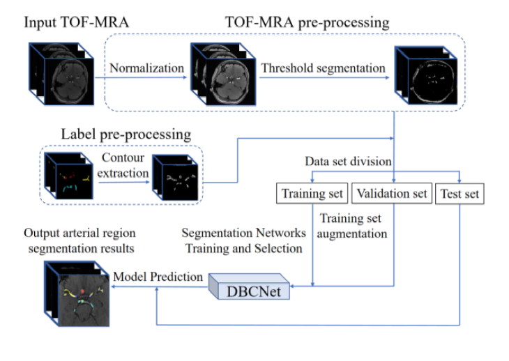
Arterial tree region segmentation from medical images of the brain is an early step in the diagnosis and evaluation of many cerebrovascular diseases. Most of the existing region segmentation methods rely on manual assistance. In this paper, we propose an automatic brain arterial tree partitioning method based on a dual branch connected network (DBCNet), which can partition the arterial tree in time of flight-magnetic resonance angiography (TOF-MRA) into six main regions. The branch feature decoupling module and the global and local feature fusion module based on Swin Transformer mechanism were used for DBCNet. The two-step training strategy of localization followed by segmentation was used for training. In this study, 111 cases of TOF-MRA data were used, of which 81 cases as the training set, 20 cases as the validation set, and 10 cases as the test set. The average Dice coefficient of the model on the test set was 74.72% and 95% Haus dorff distance (HD95) was 3.89 mm. Compared with other advanced segmentation networks, the network reported in this paper can segment each major region more accurately with robustness.
Jiajun ZHANG , Yucheng LU , Yifang BAO , Yuxin LI , Chen GENG , Fuyuan HU , Yakang DAI . An Automatic Segmentation Method of Cerebral Arterial Tree in TOF-MRA Based on DBCNet[J]. Chinese Journal of Magnetic Resonance, 2023 , 40(3) : 320 -331 . DOI: 10.11938/cjmr20223046
| [1] | GERI O, SHIRAN S I, ROTH J, et al. Vascular territorial segmentation and volumetric blood flow measurement using dynamic contrast enhanced magnetic resonance angiography of the brain[J]. J Cereb Blood Flow Metab, 2017, 37(10): 3446-3456. |
| [2] | TAHER F, PRAKASH N. Automatic cerebrovascular segmentation methods-a review[J]. IAES International Journal of Artificial Intelligence, 2021, 10(3): 576. |
| [3] | GAO X, UCHIYAMA Y, ZHOU X, et al. A fast and fully automatic method for cerebrovascular segmentation on time-of-flight (TOF) MRA image[J]. J Digit Imaging, 2011, 24(4): 609-625. |
| [4] | CHEN M, GENG C, LI Y X, et al. Automatic detection for cerebral aneurysms in TOF-MRA images based on fuzzy label and deep learning[J]. Chinese J Magn Reson, 2022, 39(3): 267-277. |
| [4] | 陈萌, 耿辰, 李郁欣, 等. 基于模糊标签和深度学习的TOF-MRA影像脑动脉瘤自动检测[J]. 波谱学杂志, 2022, 39(3): 267-277. |
| [5] | REN Y, CHEN G Z, LIU Z, et al. Reproducibility of image-based computational models of intracranial aneurysm: a comparison between 3D rotational angiography, CT angiography and MR angiography[J]. Biomed Eng Online. 2016, 15: 50. |
| [6] | MU N, LYU Z, REZAEITALESHMAHALLEH M, et al. An attention residual U-Net with differential preprocessing and geometric postprocessing: Learning how to segment vasculature including intracranial aneurysms[J]. Med Image Anal, 2023, 84: 102697. |
| [7] | LI Y, NI J, ELAZAB A, et al. Multiple self-attention network for intracranial vessel segmentation[C]// International Joint Conference on Neural Networks, Online: IEEE, 2021: 1-8. |
| [8] | BIZJAK ?, CHIEN A, BURNIK I, et al. Novel dataset and evaluation of state-of-the-art vessel segmentation methods[J]. SPIE, 2022, 12032, 120322x. |
| [9] | XIA L k, ZHANG H, WU Y, et al. 3D vessel-like structure segmentation in medical images by an edge-reinforced network[J]. Med Image Anal, 2022, 82: 102581. |
| [10] | JONES J D, CASTANHO P, BAZIRA P, et al. Anatomical variations of the circle of Willis and their prevalence, with a focus on the posterior communicating artery: A literature review and meta-analysis[J]. Clin Anat, 2021, 34(7): 978-990. |
| [11] | TAKEMURA A, SUZUKI M, HARAUCHI H, et al. Automatic segmentation method which divides a cerebral artery tree in time-of-flight MR-angiography into artery segments[J]. P Soc Photo Opt Instrum Eng, 2006, 6144: 1098-1106. |
| [12] | NOWINSKI W L, VOLKAU I, MARCHENKO Y, et al. A 3D model of human cerebrovasculature derived from 3T magnetic resonance angiography[J]. Neuroinformatics, 2009, 7(1): 23-36. |
| [13] | CHEN L, MOSSA-BASHA M, SUN J, et al. Quantification of morphometry and intensity features of intracranial arteries from 3D TOF MRA using the intracranial artery feature extraction (iCafe): A reproducibility study[J]. Magn Reson Imaging, 2019, 57: 293-302. |
| [14] | CHEN L, SUN J, HIPPE D S, et al. Quantitative assessment of the intracranial vasculature in an older adult population using iCafe[J]. Neurobiology of Aging, 2019, 79: 59-65. |
| [15] | LIU L L, CHENG J H, QUAN Q, et al. A survey on U-shaped networks in medical image segmentations[J]. Neurocomputing, 2020, 409: 244-258. |
| [16] | QIU Y, NIE S D, WEI L. Segmentation of breast tumors based on fully convolutional network and dynamic contrast enhanced magnetic resonance image[J]. Chinese J Magn Reson, 2022, 39(2): 196-207. |
| [16] | 邱玥, 聂生东, 魏珑. 基于全卷积网络的乳腺肿瘤动态增强磁共振图像分割[J]. 波谱学杂志, 2022, 39(2): 196-207. |
| [17] | FAN D P, JI G P, SUN G, et al. Camouflaged object detection[C]// Computer Vision and Pattern Recognition, 2020: 2777-2787 |
| [18] | YANG Z, SOLTANIAN-ZADEH S, FARSIU S. BiconNet: An edge-preserved connectivity-based approach for salient object detection[J]. Pattern Recogn, 2022, 121: 108231. |
| [19] | LIU Z, LIN Y T, CAO Y, et al. Swin transformer: Hierarchical vision transformer using shifted windows[C]// International Conference on Computer Vision, China:IEEE, 2021: 10012-10022. |
| [20] | YUSHKEVICH P, PIVEN J, HAZLETT H, et al. User-guided 3D active contour segmentation of anatomical structures: Significantly improved efficiency and reliability[J]. Neuroimage. 2006, 31(3): 1116-28. |
| [21] | INCI S, ERBENGI A, ?ZGEN T. Aneurysms of the distal anterior cerebral artery: report of 14 cases and a review of the literature[J]. Surg Neurol, 1998, 50(2): 130-140. |
| [22] | CANNY J. A computational approach to edge detection[J]. IEEE Trans Pattern Anal Mach Intell, 1986, 8(6): 679-98. |
| [23] | HE K M, ZHANG X Y, REN S Q, et al. Deep residual learning for image recognition[C]// Computer Vision and Pattern Recognition, USA: IEEE, 2016: 770-778. |
| [24] | WOO S H, PARK J, LEE J Y, et al. CBAM: Convolutional block attention module[J]. Computer Vision, 2018, 11211: 3-19. |
| [25] | YEUNG M, SALA E, SCH?NLIEB C B, et al. Unified focal loss: Generalising dice and cross entropy-based losses to handle class imbalanced medical image segmentation[J]. Comput Med Imag Grap, 2022, 95: 102026. |
| [26] | ISENSEE F, JAEGER P F, KOHL S A A, et al. nnU-Net: a self-configuring method for deep learning-based biomedical image segmentation[J]. Nat Methods, 2021, 18(2): 203-211. |
| [27] | ISENSEE F, KICKINGEREDER P, WICK W, et al. Brain tumor segmentation and radiomics survival prediction: Contribution to the BRATS 2017 Challenge[J]. Brainlesion: Glioma, Multiple Sclerosis, Stroke and Traumatic Brain Injuries, 2017, 10670: 287-297. |
| [28] | MILLETARI F, NAVAB N, AHMADI S A, et al. V-Net: Fully convolutional neural networks for volumetric medical image segmentation[C]// International Conference on 3d Vision, 2016: 565-571. |
| [29] | ?I?EK ?, ABDULKADIR A, LIENKAMP S S. 3D U-Net: Learning dense volumetric segmentation from sparse annotation[C]// Medical Image Computing and Computer-Assisted Intervention, 2016: 424-432. |
/
| 〈 |
|
〉 |