

Chinese Journal of Magnetic Resonance >
Application of MRI-based Finite Element Modeling and Analysis in Periacetabular Osteotomy
Received date: 2023-03-06
Online published: 2023-04-21
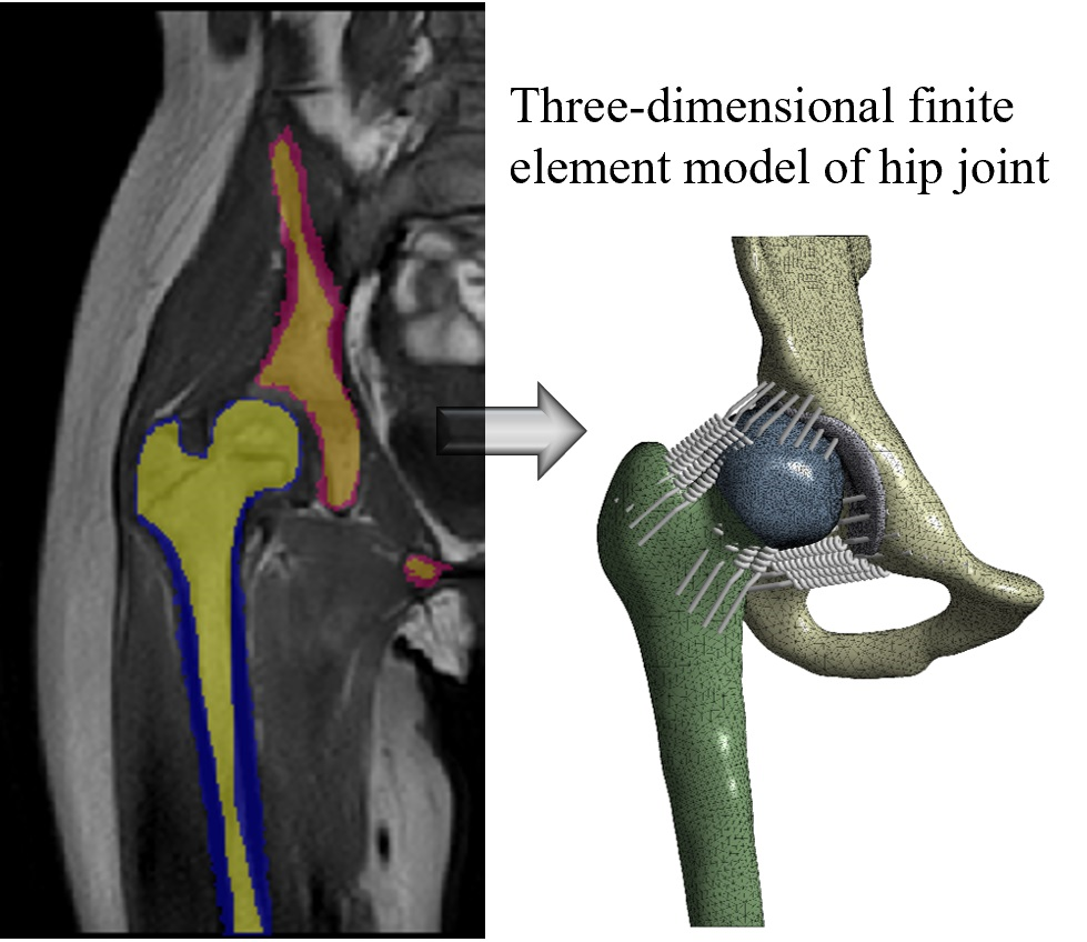
Periacetabular osteotomy (PAO) is a surgical procedure to correct acetabular orientation in developmental dysplasia of the hip (DDH). In this work, magnetic resonance imaging-based finite element models of hip joint were introduced to perform virtual surgery planning for patients with DDH. The optimal position of acetabulum following PAO was determined by analyzing the peak contact pressure and contact area between the femoral and pelvic cartilage when the acetabulum rotated from the original center edge (CE) angle in increments of 5°. Furthermore, with finite element analysis, we simulated joint movements in both the preoperative model and the planned model with the optimal position for the six cases. A comparison between the preoperative and planned models for all scenarios was made. The outcomes of finite element analysis revealed that the optimal position of acetabulum was at a CE angle of 20°. In all evaluated scenarios, the planned model showed lower peak contact pressure and larger contact area than the preoperative model did. The efficacy of virtual surgery planning was thus validated from a biomechanical perspective.
Tianli ZHOU , Dian ZHANG , Jizhi WU , Huihui JIA , Yan CHANG , Mao SHENG , Xiaodong YANG . Application of MRI-based Finite Element Modeling and Analysis in Periacetabular Osteotomy[J]. Chinese Journal of Magnetic Resonance, 2023 , 40(4) : 397 -409 . DOI: 10.11938/cjmr20233058
| [1] | YANG S, ZUSMAN N, LIEBERMAN E, et al. Developmental dysplasia of the hip[J]. Pediatrics, 2019, 143(1). |
| [2] | Wu M D, Feng J, Jia H H, et al. MRI-based morphological quantification of developmental dysplasia of the hip in children[J]. Chinese J Magn Reson, 2020, 37(4): 434-446. |
| [2] | 吴明娣, 冯洁, 贾慧惠, 等. 儿童发育性髋关节脱位的磁共振形态学定量[J]. 波谱学杂志, 2020, 37(4): 434-446. |
| [3] | GANZ R, KLAUE K, VINH T S, et al. A new periacetabular osteotomy for the treatment of hip dysplasias. Technique and preliminary results[J]. Clin Orthop Relat R, 1988, (232): 26-36. |
| [4] | HAKIM N S, KING A I. A three dimensional finite element dynamic response analysis of a vertebra with experimental verification[J]. J Biomech, 1979, 12(4): 277-285 & 287-292. |
| [5] | TANNE K, SAKUDA M. Biomechanical and clinical changes of the craniofacial complex from orthopedic maxillary protraction[J]. Angle Orthod, 1991, 61(2): 145-152. |
| [6] | GAMBOA A, COSA A, BENET F, et al. A semiautomatic segmentation method, solid tissue classification and 3D reconstruction of mandible from computed tomography imaging for biomechanical analysis[C]// Barcelona, SPAIN:9th IEEE International Symposium on Biomedical Imaging (ISBI) - From Nano to Macro, 2012: 1483-1486. |
| [7] | WANG Y C, YANG D, ZHAO L H, et al. Finite element analysis of mechanical characteristics of internal fixation for treatment of proximal femoral osteolytic lesions in children[J]. Orthop Surg, 2023, 10.1111/os.13591. |
| [8] | ZHAO X, CHOSA E, TOTORIBE K, et al. Effect of periacetabular osteotomy for acetabular dysplasia clarified by three-dimensional finite element analysis[J]. J Orthop Sci, 2010, 15(5): 632-640. |
| [9] | LEE K J, PARK S J, LEE S J, et al. Biomechanical study on the efficacy of the periacetabular osteotomy using patient-specific finite element analysis[J]. Int J Precis Eng Man, 2015, 16(4): 823-829. |
| [10] | LIU L, ECKER T M, SCHUMANN S, et al. Evaluation of constant thickness cartilage models vs. patient specific cartilage models for an optimized computer-assisted planning of periacetabular osteotomy[J]. PLoS One, 2016, 11(1): e0146452. |
| [11] | PARK S J, LEE S J, CHEN W M, et al. Computer-assisted optimization of the acetabular rotation in periacetabular osteotomy using patient’s anatomy-specific finite element analysis[J]. Appl Bionics Biomech, 2018: 9730525. |
| [12] | KITAMURA K, FUJII M, IWAMOTO M, et al. Effect of coronal plane acetabular correction on joint contact pressure in periacetabular osteotomy: a finite-element analysis[J]. BMC Musculoskelet Disord, 2022, 23(1): 48. |
| [13] | TIAN H, XU P, LU N, et al. The effect of pelvic modeling on outcome in preoperative planning for bernese acetabular osteotomy[J]. Journal of Medical Biomechanics, 2020, 35(6): 712-717. |
| [13] | 田昊, 许平, 鲁宁, 等. Bernese髋臼截骨术术前规划中骨盆建模对结果的影响[J]. 医用生物力学, 2020, 35(6): 712-717. |
| [14] | ZHANG L L, WANG X Y, CHEN X D. Study on finite element analysis method for the pre-operative planning of bernese periacetabular osteotomy[J]. Journal of Biomedical Engineering, 2016, 33(3): 455-460. |
| [14] | 张琳琳, 王旭义, 陈晓东. Bernese髋臼周围截骨术术前规划的有限元分析方法研究[J]. 生物医学工程学杂志, 2016, 33(3): 455-460. |
| [15] | STARLY B, FANG Z, SUN W, et al. Three-dimensional reconstruction for medical-CAD modeling[J]. Computer-aided Design and Applications, 2005, 2(1-4): 431-438. |
| [16] | YANG Z G, GAN L, YE J X. Construction and stability of finite element models of distal tibial fractures[J]. Chinese Journal of Tissue Engineering Research, 2016, 20(17): 2540-2545. |
| [16] | 杨志刚, 甘霖, 叶俊星. 胫骨远端骨折有限元模型的建立及稳定性分析[J]. 中国组织工程研究, 2016, 20(17): 2540-2545. |
| [17] | SOWMIANARAYANAN S, CHANDRASEKARAN A, KUMAR R K. Finite element analysis of a subtrochanteric fractured femur with dynamic hip screw, dynamic condylar screw, and proximal femur nail implants - a comparative study[J]. P I Mech Eng H, 2008, 222(1): 117-127. |
| [18] | AKRAMI M, CRAIG K, DIBAJ M, et al. A three-dimensional finite element analysis of the human hip[J]. J Med Eng Technol, 2018, 42(7): 546-552. |
| [19] | BERGMANN G, DEURETZBACHER G, HELLER M, et al. Hip contact forces and gait patterns from routine activities[J]. J Biomech, 2001, 34(7): 859-871. |
| [20] | BERGMANN G, GRAICHEN F, ROHLMANN A, et al. Hip joint forces during load carrying[J]. Clin Orthop Relat R, 1997, (335): 190-201. |
| [21] | BAY B K, HAMEL A J, OLSON S A, et al. Statically equivalent load and support conditions produce different hip joint contact pressures and periacetabular strains[J]. J Biomech, 1997, 30(2): 193-196. |
| [22] | PHILLIPS A T M, PANKAJ P, HOWIE C R, et al. Finite element modelling of the pelvis: inclusion of muscular and ligamentous boundary conditions[J]. Med Eng Phys, 2007, 29(7): 739-748. |
| [23] | ZOU Z, CHAVEZ-ARREOLA A, MANDAL P, et al. Optimization of the position of the acetabulum in a ganz periacetabular osteotomy by finite element analysis[J]. J Orthop Res, 2013, 31(3): 472-479. |
| [24] | GENDA E, IWASAKI N, LI G A, et al. Normal hip joint contact pressure distribution in single-leg standing—effect of gender and anatomic parameters[J]. J Biomech, 2001, 34(7): 895-905. |
| [25] | HARRIS M D, ANDERSON A E, HENAK C R, et al. Finite element prediction of cartilage contact stresses in normal human hips[J]. J Orthop Res, 2012, 30(7): 1133-1139. |
| [26] | AKRAMI M, QIAN Z, ZOU Z, et al. Subject-specific finite element modelling of the human foot complex during walking: sensitivity analysis of material properties, boundary and loading conditions[J]. Biomech Mode Mechan, 2018, 17(2): 559-576. |
| [27] | WU Z, CHEN S, LI Y, et al. Effect of centre-edge angle on clinical and quality of life outcomes after arthroscopic acetabular labral debridement[J]. Int Orthop, 2016, 40(7): 1427-1432. |
| [28] | KOLBER M J, CHEATHAM S W. Orthopedic management of the hip and pelvis[M]. St. Louis: Elsevier Health Sciences, 2015. |
| [29] | WU G, SIEGLER S, ALLARD P, et al. ISB recommendation on definitions of joint coordinate system of various joints for the reporting of human joint motion - part 1: ankle, hip, and spine[J]. J Biomech, 2002, 35(4): 543-548. |
| [30] | XIONG B, YANG P, LIN T, et al. Changes in hip joint contact stress during a gait cycle based on the individualized modeling method of “gait-musculoskeletal system-finite element”[J]. J Orthop Surg Res, 2022, 17(1): 267. |
| [31] | MA Y, XING C J, XIAO L. Knee joint image segmentation and model construction based on cascaded network[J]. Chinese J Magn Reson, 2022, 39(2): 184-195. |
| [31] | 马岩, 邢藏菊, 肖亮. 基于级联网络的膝关节图像分割与模型构建[J]. 波谱学杂志, 2022, 39(2): 184-195. |
/
| 〈 |
|
〉 |