

Chinese Journal of Magnetic Resonance >
Multi-source Feature Classification Model of Pancreatic Mucinous and Serous Cystic Neoplasms Based on Deep Learning
Received date: 2023-04-19
Online published: 2023-06-08
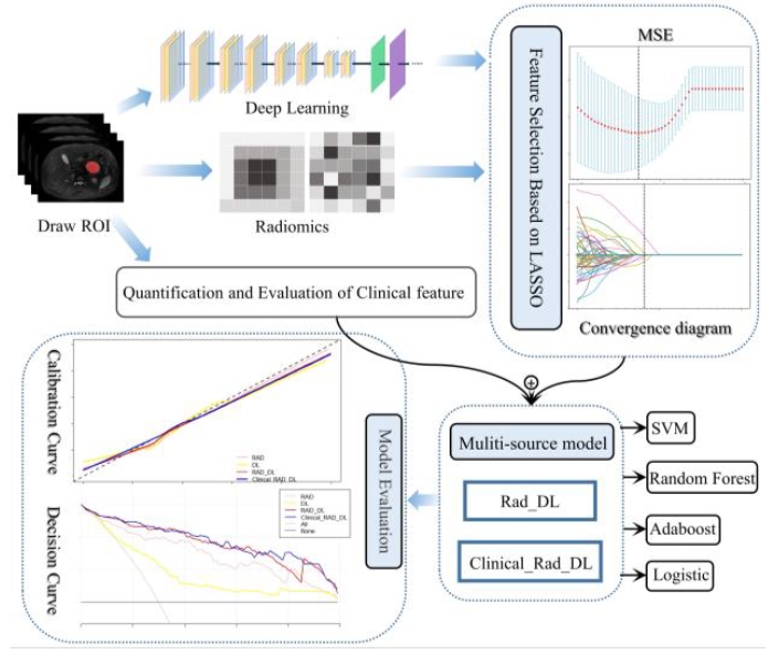
This study aims to classify and differentiate mucinous and serous cystic neoplasms of the pancreas using a multi-source feature classification model based on deep learning for preoperative auxiliary diagnosis. Deep learning features and radiomics features were extracted from segmented images using deep learning and radiomics technology, respectively. Clinical features were also evaluated and quantified. LASSO (least absolute shrinkage and selection operator) and cross-validation methods were applied to screen the features, and two multi-source feature models were constructed: the radiomics combined with deep learning (RAD_DL) model and the clinical feature combined with RAD_DL (Clinical_RAD_DL) model. Traditional radiomics (RAD) and deep learning (DL) models were used as controls. SVM (support vector machine), ADAboost (adaptive boosting), Random Forest, and Logistic were selected for classification. The Clinical_RAD_DL feature model shows the best classification performance, with the accuracy of 0.923 1, recall rate of 0.882 4, precision of 0.882 0, F1-score of 0.882 2, and AUC value of 0.912 6. The experimental results indicate that the multi-source feature classification model based on deep learning has good performance in classifying pancreatic serous cystic neoplasms and pancreatic mucinous cystic neoplasms, and can assist clinical accurate diagnosis and treatment.
Zhenshun XU , Xiaohan YUAN , Ziheng HUANG , Chengwei SHAO , Jie WU , Yun BIAN . Multi-source Feature Classification Model of Pancreatic Mucinous and Serous Cystic Neoplasms Based on Deep Learning[J]. Chinese Journal of Magnetic Resonance, 2024 , 41(1) : 19 -29 . DOI: 10.11938/cjmr20233064
| [1] | LI Z S, JIN Z D, LI X. Chinese guidelines for the diagnosis and treatment of pancreas cystic neoplasm(2022)[J]. J Clin Hepatol, 2023, 39(2): 290-298. |
| [1] | 李兆申, 金震东, 李汛. 中国胰腺囊性肿瘤诊断指南(2022年)[J]. 临床肝胆病杂志, 2023, 39(2): 290-298. |
| [2] | BIAN Y, JIANG H, CAO K, et al. The relationship between microscopic tumor size and CT tumor size in pancreatic ductal adenocarcinoma[J]. Clin Imag, 2021, 76: 30-37. |
| [3] | JAYASREE C, ABHISHEK M, LIOR G, et al. CT radiomics to predict high-risk intraductal papillary mucinous neoplasms of the pancreas[J]. Med phys, 2018, 45(11): 5019-5029. |
| [4] | AVANZO M, WEI L S, STANCANELLO J, et al. Machine and deep learning methods for radiomics[J]. Med Phys, 2020, 47(5): 185-202. |
| [5] | HAN B, XU J, WANG Y J, et al. Classification of BI-RADS 3-5 breast lesions based on MRI radiomics[J]. Chinese J Magn Reson, 2023, 40(1): 52-67. |
| [5] | 韩冰, 徐晶, 王远军, 等. 基于MRI影像组学的BI-RADS 3-5类乳腺病变三分类[J]. 波谱学杂志, 2023, 40(1): 52-67. |
| [6] | SHBOUL Z A, ALAM M, VIDYARATNE L, et al. Feature-guided deep radiomics for glioblastoma patient survival prediction[J]. Front Neurosci, 2019, 13: 966-982. |
| [7] | LI J, LIU F, FANG X, et al. CT Radiomics features in differentiation of focal-type autoimmune pancreatitis from pancreatic ductal adenocarcinoma: a propensity score analysis[J]. Acad Radiol, 2021, 29(3): 358-366. |
| [8] | PREUSS K, THACH N, LIANG X, et al. Using quantitative imaging for personalized medicine in pancreatic cancer: a review of radiomics and deep learning applications[J]. Cancers (Basel), 2022, 14(7): 1654. |
| [9] | NGUON L S, SEO K, LIM J H, et al. Deep learning-based differentiation between mucinous cystic neoplasm and serous cystic neoplasm in the pancreas using endoscopic ultrasonography[J]. Diagnostics (Basel), 2021, 11(6): 1052. |
| [10] | YANG Y F, QI Z X, NIE S D. Differentiation of benign and malignant breast lesions based on multimodal MRI and deep learning[J]. Chinese J Magn Reson, 2022, 39(4): 401-412. |
| [10] | 杨一风, 祁章璇, 聂生东. 基于多模态MRI与深度学习的乳腺病变良恶性鉴别[J]. 波谱学杂志, 2022, 39(4): 401-412. |
| [11] | ZHANG Y F, XU S S, WU J, et al. Value of CT texture analysis in differentiating pancreatic serous cystadenoma from mucinous cystadenoma[J]. Journal of Southeast University, 2022, 41(3): 308-316. |
| [11] | 张怡帆, 徐姗姗, 吴锦, 等. CT纹理分析在鉴别胰腺浆液性囊腺瘤与黏液性囊腺瘤中的价值[J]. 东南大学学报, 2022, 41(3): 308-316. |
| [12] | PARK H J, SHIN K, YOU M W, et al. Deep learning-based detection of solid and cystic pancreatic neoplasms at contrast-enhanced CT[J]. Radiology, 2023, 306(1): 140-149. |
| [13] | ISLAM W, JONES M, FAIZ R, et al. Improving performance of breast lesion classification using a ResNet50 model optimized with a novel attention mechanism[J]. Tomography, 2022, 8(5): 2411-2425. |
| [14] | LU J, WU Y, XIONG Y, et al. Breast tumor computer-aided detection system based on magnetic resonance imaging using convolutional neural network[J]. Comput Model Eng Sci, 2022, 130(1): 365-377. |
| [15] | 中国人民解放军海军军医大学第一附属医院. 胰腺外分泌功能评价模型的训练方法、系统、电子设备及介质: 中国, CN2022107020513[P], 2022-08-09. |
| [16] | LIANG W, TIAN W, WANG Y, et al. Classification prediction of pancreatic cystic neoplasms based on radiomics deep learning models[J]. BMC Cancer, 2022, 22(1): 1237-1246. |
| [17] | WANG D, HU Y, ZHAN C, et al. A nomogram based on radiomics signature and deep-learning signature for preoperative prediction of axillary lymph node metastasis in breast cancer[J]. Front Oncol, 2022, 12(20): 940-950. |
| [18] | XIE T, WANG X, ZHANG Z, et al. CT-based radiomics analysis for preoperative diagnosis of pancreatic mucinous cystic neoplasm and atypical serous cystadenomas[J]. Front Oncol, 2021, 11(6): 621520. |
| [19] | SHAO C, FENG X, YU J, et al. A nomogram for predicting pancreatic mucinous cystic neoplasm and serous cystic neoplasm[J]. Abdom Radiol (NY), 2021, 46(8): 3963-3973. |
| [20] | CHEN S. Application of CT radiomics in differential diagnosis of pancreatic serous and mucinous cystic neoplasm[J]. Chinese Journal of CT and MRI, 2022, 20(10): 92-105. |
| [20] | 陈帅. 基于CT影像组学对胰腺浆液及黏液性囊性肿瘤鉴别诊断[J]. 中国CT和MRI杂志, 2022, 20(10): 92-105. |
/
| 〈 |
|
〉 |