

Chinese Journal of Magnetic Resonance >
Application of 3D ELD_MobileNetV2 Incorporating Attention Mechanism and Dilated Convolution in Hepatic Nodules Classification
Received date: 2024-08-12
Online published: 2024-11-13
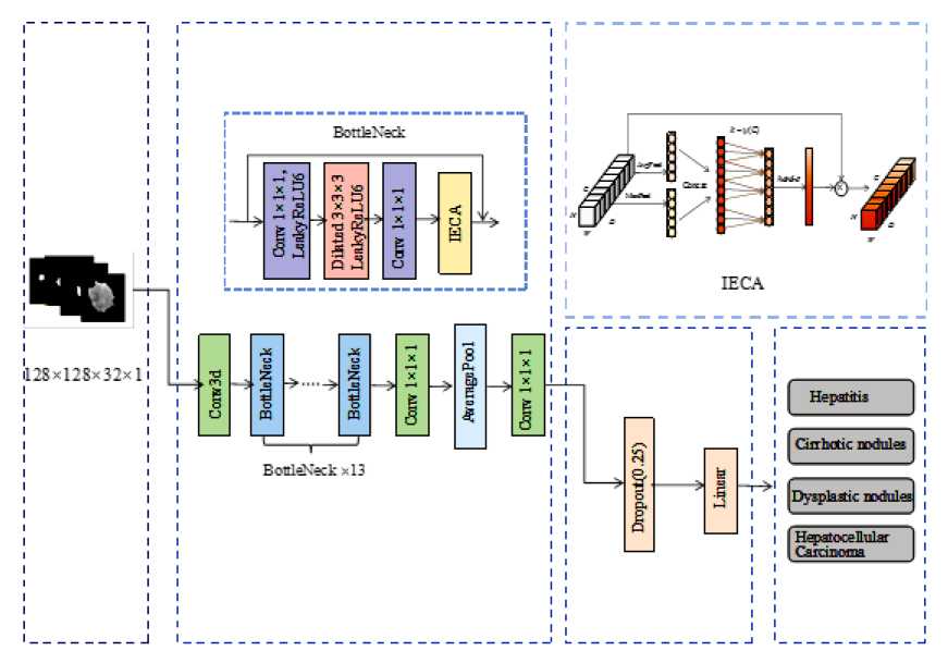
The morbidity and mortality rates of liver cancer in China remain concerning, emphasizing the urgent need for early diagnosis and treatment to improve the situation. In response to this challenge, we propose a novel 3D ELD_MobileNetV2 hepatic nodule classification model that incorporates attention mechanism and dilated convolution. This model is specifically designed to classify abdominal dynamic enhanced magnetic resonance images. Firstly, the two-dimensional network structure was extended to three dimensions to avoid the loss of spatial information during feature extraction of magnetic resonance imaging (MRI). Secondly, based on the local cross-channel interaction strategy, an efficient channel attention mechanism combining local features and global features was embedded in the bottleneck structure of MobileNetV2 network to enhance the key feature extraction capability. Then, 3D dilated structure was introduced into depthwise convolution to improve the receptive field of the convolution kernel. Meanwhile, the original activation function was replaced with Leaky ReLU6 activation function to improve the model's robustness. The model was tested and validated on a dataset comprising 120 patients (30 cases each of hepatitis, cirrhotic nodules, dysplastic nodules, and hepatocellular carcinoma). Experimental results demonstrate significant improvements over the original MobileNetV2, with an accuracy increase of 0.083. Compared to other networks, including AlexNet, VggNet16, ResNet50, ConvNeXt, the 3D ELD_MobileNetV2 achieves superior performance, with the accuracy of 0.792, F1_Score of 0.688, 0.750, 0.848, 0.872, micro-average AUC of 0.954, and macro-average AUC of 0.948. The findings highlight the effectiveness of the proposed model in classifying liver nodules across different stages. This advancement is expected to facilitate early diagnosis of liver cancer and improve clinical outcomes.
SUN Haoyun , WANG Lijia . Application of 3D ELD_MobileNetV2 Incorporating Attention Mechanism and Dilated Convolution in Hepatic Nodules Classification[J]. Chinese Journal of Magnetic Resonance, 2025 , 42(2) : 130 -142 . DOI: 10.11938/cjmr20243128
| [1] | HAN B, ZHENG R, ZENG H, et al. Cancer incidence and mortality in China, 2022[J]. J Natl Cancer Cent, 2024, 4(1): 47-53. |
| [2] | RUMGAY H, ARNOLD M, FERLAY J, et al. Global burden of primary liver cancer in 2020 and predictions to 2040[J]. J Hepatol, 2022, 77(6): 1598-1606. |
| [3] | BRAY F, LAVERSANNE M, SUNG H, et al. Global cancer statistics 2022: GLOBOCAN estimates of incidence and mortality worldwide for 36 cancers in 185 countries[J]. CA Cancer J Clin, 2024, 74(3): 229-263. |
| [4] | VOGEL A, MEYER T, SAPISOCHIN G, et al. Hepatocellular carcinoma[J]. Lancet, 2022, 400(10360): 1345-1362. |
| [5] | RENZULLI M, BRACCISCHI L, DERRICO A, et al. State-of-the-art review on the correlations between pathological and magnetic resonance features of cirrhotic nodules[J]. Histol Histopathol, 2022, 37(12): 1151-1165. |
| [6] | YOKOO T, MASAKI N, PARIKH N D, et al. Multicenter validation of abbreviated MRI for detecting early-stage hepatocellular carcinoma[J]. Radiology, 2023, 307(2): e220917. |
| [7] | BR?KER M E E, TAIMR P, DE VRIES M, et al. Performance of contrast-enhanced sonography versus MRI with a liver-specific contrast agent for diagnosis of hepatocellular adenoma and focal nodular hyperplasia[J]. AJR Am J Roentgenol, 2020, 214(1): 81-89. |
| [8] | RELA M, RAO S N, PATIL R R. Performance analysis of liver tumor classification using machine learning algorithms[J]. Int J Adv Tec Engin Explor, 2022, 9(86): 143-154. |
| [9] | LI D, HUO L, WAN M Y, et al. Application of radiomics based on new support vector machine in the classification of hepatic nodules[J]. Chinese J Magn Reson, 2022, 39(3): 278-290. |
| 李笛, 霍雷, 万梦云, 等. 基于新型支持向量LIDI机的影像组学在肝脏结节分类中的应用[J]. 波谱学杂志, 2022, 39(3): 278-290. | |
| [10] | XING Y H, HUANG Z J, HOU Y H. Application of enhanced CT-based radiomics in differentiating small hepatocellular carcinoma from hepatic dysplastic nodules[J]. Chinese Journal of CT and MRI, 2023, 21(7): 107-109. |
| 邢艳虎, 黄忠江, 侯跃宏. 基于增强CT影像组学鉴别小肝癌与肝不典型增生结节的应用研究[J]. 中国CT和MRI杂志, 2023, 21(7): 107-109. | |
| [11] | SAHA ROY S, ROY S, MUKHERJEE P, et al. An automated liver tumour segmentation and classification model by deep learning based approaches[J]. Comput Methods Biomech Biomed Engin, 2023, 11(3): 638-650. |
| [12] | CHENG N, REN Y, ZHOU J, et al. Deep learning-based classification of hepatocellular nodular lesions on whole-slide histopathologic images[J]. Gastroenterology, 2022, 162(7): 1948-1961.e7. |
| [13] | TRIVIZAKIS E, MANIKIS G C, NIKIFORAKI K, et al. Extending 2-D convolutional neural networks to 3-D for advancing deep learning cancer classification with application to MRI liver tumor differentiation[J]. IEEE J Biomed Health Inform, 2018, 23(3): 923-930. |
| [14] | ZHANG X, JIA N, WANG Y. Multi-input dense convolutional network for classification of hepatocellular carcinoma and intrahepatic cholangiocarcinoma[J]. Biomed Signal Proces, 2023, 80: 104226. |
| [15] | ZHOU J, WANG W, LEI B, et al. Automatic detection and classification of focal liver lesions based on deep convolutional neural networks: a preliminary study[J]. Front Oncol, 2021, 10: 581210. |
| [16] | KRIZHEVSKY A, SUTSKEVER I, HINTON G E. ImageNet classification with deep convolutional neural networks[J]. Communications of the ACM, 2017, 60(6): 84-90. |
| [17] | SIMONYAN K, ZISSERMAN A. Very deep convolutional networks for large-scale image recognition[J]. arXiv preprint arXiv:1409.1556, 2014. |
| [18] | ABDULSAHIB FI, AL-KHATEEB B, KóCZY LT, et al. A transfer learning approach for the classification of liver cancer[J]. J Intell Syst, 2023, 32(1): 1-14. |
| [19] | LIU Z, MAO H, WU C Y, et al. A convnet for the 2020s[C]// Proceedings of the IEEE/CVF conference on computer vision and pattern recognition. 2022: 11976-11986. |
| [20] | SANDLER M, HOWARD A, ZHU M, et al. Mobilenetv2: Inverted residuals and linear bottlenecks[C]// Proceedings of the IEEE conference on computer vision and pattern recognition. 2018: 4510-4520. |
| [21] | HE K, ZHANG X, REN S, et al. Deep residual learning for image recognition[C]// Proceedings of the IEEE conference on computer vision and pattern recognition. 2016: 770-778. |
| [22] | TRIPATHI A, SINGH T, NAIR R R, et al. Improving early detection and classification of lung diseases with innovative MobileNetV2 framework[J]. IEEE Access, 2024: 116202-116217. |
| [23] | WANG H, QI Q, SUN W, et al. Classification of skin lesions with generative adversarial networks and improved MobileNetV2[J]. Int J Imaging Syst Technol, 2023, 33(5): 1561-1576. |
| [24] | XU L, MOHAMMADI M. Brain tumor diagnosis from MRI based on Mobilenetv2 optimized by contracted fox optimization algorithm[J]. Heliyon, 2023, 10(1): e23866. |
| [25] | WANG Q, WU B, ZHU P, et al. ECA-Net: Efficient channel attention for deep convolutional neural networks[C]// Proceedings of the IEEE/CVF conference on computer vision and pattern recognition. 2020: 11534-11542. |
| [26] | TU C, LIU W, JIANG W, et al. Hyperspectral image classification based on residual dense and dilated convolution[J]. Infrared Phys Techn, 2023, 131: 104706. |
| [27] | DAUBECHIES I, DEVORE R, FOUCART S, et al. Nonlinear approximation and (deep) ReLU networks[J]. Constr Approx, 2022, 55(1): 127-172. |
| [28] | ZHAO L, WANG N, ZHU X, et al. Establishment and validation of an artificial intelligence-based model for real-time detection and classification of colorectal adenoma[J]. Sci Rep, 2024, 14(1): 10750. |
| [29] | XU J, LI Z, DU B, et al. Reluplex made more practical: Leaky ReLU[C]// 2020 IEEE Symposium on Computers and communications (ISCC). IEEE, 2020: 1-7. |
| [30] | GARCEA F, SERRA A, LAMBERTI F, et al. Data augmentation for medical imaging: A systematic literature review[J]. Comput Biol Med, 2023, 152: 106391. |
| [31] | CARTER J V, PAN J, RAI S N, et al. ROC-ing along: Evaluation and interpretation of receiver operating characteristic curves[J]. Surgery, 2016, 159(6): 1638-1645. |
| [32] | TIAN H, WU J, BIAN Y, et al. Classification of pancreatic cystic tumors based on DenseNet and transfer learning[J]. Chinese J Magn Reson, 2023, 40(3): 270-279. |
| 田慧, 武杰, 边云, 等. 基于DenseNet结合迁移学习的胰腺囊性肿瘤分类方法[J]. 波谱学杂志, 2023, 40(3): 270-279. |
/
| 〈 |
|
〉 |