

Chinese Journal of Magnetic Resonance >
An Intelligent Diagnosis Method for NIID Based on Cross Self-supervision and DWI
Received date: 2024-11-05
Online published: 2024-12-10
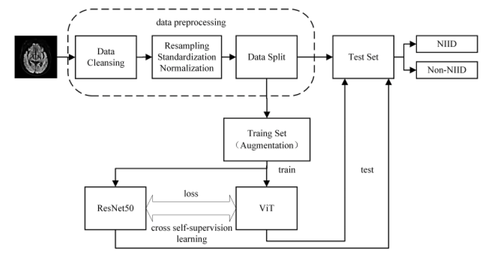
Neuronal intranuclear inclusion disease (NIID) is a rare neurodegenerative disease primarily diagnosed through diffusion-weighted imaging (DWI). However, the limitation of human visual interpretation and clinical experience can lead to inaccuracies in diagnosis. This research proposes a deep learning method based on cross self-supervision, alongside the construction of Co-ResNet50 and CO-ViT models for intelligent auxiliary diagnosis of NIID. This method uses self-supervised learning and effectively combines the characteristics of ResNet50 and ViT networks to improve the model’s feature extraction capabilities. The experiment preprocessed 249 DWI data and divided them into 204 training sets and 45 test sets. The results reveal that the CO-ResNet50 model has the best performance, with an accuracy of 95.49%, precision of 95.51%, recall of 95.44%, F1 score of 0.954 7, and AUC of 0.989 7. These findings underscore the model's potential to provide support for clinical NIID diagnosis.
Key words: MRI; NIID; self-supervised learning; intelligent diagnosis; deep learning
CAO Fei , XU Qianqian , CHEN Hao , ZU Jie , LI Xiaowen , TIAN Jin , BAO Lei . An Intelligent Diagnosis Method for NIID Based on Cross Self-supervision and DWI[J]. Chinese Journal of Magnetic Resonance, 2025 , 42(2) : 154 -163 . DOI: 10.11938/cjmr20243136
| [1] | WEI Y, WANG C H, ZHANG Y, et al. MRI manifestations of adult-onset neuronal intranuclear inclusion disease[J]. Journal of Zhengzhou University (Medical Sciences), 2024, 59(3): 405-408. |
| 魏莹, 王彩鸿, 张勇, 等. 成人型神经元核内包涵体病脑MRI表现[J]. 郑州大学学报(医学版), 2024, 59(3): 405-408. | |
| [2] | HE W G, WANG W P, LI Y J, et al. MR findings of adult neuronal intranuclear inclusion disease: report of 8 cases and literature review[J]. Radiol Practice, 2022, 37(11): 1343-1346. |
| 贺伟光, 王维平, 李又洁, 等. 成人型神经元核内包涵体病的颅脑MR表现[J]. 放射学实践, 2022, 37(11): 1343-1346. | |
| [3] | CAO Y, WU J, YUE Y, et al. Expanding the clinical spectrum of adult-onset neuronal intranuclear inclusion disease[J]. Acta Neurol Belg, 2022, 122(3): 647-658. |
| [4] | SONE J, MORI K, INAGAKI T, et al. Clinicopathological features of adult-onset neuronal intranuclear inclusion disease[J]. Brain, 2016, 139: 3170-3186. |
| [5] | SONE J, TANAKA F, KOIKE H, et al. Skin biopsy is useful for the antemortem diagnosis of neuronal intranuclear inclusion disease[J]. Neurology, 2011, 76: 1372-1376. |
| [6] | ISHIURA H, SHIBATA S, YOSHIMURA J, et al. Noncoding CGG repeat expansions in neuronal intranuclear inclusion disease, oculopharyngodistal myopathy and an overlapping disease[J]. Nat Genet, 2019, 51: 1222-1232. |
| [7] | ZHU R, QU J, XU G, et al. Clinical and multimodal imaging features of adult-onset neuronal intranuclear inclusion disease[J]. Neurol Sci, 2024, 45: 5795-5805. |
| [8] | CHEN M, GENG C, LI Y X, et al. Automatic detection for cerebral aneurysms in TOF-MRA images based on fuzzy label and deep learning[J]. Chinese J Magn Reson, 2022, 39(3): 267-277. |
| 陈萌, 耿辰, 李郁欣, 等. 基于模糊标签和深度学习的TOF-MRA影像脑动脉瘤自动检测[J]. 波谱学杂志, 2022, 39(3): 267-277. | |
| [9] | QIAN C Y, WANG Y J. Research progress on imaging classification of Alzheimer’s disease based on deep learning[J]. Chinese J Magn Reson, 2023, 40(2): 220-238. |
| 钱程一, 王远军. 基于深度学习的阿尔兹海默症影像学分类研究进展[J]. 波谱学杂志, 2023, 40(2): 220-238. | |
| [10] | CALABRESE E, RUDIE J D, RAUSCHECKER A M, et al. Combining radiomics and deep convolutional neural network features from preoperative MRI for predicting clinically relevant genetic biomarkers in glioblastoma[J]. Neurooncol Adv, 2022, 4(1): vdac060. |
| [11] | XUE P Y, GENG C, LI Y X, et al. A Classification Method for Cerebral Aneurysms in TOF-MRA Based on Improved 3D ResNet50 Model[J]. Chinese J Magn Reson, 2025, 42(1): 56-66. |
| 薛培阳, 耿辰, 李郁欣, 等. 基于改进的3D ResNet50模型的TOF-MRA脑动脉瘤分类方法[J]. 波谱学杂志, 2025, 42(1): 56-66. | |
| [12] | AZIZI S, MUSTAFA B, RYAN F, et al. Big self-supervised models advance medical image classification[C]// International Conference on Computer Vision, 2021: 3458-3468. |
| [13] | GHESU F C, GEORGESCU B, MANSOOR A, et al. Contrastive self-supervised learning from 100 million medical images with optional supervision[J]. J Med Imaging (Bellingham), 2022, 9(6): 064503. |
| [14] | ZHU Z Y, ZHAO S G. Research on brain tumor medical image classification based on diffusion self-supervised representation learning[J]. Computer Science and Application, 2024, 14(4): 133-140. |
| 朱泽宇, 赵曙光. 基于扩散模型自监督表征学习的脑瘤医学图像分类研究[J]. 计算机科学与应用, 2024, 14(4): 133-140. | |
| [15] | WANG N, LIN S H, QI F L, et al. Self-supervised Learning for Multi-modal Medical Image Segmentation[J]. Journal of Computer-Aided Design & Computer Graphics, 2025, 37(3): 474-483. |
| 王楠, 林绍辉, 齐福霖, 等. 基于自监督学习的医学影像异常检测[J]. 计算机辅助设计与图形学学报, 2025, 37(3): 474-483. | |
| [16] | XIE Y, ZHANG J, XIA Y, et al. UniMiSS: universal medical self-supervised learning via breaking dimensionality barrier[C]// European Conference on Computer Vision, 2022: 558-575. |
| [17] | GONG Y, KHURANA S, ROUDITCHENKO A, et al. CMKD: CNN/Transformerbased cross-model knowledge distillation for audio classification[J]. arXiv: abs/2203.06760, 2022. |
| [18] | SINGH P, SIZIKOVA E, CIRRONE J. CASS: Cross architectural self-supervision for medical image analysis[J]. arXiv: abs/2206.04170, 2022. |
| [19] | HE K, ZHANG X, REN S, et al. Deep residual learning for image recognition[C]// Conference Vision and Pattern Recognition, 2016: 770-778. |
| [20] | DOSOVITSKIY A, BEYER L, KOLESNIKOV A, et al. An image is worth 16x16 words: Transformers for image recognition at scale[C]// International Conference on Learning Representations, 2020. |
| [21] | HE K, CHEN X, XIE S, et al. Masked autoencoders are scalable vision learners[C]// Conference Vision and Pattern Recognition, 2021: 16000-16009. |
/
| 〈 |
|
〉 |