

Chinese Journal of Magnetic Resonance >
Application of Estrogen and Tumor Markers Combined with DCE-MRI in Diagnosis and Clinical Staging of Cervical Cancer
Received date: 2024-10-16
Online published: 2025-01-16
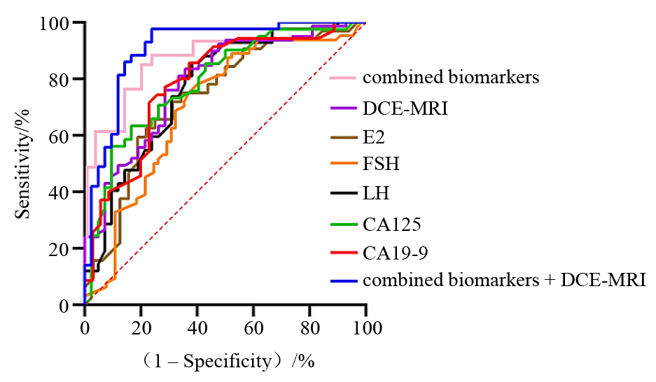
Accurate and reliable diagnosis of cervical cancer is crucial for effective clinical treatment and prognosis evaluation. Given the low sensitivity of existing serum estrogen and tumor markers in assessing lesion features like parauterine invasion and lymph node metastasis, this study proposes a diagnostic method integrating multi-phase dynamic enhanced magnetic resonance imaging (DCE-MRI). Firstly, serum estradiol (E2), follicle-stimulating hormone (FSH), luteinizing hormone (LH), glycochain antigen 125 (CA125), glycochain antigen 19-9 (CA19-9) and DCE-MRI were detected to analyze the correlation between cervical cancer stage and serum levels of E2, FSH, LH, CA125 and CA19-9. Then, cervical biopsy results were used as the gold standard to analyze the diagnostic accuracy of DCE-MRI for cervical deep muscle invasion and lymph node metastasis. Finally, the serum levels of E2, FSH, LH, CA125, CA19-9, DCE-MRI diagnosis and combined diagnosis of cervical cancer were evaluated based on the receiver operating characteristic (ROC) curve. The experimental results showed that the AUC of combined diagnosis was 0.908, indicating that the combined diagnosis method effectively distinguished benign and malignant cervical lesions. Furthermore, DCE-MRI parameters Ktrans and Kep enriched the feature information, thereby improving the detection accuracy of lesions. This provides a novel approach for differentiating between benign and malignant cervical cancer and for determining its clinical stage.
ZUO Bingyu , SHI Lili , SONG Jia , ZHAO Yang , LI Qian . Application of Estrogen and Tumor Markers Combined with DCE-MRI in Diagnosis and Clinical Staging of Cervical Cancer[J]. Chinese Journal of Magnetic Resonance, 2025 , 42(2) : 164 -173 . DOI: 10.11938/cjmr20243133
| [1] | MILLER E A, PINSKY P F. Cervical cancer screening and predictors of screening by diabetes status[J]. Cancer Causes Control, 2022, 33(10): 1305-1312. |
| [2] | SHEN S, ZHANG S, LIU P, et al. Potential role of microRNAs in the treatment and diagnosis of cervical cancer[J]. Cancer Genet, 2020, 248-249: 25-30. |
| [3] | LIN S, GAO K, GU S, et al. Worldwide trends in cervical cancer incidence and mortality, with predictions for the next 15 years[J]. Cancer, 2021, 127(21): 4030-4039. |
| [4] | YANG X, WANG J, LI WP, et al. Desmocollin 3 mediates follicle stimulating hormone-induced ovarian epithelial cancer cell proliferation by activating the EGFR/Akt signaling pathway[J]. Int J Clin Exp Pathol, 2015, 8(6): 6716-23. |
| [5] | HUANG Z Y, SONG X Y, LI Z Y. Study on the relationship between serum 25-hydroxy-vitamin D and estradiol levels and the occurrence of cervical cancer[J]. Chinese Journal of Health Inspection, 2015, 25(11): 1867-1868. |
| 黄朝阳, 宋秀宇, 李志勇. 血清25-羟-维生素D及雌二醇水平与宫颈癌发生的关系研究[J]. 中国卫生检验杂志, 2015, 25(11): 1867-1868. | |
| [6] | ZHANG L, ZHANG H, HUANG Y, et al. Expression of immune cell markers and tumor markers in patients with cervical cancer[J]. Int J Gynecol Cancer, 2020, 30(7): 969-974. |
| [7] | QIU Y, NIE S D, WEI L. Segmentation of breast tumors based on fully convolutional network and dynamic contrast enhanced magnetic resonance image[J]. Chinese J Magn Reson, 2022, 39(2): 196-207. |
| 邱玥, 聂生东, 魏珑. 基于全卷积网络的乳腺肿瘤动态增强磁共振图像分割[J]. 波谱学杂志, 2022, 39(2): 196-207. | |
| [8] | ZHENG C, QING S, WANG J, et al. Diagnosis of cervical squamous cell carcinoma and cervical adenocarcinoma based on Raman spectroscopy and support vector machine[J]. Photodiagnosis Photodyn Ther, 2019, 27: 156-161. |
| [9] | SHU W Q. Relationship between serum SCC-Ag and tissue ATF3 and clinical stage and tumor diameter in patients with cervical squamous cell carcinoma[J]. Chinese Journal of Modern Medicine, 2016, 18(10): 5-7. |
| 舒维清. 子宫颈鳞癌患者血清SCC-Ag及组织ATF3与临床分期及肿瘤直径的关系[J]. 中国现代医药杂志, 2016, 18(10): 5-7. | |
| [10] | 崔蕾. 浆液性卵巢癌患者血清中FSH、LH、PRL水平、临床意义及其联合检测价值[D]. 天津: 天津医科大学, 2017. |
| [11] | LIN L J, LV F B, XIE S M. Application value of dynamic enhanced magnetic resonance imaging combined with diffusion-weighted magnetic resonance imaging in staging diagnosis of cervical cancer[J]. China Maternal and Child Health Care, 2021, 36(21): 5099-5101. |
| 林丽君, 吕芳滨, 谢森淼. 动态增强磁共振成像联合磁共振弥散加权成像在宫颈癌患者分期诊断中的应用价值[J]. 中国妇幼保健, 2021, 36(21): 5099-5101. | |
| [12] | NG J, CHEUNG B, LEE D, et al. Detection rates and factors affecting thereof in endometrial hyperplasia, endometrial carcinoma, and cervical glandular lesions on cervical smear[J]. Cancer Med, 2023, 12(17): 17581-17591. |
| [13] | LEE A, TYRER J, DOHERTY J, et al. Evaluating the ovarian cancer gonadotropin hypothesis: a candidate gene study[J]. Gynecol Oncol, 2015, 136(3): 542-548. |
| [14] | CHEN Q H, LI Y J, WANG X M, et al. Analysis of diagnostic accuracy of pancreatic cancer by enhanced magnetic resonance imaging combined with serum carbohydrate antigen 19-9, carbohydrate antigen 125 and carcinoembryonic antigen detection[J]. Chinese Journal of Health Inspection, 2020, 30(13): 1616-1619. |
| 陈庆华, 李彦君, 王向明, 等. 增强磁共振成像联合血清糖类抗原19-9、糖类抗原125、癌胚抗原检测对胰腺癌诊断准确性的分析[J]. 中国卫生检验杂志, 2020, 30(13): 1616-1619. | |
| [15] | YANG Y F, QI Z X, NIE S D. Identification of benign and malignant breast lesions based on multimodal MRI and deep learning[J]. Chinese J Magn Reson, 2022, 39(4): 401-412. |
| 杨一风, 祁章璇, 聂生东. 基于多模态MRI与深度学习的乳腺病变良恶性鉴别[J]. 波谱学杂志, 2022, 39(4): 401-412. | |
| [16] | LOMBARDI A, VICENTE C, PORTO C. Estrogen receptors promote migration, invasion and colony formation of the androgen-independent prostate cancer cells PC-3 through β-catenin pathway[J]. Front Endocrinol (Lausanne), 2020, 11: 184. |
| [17] | DOORBAR J, EGAWA N, GRIFFIN H, et al. Human papillomavirus molecular biology and disease association[J]. Rev Med Virol, 2015, 25: 2-23. |
| [18] | LI Y J, LIU N, LEI T, et al. Analysis of relevant test indicators in differential diagnosis of prostate cancer[J]. Marker Immunoassay and Clinical Analysis, 2021, 28(10): 1750-1753. |
| 李亦君, 刘娜, 雷婷, 等. 前列腺癌鉴别诊断的相关检验指标分析[J]. 标记免疫分析与临床, 2021, 28(10): 1750-1753. | |
| [19] | CHEN L J, ZHAI M J. Correlation between Galectin-3, CA19-9 levels and clinicopathological characteristics of cervical cancer[J]. China Maternal and Child Health Care, 2019, 34(22): 5298-5301. |
| 陈立江, 翟梅娟. 宫颈癌组织中Galectin-3、CA19-9水平与临床病理特征的相关性[J]. 中国妇幼保健, 2019, 34(22): 5298-5301. | |
| [20] | LI X F. Clinical value of combined detection of serum tumor marker CA125 and CEA in diagnosis of cervical cancer[J]. Imaging Research and Medical Application, 2017, 1(11): 233-234. |
| 李晓帆. 血清肿瘤标志物CA125、CEA联合检测在宫颈癌诊断中的临床价值[J]. 影像研究与医学应用, 2017, 1(11): 233-234. | |
| [21] | HUO L, HU X X, XIAO Q, et al. Automatic segmentation of breast and gland in breast DCE-MR images based on nnU-Net[J]. Chinese J Magn Reson, 2021, 38(3): 367-380. |
| 霍璐, 胡晓欣, 肖勤, 等. 基于nnU-Net的乳腺DCE-MR图像中乳房和腺体自动分割[J]. 波谱学杂志, 2021, 38(3): 367-380. | |
| [22] | LI D, HUO L, WAN M Y, et al. Application of image omics based on novel support vector machine to liver nodule classification[J]. Chinese J Magn Reson, 2022, 39(3): 278-290. |
| 李笛, 霍雷, 万梦云, 等. 基于新型支持向量机的影像组学在肝脏结节分类中的应用[J]. 波谱学杂志, 2022, 39(3): 278-290. | |
| [23] | WEI R F, CHEN X P. Significance of MRI combined with serum SCCA and CA125 in the diagnosis of cervical cancer[J]. Chinese Journal of CT and MRI, 2020, 18(7): 97-99. |
| 魏荣富, 陈晓萍. MRI联合血清SCCA、CA125对宫颈癌的诊断意义[J]. 中国CT和MRI杂志, 2020, 18(7): 97-99 | |
| [24] | HUANG G, CUI Y, WANG P, et al. Multi-parametric magnetic resonance imaging-based radiomics analysis of cervical cancer for preoperative prediction of lymphovascular space invasion[J]. Front Oncol, 2022, 11: 663370. |
| [25] | LI J. The value of detection of FT_4, FT_3, E_2 and progesterone levels in the diagnosis of breast cancer[J]. Chinese Journal of Medical Sciences, 2023, 35(14): 117-119. |
| 李洁. FT_4、FT_3、E_2、孕酮水平检测在乳腺癌诊断中的价值[J]. 中国民康医学, 2023, 35(14): 117-119. | |
| [26] | JIANG X D. The diagnostic value of MRI combined with tumor markers in ovarian cancer[J]. Chinese Journal of CT and MRI, 2023, 21(8): 129-131+145. |
| 蒋小冬. MRI联合肿瘤标志物对卵巢癌的诊断价值分析[J]. 中国CT和MRI杂志, 2023, 21(8): 129-131+145. |
/
| 〈 |
|
〉 |