

Chinese Journal of Magnetic Resonance >
Study on Pancreas Automatic Segmentation, Regional Quantification, and Diabetes Assessment
Received date: 2025-03-27
Online published: 2025-04-23
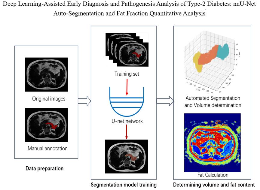
Pancreatic health is closely linked to diabetes, making accurate fat quantification crucial for early diagnosis. This study proposes a deep learning-based method for automatic pancreatic segmentation and fat quantification. The nnU-Net model achieves high-precision segmentation on m-Dixon Imaging, with a Dice similarity coefficient (DSC) of 0.92. A novel sub-region partitioning and quantification method enables precise delineation of the pancreatic head, body, and tail. Analysis of 256 subjects (healthy, prediabetic, diabetic) reveals a significant association between pancreatic tail fat and type 2 diabetes (p < 0.05). Using random forest classifiers, diabetes risk was effectively predicted based on tail fat content and a composite fat index, yielding an area under the curve (AUC) of 0.68 and 0.73, respectively. This method offers a promising tool for the early diagnosis of diabetes.
LI Yinghao , WANG Lihui , WANG Sucheng , ZHU Zhongqi , HUANG Changdong , LI Renfeng , CAO Kaiming , HU Haiyang , JIA Yiming , LIANG Songtao , YANG Guang , LU Qing , WANG Hongzhi . Study on Pancreas Automatic Segmentation, Regional Quantification, and Diabetes Assessment[J]. Chinese Journal of Magnetic Resonance, 2025 , 42(4) : 378 -389 . DOI: 10.11938/cjmr20253155
| [1] | ONG K L, STAFFORD L K, MCLAUGHLIN S A, et al. Global, regional, and national burden of diabetes from 1990 to 2021, with projections of prevalence to 2050: a systematic analysis for the Global Burden of Disease Study 2021[J]. Lancet, 2023, 402(10397): 203-234. |
| [2] | TURNER C D, BAGNARA J T. General endocrinology[M]. 6th ed. Philadelphia: W. B. Saunders Company, 1976. |
| [3] | TAYLOR R. Understanding the cause of type 2 diabetes[J]. Lancet Diabetes Endocrinol, 2024, 12(9): 664-673. |
| [4] | WANG X, MISAWA R, ZIELINSKI M C, et al. Regional differences in islet distribution in the human pancreas-preferential beta-cell loss in the head region in patients with type 2 diabetes[J]. PloS one, 2013, 8(6): e67454. |
| [5] | SARMA M K, SAUCEDO A, DARWIN C H, et al. Noninvasive assessment of abdominal adipose tissues and quantification of hepatic and pancreatic fat fractions in type 2 diabetes mellitus[J]. Magn Reson Imaging, 2020, 72: 95-102. |
| [6] | NADARAJAH C, FANANAPAZIR G, CUI E, et al. Association of pancreatic fat content with type II diabetes mellitus[J]. Clin Radiol, 2020, 75(1): 51-56. |
| [7] | GO VLW, DIMAGNO E P, GARDNER J D, et al. The Pancreas: An Integrated Textbook of Basic Science, Medicine, and Surgery[M]. 2nd ed. Hoboken: Blackwell Publishing, 2004. |
| [8] | SHEN J, BAUM T, CORDES C, et al. Automatic segmentation of abdominal organs and adipose tissue compartments in water-fat MRI: application to weight-loss in obesity[J]. Eur J Radiol, 2016, 85(9): 1613-1621. |
| [9] | WOLZ R, CHU C, MISAWA K, et al. Automated abdominal multi-organ segmentation with subject-specific atlas generation[J]. IEEE Trans Med Imaging, 2013, 32(9): 1723-1730. |
| [10] | CHU C, ODA M, KITASAKAI T, et al. Multi-organ segmentation based on spatially-divided probabilistic atlas from 3D abdominal CT images[C]// Medical Image Computing and Computer-Assisted Intervention-MICCAI 2013: 16th International Conference, Nagoya, Japan, September 22-26, 2013, Proceedings, Part II 16. Springer Berlin Heidelberg, 2013: 165-172. |
| [11] | SAITO A, NAWANO S, SHIMIZU A. Joint optimization of segmentation and shape prior from level-set-based statistical shape model, and its application to the automated segmentation of abdominal organs[J]. Med Image Anal, 2016, 28: 46-65. |
| [12] | 王鑫. 基于统计模型的胰腺分割算法的研究与实现[D]. 沈阳: 东北大学, 2013. |
| [13] | DAI J L, HE C, WU J, et al. Pancreatic cystic neoplasms segmentation network combining dual decoding and global attention upsampling modules[J]. Chinese J Magn Reson, 2024, 41(2): 151-161. |
| 戴俊龙, 何聪, 武杰, 等. 融合双解码和全局注意力上采样模块的胰腺囊性肿瘤分割网络[J]. 波谱学杂志, 2024, 41(2): 151-161. | |
| [14] | CHEN L, WAN L. CTUNet: automatic pancreas segmentation using a channel-wise transformer and 3D U-Net[J]. Vis Comput, 2023, 39(11): 5229-5243. |
| [15] | PAITHANE P, KAKARWA S. LMNS-Net: Lightweight multiscale novel semantic-net deep learning approach used for automatic pancreas image segmentation in CT scan images[J]. Expert Syst Appl, 2023, 234: 121064. |
| [16] | GHORPADE H, JAGTA J, PATIL S, et al. Automatic segmentation of pancreas and pancreatic tumor: a review of a decade of research[J]. IEEE Access, 2023, 11: 108727-108745. |
| [17] | ISENSEE F, JAEGER P F, KOHL S A A, et al. nnU-Net: a self-configuring method for deep learning-based biomedical image segmentation[J]. Nat Methods, 2021, 18(2): 203-211. |
| [18] | SULOCHANA S, SIVAKAMI T. A gross morphological study of the pancreas in human cadavers[J]. Natl J Clin Anat, 2012, 1(2): 55-60. |
| [19] | KUKU G M, HITTATIYA K, SPRINKART A M, et al. Comparison between modified Dixon MRI techniques, MR spectroscopic relaxometry, and different histologic quantification methods in the assessment of hepatic steatosis[J]. Eur Radiol, 2015, 25: 2869-2879. |
| [20] | FEDOROV A, BEICHEL R, KALPATHY-CRAMER J, et al. 3D Slicer as an image computing platform for the Quantitative Imaging Network[J]. Magn Reson Imaging, 2012, 30(9): 1323-1341. |
| [21] | BI X L, LU M, XIAO B, et al. Pancreas segmentation based on dual-decoder U-Net convolutional neural network[J]. J Softw, 2022, 33(5): 1947-1958. |
| 毕秀丽, 陆猛, 肖斌, 等. 基于双解码U型卷积神经网络的胰腺分割[J]. 软件学报, 2022, 33(5): 1947-1958. | |
| [22] | ZHANG Z, KELES E, DURAK G, et al. Large-scale multi-center CT and MRI segmentation of pancreas with deep learning[J]. Med Image Anal, 2025, 99: 103382. |
| [23] | QU T, LI X, WANG X, et al. Transformer guided progressive fusion network for 3D pancreas and pancreatic mass segmentation[J]. Med Image Anal, 2023, 86: 102801. |
| [24] | CHEN J, CHEN W, ZHU Z, et al. MAFE-Net: A multi-level attention feature extraction network for pancreas segmentation[J]. J Mach Learn, 2024,10: 1-19. |
| [25] | SKUDDE-HILL L, SEQUEIRA I R, CHO J, et al. Fat distribution within the pancreas according to diabetes status and insulin traits[J]. Diabetes, 2022, 71(6): 1182-1192. |
/
| 〈 |
|
〉 |