),胡伏原1,§(),戴亚康1,3,*()
),HU Fuyuan1,§(),DAI Yakang1,3,*()
),胡伏原1,§(),戴亚康1,3,*()
),HU Fuyuan1,§(),DAI Yakang1,3,*()
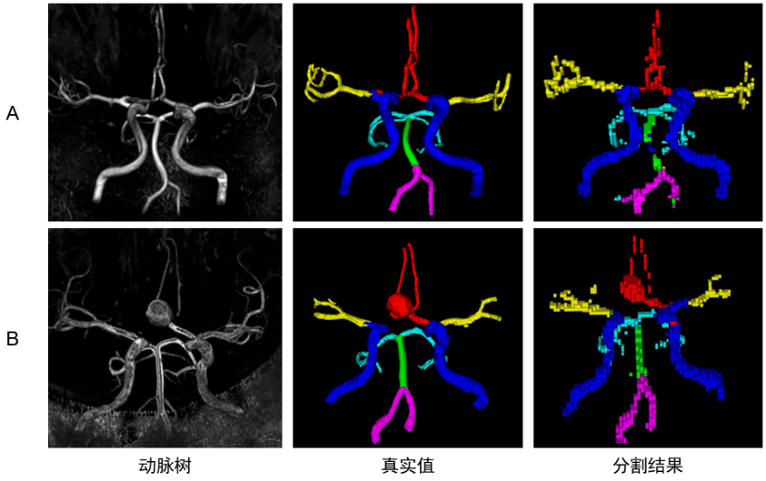
图6. DBCNet颅内动脉树区域分割预测结果的三维重建展示图. A行为1例健康人的动脉树三维重建(对TOF-MRA进行阈值分割得到)、标注真实值和分割结果展示;B行为1例颅内动脉瘤患者的动脉树三维重建、标注真实值和分割结果展示,患者动脉瘤在大脑前动脉(ACA)区域.真实值和模型分割结果视觉效果不同,是因为标注真实值为人工使用实心小球绘制,而模型分割结果是由体素级分割后上采样回原图像大小得到的. 颈内动脉:ICA,蓝色;基底动脉:BA,绿色;椎动脉:VA,紫色;大脑中动脉:MCA,黄色;大脑前动脉:ACA,红色大脑后动脉:PCA,青色
Fig. 6. 3D reconstruction for DBCNet intracranial arterial tree region segmentation prediction results. Row A shows the arterial tree 3D reconstruction (threshold segmentation result of TOF-MRA), labeled real values and segmentation results of a healthy person; row B shows the arterial tree 3D reconstruction, labeled real values and segmentation results of a patient with intracranial aneurysm, the patient’s aneurysm is in the anterior cerebral artery (ACA) region. The visual effect of the real values and model segmentation results is different because the labeled real values are drawn manually using solid spheres, while the model segmentation results are obtained by up-sampling back to the original image size after voxel-level segmentation. Internal carotid arteries (ICA, blue), basilar artery (BA, green), vertebral artery (VA, purple), middle cerebral artery (MCA, yellow), anterior (ACA, red) and posterior cerebral artery (PCA, cyan)