改进约束球面反卷积模型的脑灰质微结构成像
Improved Constrained Spherical Deconvolution for Microstructural Imaging of Brain Gray Matter
改进约束球面反卷积模型的脑灰质微结构成像 |
| 杨佳铖, 王远军 |
|
Improved Constrained Spherical Deconvolution for Microstructural Imaging of Brain Gray Matter |
| YANG Jiacheng, WANG Yuanjun |
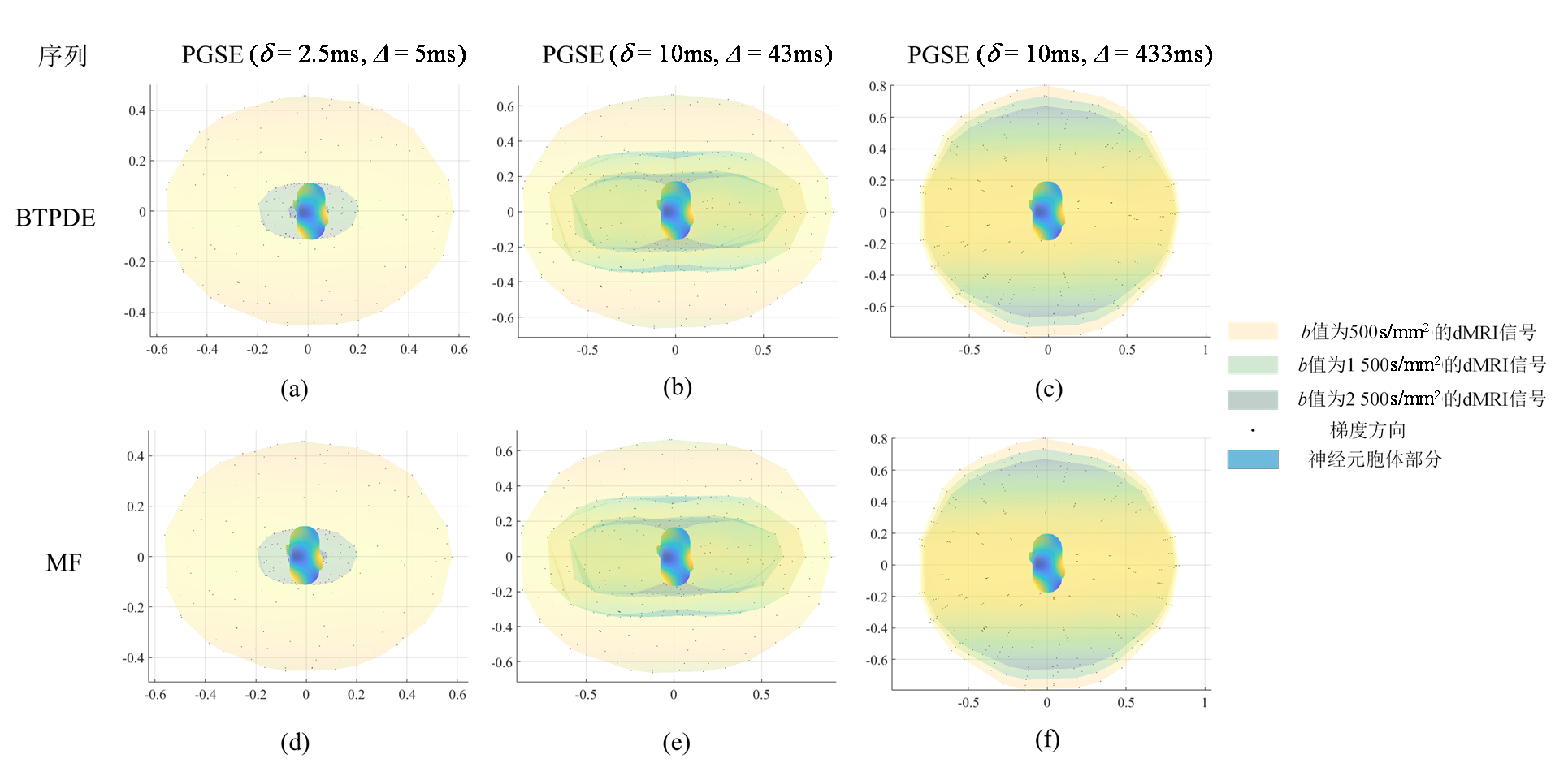
| 图4 使用BTPDE和MF方法仿真的纺锤体神经元的胞体归一化dMRI信号.(a)、(d)对应PGSE(δ=2.5 ms,Δ=5 ms)时不同b值生成的信号;(b)、(e)对应PGSE(δ=10 ms,Δ=43 ms)时不同b值生成的信号;(c)、(f)对应PGSE(δ=10 ms,Δ=433 ms)时不同b值生成的信号. 图中横纵坐标代表了归一化信号的大小|S/S0|;图中最外圈信号值对应的b值为500 s/mm2,中间对应的b值为1 500 s/mm2,最内圈对应的b值为2 500 s/mm2. |
| Fig. 4 dMRI signals of spindle neuron soma simulated using BTPDE and MF methods. (a) and (d) correspond to signals generated at different b values at PGSE (δ=2.5 ms, Δ=5 ms); (b) and (e) correspond to signals generated at different b values at PGSE (δ=10 ms, Δ=43 ms); (c) and (f) correspond to signals generated at different b values at PGSE (δ=10 ms, Δ=433 ms). The horizontal and vertical coordinates of the figure represent the normalized magnitude of the signals |S/S0|; the signal value in the outermost circle of the figure corresponds to a b value of 500 s/mm2, the one in the middle corresponds to a b value of 1 500 s/mm2, and the one in the innermost circle corresponds to a b value of 2 500 s/mm2. |
|
|