雌激素、肿瘤标志物联合DCE-MRI在宫颈癌诊断及临床分期中的应用
Application of Estrogen and Tumor Markers Combined with DCE-MRI in Diagnosis and Clinical Staging of Cervical Cancer
雌激素、肿瘤标志物联合DCE-MRI在宫颈癌诊断及临床分期中的应用 |
| 左冰玉, 石丽莉, 宋佳, 赵阳, 李倩 |
|
Application of Estrogen and Tumor Markers Combined with DCE-MRI in Diagnosis and Clinical Staging of Cervical Cancer |
| ZUO Bingyu, SHI Lili, SONG Jia, ZHAO Yang, LI Qian |
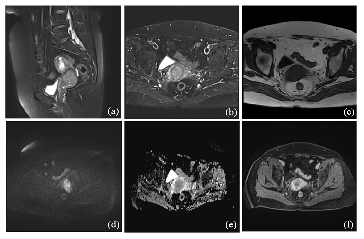
| 图2 宫颈癌的DCE-MRI影像学表现. (a)矢状位T2WI抑脂序列示宫颈区肿块;(b) T2WI示肿块呈高信号,低信号基质环局部中断;(c) T1WI示肿块呈等信号;(d) DWI示肿块弥散受限呈高信号;(e) ADC呈低信号,值约0.73×10⁻³ mm2/s;(f)横断位T1WI脂肪抑制增强示肿块明显不均匀强化,静脉期及延迟期呈持续性强化,延迟期强化程度低于子宫肌层 |
| Figure 2 DCE-MRI imaging findings of cervical cancer. (a) Sagittal T2WI lipid suppression sequence showed cervical mass; (b) The mass on T2WI showed high signal and local interruption of stromal ring with low signal; (c) T1WI showed equal-signal mass; (d) DWI showed limited mass diffusion with high signal; (e) The ADC is a low signal with a value of about 0.73×10⁻³ mm2/s; (f) Enhanced fat inhibition at transversal T1WI showed significant uneven enhancement of the mass, sustained enhancement in the venous phase and the delayed phase, and the degree of enhancement in the delayed phase was lower than that in the myometrium |
|
|