Research progress on the impact of exercise intervention on brain executive functions in children with attention deficit hyperactivity disorder: a neuroimaging perspective
2
2023
... 本文系统总结了MRI技术在儿童运动干预研究中的应用,涵盖多种MRI模态及其研究进展.结构磁共振成像(structural magnetic resonance imaging,sMRI)主要聚焦运动干预对脑灰质体积、密度和皮层厚度的影响,总结了引起这些变化的可能机制.扩散张量成像(diffusion tensor imaging,DTI)着重分析白质纤维束的完整性和连通性变化,探讨水分子扩散特性的变化及其与神经可塑性的关系.静息态和任务态功能磁共振成像(functional magnetic resonance imaging,fMRI)关注运动干预对脑功能连接及大脑激活模式的影响,探讨功能改变与认知和执行功能的关联.磁共振波谱(magnetic resonance spectroscopy,MRS)涉及运动干预对脑代谢物变化的研究,揭示其与认知功能改善的潜在机制.多模态磁共振技术则整合不同模态的优势,从多维度评估脑结构与功能的改善.现阶段儿童运动干预的系统综述大多只针对单一病症,例如注意力缺陷多动障碍和发展性协调障碍[1 ,2 ] .目前也有部分综述探讨儿童体育活动与脑结构、认知能力或肥胖等因素之间的关系[3 ,4 ] .集中关注神经影像方法的儿童运动干预系统性综述尚较有限.本综述旨在通过总结不同MRI技术在儿童运动干预研究领域的应用,对比不同成像方法的特点,为儿童运动干预的神经影像学研究提供有益的参考. ...
... 部分实验结果[7 ,27 ,32 ,33 ] 没有发现显著差异,这可能是多种原因导致的.首先,干预措施没有持续足够的时间,导致大脑未产生明显变化,有研究认为6周是获得最小程度改善的最短干预时间,更显著的改变需要更长时间和更强的干预[41 ] .其次,实验中需要选择合适的干预措施,不同的干预措施可能对不同的脑功能有影响,可能需要更具针对性的干预才能产生明显的效果.Kong等人[1 ] 的综述提到不同的运动干预方案对注意缺陷多动障碍儿童执行功能的各个子功能及整体的改善具有差异,例如定向运动干预可以改善患儿的转换功能,而长期的、以开放式动作技能为主的中高强度的运动干预可以提高患儿的抑制功能.此外,个体差异也需要考虑,受试儿童的年龄差异和大脑受损程度会影响实验结果,每个个体对于干预措施的响应程度不同,这种个体差异可能导致实验结果在整体上看不到明显的干预效果[27 ] . ...
运动干预对注意缺陷多动障碍儿童脑执行功能影响的神经影像学研究进展
2
2023
... 本文系统总结了MRI技术在儿童运动干预研究中的应用,涵盖多种MRI模态及其研究进展.结构磁共振成像(structural magnetic resonance imaging,sMRI)主要聚焦运动干预对脑灰质体积、密度和皮层厚度的影响,总结了引起这些变化的可能机制.扩散张量成像(diffusion tensor imaging,DTI)着重分析白质纤维束的完整性和连通性变化,探讨水分子扩散特性的变化及其与神经可塑性的关系.静息态和任务态功能磁共振成像(functional magnetic resonance imaging,fMRI)关注运动干预对脑功能连接及大脑激活模式的影响,探讨功能改变与认知和执行功能的关联.磁共振波谱(magnetic resonance spectroscopy,MRS)涉及运动干预对脑代谢物变化的研究,揭示其与认知功能改善的潜在机制.多模态磁共振技术则整合不同模态的优势,从多维度评估脑结构与功能的改善.现阶段儿童运动干预的系统综述大多只针对单一病症,例如注意力缺陷多动障碍和发展性协调障碍[1 ,2 ] .目前也有部分综述探讨儿童体育活动与脑结构、认知能力或肥胖等因素之间的关系[3 ,4 ] .集中关注神经影像方法的儿童运动干预系统性综述尚较有限.本综述旨在通过总结不同MRI技术在儿童运动干预研究领域的应用,对比不同成像方法的特点,为儿童运动干预的神经影像学研究提供有益的参考. ...
... 部分实验结果[7 ,27 ,32 ,33 ] 没有发现显著差异,这可能是多种原因导致的.首先,干预措施没有持续足够的时间,导致大脑未产生明显变化,有研究认为6周是获得最小程度改善的最短干预时间,更显著的改变需要更长时间和更强的干预[41 ] .其次,实验中需要选择合适的干预措施,不同的干预措施可能对不同的脑功能有影响,可能需要更具针对性的干预才能产生明显的效果.Kong等人[1 ] 的综述提到不同的运动干预方案对注意缺陷多动障碍儿童执行功能的各个子功能及整体的改善具有差异,例如定向运动干预可以改善患儿的转换功能,而长期的、以开放式动作技能为主的中高强度的运动干预可以提高患儿的抑制功能.此外,个体差异也需要考虑,受试儿童的年龄差异和大脑受损程度会影响实验结果,每个个体对于干预措施的响应程度不同,这种个体差异可能导致实验结果在整体上看不到明显的干预效果[27 ] . ...
Neural basis and motor imagery intervention methodology based on neuroimaging studies in children with developmental coordination disorders: a review
1
2021
... 本文系统总结了MRI技术在儿童运动干预研究中的应用,涵盖多种MRI模态及其研究进展.结构磁共振成像(structural magnetic resonance imaging,sMRI)主要聚焦运动干预对脑灰质体积、密度和皮层厚度的影响,总结了引起这些变化的可能机制.扩散张量成像(diffusion tensor imaging,DTI)着重分析白质纤维束的完整性和连通性变化,探讨水分子扩散特性的变化及其与神经可塑性的关系.静息态和任务态功能磁共振成像(functional magnetic resonance imaging,fMRI)关注运动干预对脑功能连接及大脑激活模式的影响,探讨功能改变与认知和执行功能的关联.磁共振波谱(magnetic resonance spectroscopy,MRS)涉及运动干预对脑代谢物变化的研究,揭示其与认知功能改善的潜在机制.多模态磁共振技术则整合不同模态的优势,从多维度评估脑结构与功能的改善.现阶段儿童运动干预的系统综述大多只针对单一病症,例如注意力缺陷多动障碍和发展性协调障碍[1 ,2 ] .目前也有部分综述探讨儿童体育活动与脑结构、认知能力或肥胖等因素之间的关系[3 ,4 ] .集中关注神经影像方法的儿童运动干预系统性综述尚较有限.本综述旨在通过总结不同MRI技术在儿童运动干预研究领域的应用,对比不同成像方法的特点,为儿童运动干预的神经影像学研究提供有益的参考. ...
Physical activity, fitness, cognitive function, and academic achievement in children: a systematic review
1
2016
... 本文系统总结了MRI技术在儿童运动干预研究中的应用,涵盖多种MRI模态及其研究进展.结构磁共振成像(structural magnetic resonance imaging,sMRI)主要聚焦运动干预对脑灰质体积、密度和皮层厚度的影响,总结了引起这些变化的可能机制.扩散张量成像(diffusion tensor imaging,DTI)着重分析白质纤维束的完整性和连通性变化,探讨水分子扩散特性的变化及其与神经可塑性的关系.静息态和任务态功能磁共振成像(functional magnetic resonance imaging,fMRI)关注运动干预对脑功能连接及大脑激活模式的影响,探讨功能改变与认知和执行功能的关联.磁共振波谱(magnetic resonance spectroscopy,MRS)涉及运动干预对脑代谢物变化的研究,揭示其与认知功能改善的潜在机制.多模态磁共振技术则整合不同模态的优势,从多维度评估脑结构与功能的改善.现阶段儿童运动干预的系统综述大多只针对单一病症,例如注意力缺陷多动障碍和发展性协调障碍[1 ,2 ] .目前也有部分综述探讨儿童体育活动与脑结构、认知能力或肥胖等因素之间的关系[3 ,4 ] .集中关注神经影像方法的儿童运动干预系统性综述尚较有限.本综述旨在通过总结不同MRI技术在儿童运动干预研究领域的应用,对比不同成像方法的特点,为儿童运动干预的神经影像学研究提供有益的参考. ...
The developing brain: considering the multifactorial effects of obesity, physical activity & mental wellbeing in childhood and adolescence
1
2022
... 本文系统总结了MRI技术在儿童运动干预研究中的应用,涵盖多种MRI模态及其研究进展.结构磁共振成像(structural magnetic resonance imaging,sMRI)主要聚焦运动干预对脑灰质体积、密度和皮层厚度的影响,总结了引起这些变化的可能机制.扩散张量成像(diffusion tensor imaging,DTI)着重分析白质纤维束的完整性和连通性变化,探讨水分子扩散特性的变化及其与神经可塑性的关系.静息态和任务态功能磁共振成像(functional magnetic resonance imaging,fMRI)关注运动干预对脑功能连接及大脑激活模式的影响,探讨功能改变与认知和执行功能的关联.磁共振波谱(magnetic resonance spectroscopy,MRS)涉及运动干预对脑代谢物变化的研究,揭示其与认知功能改善的潜在机制.多模态磁共振技术则整合不同模态的优势,从多维度评估脑结构与功能的改善.现阶段儿童运动干预的系统综述大多只针对单一病症,例如注意力缺陷多动障碍和发展性协调障碍[1 ,2 ] .目前也有部分综述探讨儿童体育活动与脑结构、认知能力或肥胖等因素之间的关系[3 ,4 ] .集中关注神经影像方法的儿童运动干预系统性综述尚较有限.本综述旨在通过总结不同MRI技术在儿童运动干预研究领域的应用,对比不同成像方法的特点,为儿童运动干预的神经影像学研究提供有益的参考. ...
Effects of exercise intervention on executive functions and gray matter volume in deaf children
2
2018
... sMRI通常包括T 1 加权成像、T 2 加权成像、液体衰减反转恢复序列成像(FLAIR)等,儿童运动干预的脑结构研究主要采用三维容积高空间分辨率T 1 加权图像,常用于研究灰质体积与密度、皮层厚度等变化.主要采用的方法有基于体素的形态学分析和基于皮层的形态学分析,可用于探索儿童运动干预前后的局部脑区灰质体积或密度,以及皮质厚度、表面积和褶皱度等指标,基于形变的形态学分析可以比较个体或群体间的形变场来评估组织体积的变化.Chen等人[5 ] 发现有氧运动干预可以使聋哑儿童执行功能得到改善,右侧小脑前部的灰质体积减小.有研究表明有氧干预可以增加脑瘤儿童皮质厚度,并使用了基于形变的形态学分析方法,发现患儿右侧运动和躯体感觉皮层下白质体积增加[6 ] .但Bunketorp等人[7 ] 发现儿童在校内外体育活动后海马体体积无明显变化,他们认为这可能是干预措施不够有效导致的. ...
... Results of different examination methods in pediatric exercise intervention experiments
Table 1 文献 检查技术 干预方法 干预时间 实验对象 年龄/岁 干预结果 [5 ] sMRI 中等强度组合方案 11周 聋哑儿童 11.01±0.64 右侧小脑前部灰质体积减小 [6 ] sMRI 团体有氧运动 12周 脑瘤儿童 11.19±2.98 皮质厚度和右侧运动和躯体感觉皮层下白质体积增加 [7 ] sMRI 校内外体育活动 4年 健康儿童 10.1±2.1 海马体体积无明显变化 [10 ] DTI 有氧运动 8个月 超重儿童 9.9±0.6 额颞白质FA增加,RD减少 [11 ] DTI 下肢选择性动作 1个月 早产脑瘫 11.5±2.8 运动相关的白质区域RD和MD显著降低 [12 ] DTI 小篮球运动 12周 孤独症儿童 5.13±0.61 白质完整性得到改善 [13 ] DTI 调查体育活动水平* / 健康儿童 9.71±0.28 皮质脊髓束、上纵束和下纵束等表现出更大的FA [14 ] DTI 测量有氧运动能力* / 健康儿童 9.9±0.6 胼胝体、放射冠、上级纵束表现出更大的FA [15 ] DTI 测量有氧运动能力* / 健康儿童 14.3±0.9 胼胝体、双侧上放射冠为主的白质纤维束表现出更大的FA [17 ] 静息态fMRI 小篮球运动 12周 孤独症儿童 6.40±2.07 感觉运动网络功能连接减少,左颞下回和左尾状核为中心的两个子网络形态连通性降低 [18 ] 静息态fMRI 小篮球运动 12周 孤独症儿童 5.07±0.59 改善执行控制网络功能连接 [19 ] 静息态fMRI 小篮球运动 12周 孤独症儿童 5.13±0.64 重塑脑功能网络特征 [20 ] 静息态fMRI 小篮球运动 12周 孤独症儿童 5.06±0.63 执行功能相关脑区功能局部一致性改变 [21 ] 静息态fMRI 小篮球运动 12周 孤独症儿童 5.00 改善了默认模式网络的功能连接 [22 ] 静息态fMRI 中等强度组合方案 11周 聋哑儿童 11.01±0.64 改善执行控制网络功能连接 [23 ] 静息态fMRI 中等强度有氧运动 短时 健康儿童 10.00 增加了静息状态下脑功能局部一致性 [24 ] 静息态fMRI 心血管健康和大运动技能* / 健康儿童 9.13±0.62 与更好的神经认知功能相关 [25 ] 任务态fMRI 中等强度组合方案 14周 聋哑儿童 10.14±1.03 改善了工作记忆的脑激活模式 [26 ] 任务态fMRI 中等强度有氧急性运动 短时 健康儿童 10.00 改善多个脑区工作记忆脑激活模式 [27 ] 任务态fMRI 有氧运动 14周 健康儿童 9.22±0.72 大脑激活无显著影响 [28 ] sMRI+ 中等强度组合方案 11周 聋哑儿童 11.26±1.24 灰质体积增加,执行控制相关脑区功能连接重组 [29 ] sMRI+ 中等强度组合方案 11周 聋哑儿童 11.26±1.24 增强聋哑儿童脑结构和脑功能协变网络的小世界属性 [30 ] 静息态MRI+ 课外运动 11周 聋哑儿童 10.136±1.221 左侧海马与其他脑区功能连接增强 [31 ] 静息态MRI+ 有手部抓握训练 / 健康儿童 12.25±1.87 脑区激活程度更强,激活区域更广 [32 ] DTI+ 有氧运动和认知 14周 健康儿童 9.20±0.68 未发现FA、MD和认知功能的改变 [33 ] sMRI+ 运动课程 10周 超重儿童 10.0±1.1 脑结构以及海马体和前额皮质之间功能连接无显著改变 [37 ] MRS 有氧运动 8周 超重儿童 12.24±1.08 位于额叶的感兴趣区脑代谢提高 [38 ] MRS 有氧运动 / 超重儿童 12.24±1.08 影响外围器官与中枢神经系统的双向神经信息交流模式,逆转额叶代谢下降
*标注为横断面研究 ...
运动干预对聋哑儿童执行功能及脑灰质体积的影响
2
2018
... sMRI通常包括T 1 加权成像、T 2 加权成像、液体衰减反转恢复序列成像(FLAIR)等,儿童运动干预的脑结构研究主要采用三维容积高空间分辨率T 1 加权图像,常用于研究灰质体积与密度、皮层厚度等变化.主要采用的方法有基于体素的形态学分析和基于皮层的形态学分析,可用于探索儿童运动干预前后的局部脑区灰质体积或密度,以及皮质厚度、表面积和褶皱度等指标,基于形变的形态学分析可以比较个体或群体间的形变场来评估组织体积的变化.Chen等人[5 ] 发现有氧运动干预可以使聋哑儿童执行功能得到改善,右侧小脑前部的灰质体积减小.有研究表明有氧干预可以增加脑瘤儿童皮质厚度,并使用了基于形变的形态学分析方法,发现患儿右侧运动和躯体感觉皮层下白质体积增加[6 ] .但Bunketorp等人[7 ] 发现儿童在校内外体育活动后海马体体积无明显变化,他们认为这可能是干预措施不够有效导致的. ...
... Results of different examination methods in pediatric exercise intervention experiments
Table 1 文献 检查技术 干预方法 干预时间 实验对象 年龄/岁 干预结果 [5 ] sMRI 中等强度组合方案 11周 聋哑儿童 11.01±0.64 右侧小脑前部灰质体积减小 [6 ] sMRI 团体有氧运动 12周 脑瘤儿童 11.19±2.98 皮质厚度和右侧运动和躯体感觉皮层下白质体积增加 [7 ] sMRI 校内外体育活动 4年 健康儿童 10.1±2.1 海马体体积无明显变化 [10 ] DTI 有氧运动 8个月 超重儿童 9.9±0.6 额颞白质FA增加,RD减少 [11 ] DTI 下肢选择性动作 1个月 早产脑瘫 11.5±2.8 运动相关的白质区域RD和MD显著降低 [12 ] DTI 小篮球运动 12周 孤独症儿童 5.13±0.61 白质完整性得到改善 [13 ] DTI 调查体育活动水平* / 健康儿童 9.71±0.28 皮质脊髓束、上纵束和下纵束等表现出更大的FA [14 ] DTI 测量有氧运动能力* / 健康儿童 9.9±0.6 胼胝体、放射冠、上级纵束表现出更大的FA [15 ] DTI 测量有氧运动能力* / 健康儿童 14.3±0.9 胼胝体、双侧上放射冠为主的白质纤维束表现出更大的FA [17 ] 静息态fMRI 小篮球运动 12周 孤独症儿童 6.40±2.07 感觉运动网络功能连接减少,左颞下回和左尾状核为中心的两个子网络形态连通性降低 [18 ] 静息态fMRI 小篮球运动 12周 孤独症儿童 5.07±0.59 改善执行控制网络功能连接 [19 ] 静息态fMRI 小篮球运动 12周 孤独症儿童 5.13±0.64 重塑脑功能网络特征 [20 ] 静息态fMRI 小篮球运动 12周 孤独症儿童 5.06±0.63 执行功能相关脑区功能局部一致性改变 [21 ] 静息态fMRI 小篮球运动 12周 孤独症儿童 5.00 改善了默认模式网络的功能连接 [22 ] 静息态fMRI 中等强度组合方案 11周 聋哑儿童 11.01±0.64 改善执行控制网络功能连接 [23 ] 静息态fMRI 中等强度有氧运动 短时 健康儿童 10.00 增加了静息状态下脑功能局部一致性 [24 ] 静息态fMRI 心血管健康和大运动技能* / 健康儿童 9.13±0.62 与更好的神经认知功能相关 [25 ] 任务态fMRI 中等强度组合方案 14周 聋哑儿童 10.14±1.03 改善了工作记忆的脑激活模式 [26 ] 任务态fMRI 中等强度有氧急性运动 短时 健康儿童 10.00 改善多个脑区工作记忆脑激活模式 [27 ] 任务态fMRI 有氧运动 14周 健康儿童 9.22±0.72 大脑激活无显著影响 [28 ] sMRI+ 中等强度组合方案 11周 聋哑儿童 11.26±1.24 灰质体积增加,执行控制相关脑区功能连接重组 [29 ] sMRI+ 中等强度组合方案 11周 聋哑儿童 11.26±1.24 增强聋哑儿童脑结构和脑功能协变网络的小世界属性 [30 ] 静息态MRI+ 课外运动 11周 聋哑儿童 10.136±1.221 左侧海马与其他脑区功能连接增强 [31 ] 静息态MRI+ 有手部抓握训练 / 健康儿童 12.25±1.87 脑区激活程度更强,激活区域更广 [32 ] DTI+ 有氧运动和认知 14周 健康儿童 9.20±0.68 未发现FA、MD和认知功能的改变 [33 ] sMRI+ 运动课程 10周 超重儿童 10.0±1.1 脑结构以及海马体和前额皮质之间功能连接无显著改变 [37 ] MRS 有氧运动 8周 超重儿童 12.24±1.08 位于额叶的感兴趣区脑代谢提高 [38 ] MRS 有氧运动 / 超重儿童 12.24±1.08 影响外围器官与中枢神经系统的双向神经信息交流模式,逆转额叶代谢下降
*标注为横断面研究 ...
Repairing the brain with physical exercise: cortical thickness and brain volume increases in long-term pediatric brain tumor survivors in response to a structured exercise intervention
3
2018
... sMRI通常包括T 1 加权成像、T 2 加权成像、液体衰减反转恢复序列成像(FLAIR)等,儿童运动干预的脑结构研究主要采用三维容积高空间分辨率T 1 加权图像,常用于研究灰质体积与密度、皮层厚度等变化.主要采用的方法有基于体素的形态学分析和基于皮层的形态学分析,可用于探索儿童运动干预前后的局部脑区灰质体积或密度,以及皮质厚度、表面积和褶皱度等指标,基于形变的形态学分析可以比较个体或群体间的形变场来评估组织体积的变化.Chen等人[5 ] 发现有氧运动干预可以使聋哑儿童执行功能得到改善,右侧小脑前部的灰质体积减小.有研究表明有氧干预可以增加脑瘤儿童皮质厚度,并使用了基于形变的形态学分析方法,发现患儿右侧运动和躯体感觉皮层下白质体积增加[6 ] .但Bunketorp等人[7 ] 发现儿童在校内外体育活动后海马体体积无明显变化,他们认为这可能是干预措施不够有效导致的. ...
... sMRI表明有氧运动干预会引起儿童大脑灰质体积的变化.灰质的主要构成部分为神经元,合适的运动会刺激运动相关的神经,使得经常被用到的神经连接被巩固,而无用的神经连接被退化淘汰,从而引起部分脑区灰质体积增加或减少[8 ] .部分实验[6 ] 报道了运动干预后白质的体积变化,这被认为是干预导致皮层区域神经活动的增加,诱发活动依赖性髓鞘的形成和轴突的萌发,进而导致白质体积的增加. ...
... Results of different examination methods in pediatric exercise intervention experiments
Table 1 文献 检查技术 干预方法 干预时间 实验对象 年龄/岁 干预结果 [5 ] sMRI 中等强度组合方案 11周 聋哑儿童 11.01±0.64 右侧小脑前部灰质体积减小 [6 ] sMRI 团体有氧运动 12周 脑瘤儿童 11.19±2.98 皮质厚度和右侧运动和躯体感觉皮层下白质体积增加 [7 ] sMRI 校内外体育活动 4年 健康儿童 10.1±2.1 海马体体积无明显变化 [10 ] DTI 有氧运动 8个月 超重儿童 9.9±0.6 额颞白质FA增加,RD减少 [11 ] DTI 下肢选择性动作 1个月 早产脑瘫 11.5±2.8 运动相关的白质区域RD和MD显著降低 [12 ] DTI 小篮球运动 12周 孤独症儿童 5.13±0.61 白质完整性得到改善 [13 ] DTI 调查体育活动水平* / 健康儿童 9.71±0.28 皮质脊髓束、上纵束和下纵束等表现出更大的FA [14 ] DTI 测量有氧运动能力* / 健康儿童 9.9±0.6 胼胝体、放射冠、上级纵束表现出更大的FA [15 ] DTI 测量有氧运动能力* / 健康儿童 14.3±0.9 胼胝体、双侧上放射冠为主的白质纤维束表现出更大的FA [17 ] 静息态fMRI 小篮球运动 12周 孤独症儿童 6.40±2.07 感觉运动网络功能连接减少,左颞下回和左尾状核为中心的两个子网络形态连通性降低 [18 ] 静息态fMRI 小篮球运动 12周 孤独症儿童 5.07±0.59 改善执行控制网络功能连接 [19 ] 静息态fMRI 小篮球运动 12周 孤独症儿童 5.13±0.64 重塑脑功能网络特征 [20 ] 静息态fMRI 小篮球运动 12周 孤独症儿童 5.06±0.63 执行功能相关脑区功能局部一致性改变 [21 ] 静息态fMRI 小篮球运动 12周 孤独症儿童 5.00 改善了默认模式网络的功能连接 [22 ] 静息态fMRI 中等强度组合方案 11周 聋哑儿童 11.01±0.64 改善执行控制网络功能连接 [23 ] 静息态fMRI 中等强度有氧运动 短时 健康儿童 10.00 增加了静息状态下脑功能局部一致性 [24 ] 静息态fMRI 心血管健康和大运动技能* / 健康儿童 9.13±0.62 与更好的神经认知功能相关 [25 ] 任务态fMRI 中等强度组合方案 14周 聋哑儿童 10.14±1.03 改善了工作记忆的脑激活模式 [26 ] 任务态fMRI 中等强度有氧急性运动 短时 健康儿童 10.00 改善多个脑区工作记忆脑激活模式 [27 ] 任务态fMRI 有氧运动 14周 健康儿童 9.22±0.72 大脑激活无显著影响 [28 ] sMRI+ 中等强度组合方案 11周 聋哑儿童 11.26±1.24 灰质体积增加,执行控制相关脑区功能连接重组 [29 ] sMRI+ 中等强度组合方案 11周 聋哑儿童 11.26±1.24 增强聋哑儿童脑结构和脑功能协变网络的小世界属性 [30 ] 静息态MRI+ 课外运动 11周 聋哑儿童 10.136±1.221 左侧海马与其他脑区功能连接增强 [31 ] 静息态MRI+ 有手部抓握训练 / 健康儿童 12.25±1.87 脑区激活程度更强,激活区域更广 [32 ] DTI+ 有氧运动和认知 14周 健康儿童 9.20±0.68 未发现FA、MD和认知功能的改变 [33 ] sMRI+ 运动课程 10周 超重儿童 10.0±1.1 脑结构以及海马体和前额皮质之间功能连接无显著改变 [37 ] MRS 有氧运动 8周 超重儿童 12.24±1.08 位于额叶的感兴趣区脑代谢提高 [38 ] MRS 有氧运动 / 超重儿童 12.24±1.08 影响外围器官与中枢神经系统的双向神经信息交流模式,逆转额叶代谢下降
*标注为横断面研究 ...
Effects of a curricular physical activity intervention on children's school performance, wellness, and brain development
3
2015
... sMRI通常包括T 1 加权成像、T 2 加权成像、液体衰减反转恢复序列成像(FLAIR)等,儿童运动干预的脑结构研究主要采用三维容积高空间分辨率T 1 加权图像,常用于研究灰质体积与密度、皮层厚度等变化.主要采用的方法有基于体素的形态学分析和基于皮层的形态学分析,可用于探索儿童运动干预前后的局部脑区灰质体积或密度,以及皮质厚度、表面积和褶皱度等指标,基于形变的形态学分析可以比较个体或群体间的形变场来评估组织体积的变化.Chen等人[5 ] 发现有氧运动干预可以使聋哑儿童执行功能得到改善,右侧小脑前部的灰质体积减小.有研究表明有氧干预可以增加脑瘤儿童皮质厚度,并使用了基于形变的形态学分析方法,发现患儿右侧运动和躯体感觉皮层下白质体积增加[6 ] .但Bunketorp等人[7 ] 发现儿童在校内外体育活动后海马体体积无明显变化,他们认为这可能是干预措施不够有效导致的. ...
... Results of different examination methods in pediatric exercise intervention experiments
Table 1 文献 检查技术 干预方法 干预时间 实验对象 年龄/岁 干预结果 [5 ] sMRI 中等强度组合方案 11周 聋哑儿童 11.01±0.64 右侧小脑前部灰质体积减小 [6 ] sMRI 团体有氧运动 12周 脑瘤儿童 11.19±2.98 皮质厚度和右侧运动和躯体感觉皮层下白质体积增加 [7 ] sMRI 校内外体育活动 4年 健康儿童 10.1±2.1 海马体体积无明显变化 [10 ] DTI 有氧运动 8个月 超重儿童 9.9±0.6 额颞白质FA增加,RD减少 [11 ] DTI 下肢选择性动作 1个月 早产脑瘫 11.5±2.8 运动相关的白质区域RD和MD显著降低 [12 ] DTI 小篮球运动 12周 孤独症儿童 5.13±0.61 白质完整性得到改善 [13 ] DTI 调查体育活动水平* / 健康儿童 9.71±0.28 皮质脊髓束、上纵束和下纵束等表现出更大的FA [14 ] DTI 测量有氧运动能力* / 健康儿童 9.9±0.6 胼胝体、放射冠、上级纵束表现出更大的FA [15 ] DTI 测量有氧运动能力* / 健康儿童 14.3±0.9 胼胝体、双侧上放射冠为主的白质纤维束表现出更大的FA [17 ] 静息态fMRI 小篮球运动 12周 孤独症儿童 6.40±2.07 感觉运动网络功能连接减少,左颞下回和左尾状核为中心的两个子网络形态连通性降低 [18 ] 静息态fMRI 小篮球运动 12周 孤独症儿童 5.07±0.59 改善执行控制网络功能连接 [19 ] 静息态fMRI 小篮球运动 12周 孤独症儿童 5.13±0.64 重塑脑功能网络特征 [20 ] 静息态fMRI 小篮球运动 12周 孤独症儿童 5.06±0.63 执行功能相关脑区功能局部一致性改变 [21 ] 静息态fMRI 小篮球运动 12周 孤独症儿童 5.00 改善了默认模式网络的功能连接 [22 ] 静息态fMRI 中等强度组合方案 11周 聋哑儿童 11.01±0.64 改善执行控制网络功能连接 [23 ] 静息态fMRI 中等强度有氧运动 短时 健康儿童 10.00 增加了静息状态下脑功能局部一致性 [24 ] 静息态fMRI 心血管健康和大运动技能* / 健康儿童 9.13±0.62 与更好的神经认知功能相关 [25 ] 任务态fMRI 中等强度组合方案 14周 聋哑儿童 10.14±1.03 改善了工作记忆的脑激活模式 [26 ] 任务态fMRI 中等强度有氧急性运动 短时 健康儿童 10.00 改善多个脑区工作记忆脑激活模式 [27 ] 任务态fMRI 有氧运动 14周 健康儿童 9.22±0.72 大脑激活无显著影响 [28 ] sMRI+ 中等强度组合方案 11周 聋哑儿童 11.26±1.24 灰质体积增加,执行控制相关脑区功能连接重组 [29 ] sMRI+ 中等强度组合方案 11周 聋哑儿童 11.26±1.24 增强聋哑儿童脑结构和脑功能协变网络的小世界属性 [30 ] 静息态MRI+ 课外运动 11周 聋哑儿童 10.136±1.221 左侧海马与其他脑区功能连接增强 [31 ] 静息态MRI+ 有手部抓握训练 / 健康儿童 12.25±1.87 脑区激活程度更强,激活区域更广 [32 ] DTI+ 有氧运动和认知 14周 健康儿童 9.20±0.68 未发现FA、MD和认知功能的改变 [33 ] sMRI+ 运动课程 10周 超重儿童 10.0±1.1 脑结构以及海马体和前额皮质之间功能连接无显著改变 [37 ] MRS 有氧运动 8周 超重儿童 12.24±1.08 位于额叶的感兴趣区脑代谢提高 [38 ] MRS 有氧运动 / 超重儿童 12.24±1.08 影响外围器官与中枢神经系统的双向神经信息交流模式,逆转额叶代谢下降
*标注为横断面研究 ...
... 部分实验结果[7 ,27 ,32 ,33 ] 没有发现显著差异,这可能是多种原因导致的.首先,干预措施没有持续足够的时间,导致大脑未产生明显变化,有研究认为6周是获得最小程度改善的最短干预时间,更显著的改变需要更长时间和更强的干预[41 ] .其次,实验中需要选择合适的干预措施,不同的干预措施可能对不同的脑功能有影响,可能需要更具针对性的干预才能产生明显的效果.Kong等人[1 ] 的综述提到不同的运动干预方案对注意缺陷多动障碍儿童执行功能的各个子功能及整体的改善具有差异,例如定向运动干预可以改善患儿的转换功能,而长期的、以开放式动作技能为主的中高强度的运动干预可以提高患儿的抑制功能.此外,个体差异也需要考虑,受试儿童的年龄差异和大脑受损程度会影响实验结果,每个个体对于干预措施的响应程度不同,这种个体差异可能导致实验结果在整体上看不到明显的干预效果[27 ] . ...
Structural brain development between childhood and adulthood: Convergence across four longitudinal samples
1
2016
... sMRI表明有氧运动干预会引起儿童大脑灰质体积的变化.灰质的主要构成部分为神经元,合适的运动会刺激运动相关的神经,使得经常被用到的神经连接被巩固,而无用的神经连接被退化淘汰,从而引起部分脑区灰质体积增加或减少[8 ] .部分实验[6 ] 报道了运动干预后白质的体积变化,这被认为是干预导致皮层区域神经活动的增加,诱发活动依赖性髓鞘的形成和轴突的萌发,进而导致白质体积的增加. ...
Research progress on denoising algorithms for diffusion tensor imaging
1
2024
... DTI是扩散加权成像的衍生技术,通过评估水分子在组织中扩散的各向异性和程度,间接推断生物组织的微结构完整性[9 ] .DTI常见的指标有分数各向异性(FA),数值越大表示各向异性越高;平均扩散率(MD)可表征水分子的平均扩散程度,还有径向扩散率(RD)、轴向扩散率(AD),分别刻画水分子沿神经纤维方向和垂直于神经纤维方向的扩散程度.研究表明,有氧运动会使超重儿童额颞白质的FA增加、RD减少[10 ] ,指导性运动疗法和下肢选择性动作控制干预可以显著改善脑瘫患儿皮质脊髓束的通路连接[11 ] . Cai等人[12 ] 的研究指出,小篮球运动干预对学龄前孤独症儿童的社交障碍和白质完整性均有积极改善作用. 横断面研究发现儿童总体体育活动水平与皮质脊髓束、上纵束和下纵束等的FA呈正相关[13 ] ,儿童的有氧运动能力与胼胝体、放射冠等白质纤维束FA指标正相关[14 ,15 ] . ...
扩散张量图像去噪算法研究进展
1
2024
... DTI是扩散加权成像的衍生技术,通过评估水分子在组织中扩散的各向异性和程度,间接推断生物组织的微结构完整性[9 ] .DTI常见的指标有分数各向异性(FA),数值越大表示各向异性越高;平均扩散率(MD)可表征水分子的平均扩散程度,还有径向扩散率(RD)、轴向扩散率(AD),分别刻画水分子沿神经纤维方向和垂直于神经纤维方向的扩散程度.研究表明,有氧运动会使超重儿童额颞白质的FA增加、RD减少[10 ] ,指导性运动疗法和下肢选择性动作控制干预可以显著改善脑瘫患儿皮质脊髓束的通路连接[11 ] . Cai等人[12 ] 的研究指出,小篮球运动干预对学龄前孤独症儿童的社交障碍和白质完整性均有积极改善作用. 横断面研究发现儿童总体体育活动水平与皮质脊髓束、上纵束和下纵束等的FA呈正相关[13 ] ,儿童的有氧运动能力与胼胝体、放射冠等白质纤维束FA指标正相关[14 ,15 ] . ...
An 8-month exercise intervention alters frontotemporal white matter integrity in overweight children
2
2014
... DTI是扩散加权成像的衍生技术,通过评估水分子在组织中扩散的各向异性和程度,间接推断生物组织的微结构完整性[9 ] .DTI常见的指标有分数各向异性(FA),数值越大表示各向异性越高;平均扩散率(MD)可表征水分子的平均扩散程度,还有径向扩散率(RD)、轴向扩散率(AD),分别刻画水分子沿神经纤维方向和垂直于神经纤维方向的扩散程度.研究表明,有氧运动会使超重儿童额颞白质的FA增加、RD减少[10 ] ,指导性运动疗法和下肢选择性动作控制干预可以显著改善脑瘫患儿皮质脊髓束的通路连接[11 ] . Cai等人[12 ] 的研究指出,小篮球运动干预对学龄前孤独症儿童的社交障碍和白质完整性均有积极改善作用. 横断面研究发现儿童总体体育活动水平与皮质脊髓束、上纵束和下纵束等的FA呈正相关[13 ] ,儿童的有氧运动能力与胼胝体、放射冠等白质纤维束FA指标正相关[14 ,15 ] . ...
... Results of different examination methods in pediatric exercise intervention experiments
Table 1 文献 检查技术 干预方法 干预时间 实验对象 年龄/岁 干预结果 [5 ] sMRI 中等强度组合方案 11周 聋哑儿童 11.01±0.64 右侧小脑前部灰质体积减小 [6 ] sMRI 团体有氧运动 12周 脑瘤儿童 11.19±2.98 皮质厚度和右侧运动和躯体感觉皮层下白质体积增加 [7 ] sMRI 校内外体育活动 4年 健康儿童 10.1±2.1 海马体体积无明显变化 [10 ] DTI 有氧运动 8个月 超重儿童 9.9±0.6 额颞白质FA增加,RD减少 [11 ] DTI 下肢选择性动作 1个月 早产脑瘫 11.5±2.8 运动相关的白质区域RD和MD显著降低 [12 ] DTI 小篮球运动 12周 孤独症儿童 5.13±0.61 白质完整性得到改善 [13 ] DTI 调查体育活动水平* / 健康儿童 9.71±0.28 皮质脊髓束、上纵束和下纵束等表现出更大的FA [14 ] DTI 测量有氧运动能力* / 健康儿童 9.9±0.6 胼胝体、放射冠、上级纵束表现出更大的FA [15 ] DTI 测量有氧运动能力* / 健康儿童 14.3±0.9 胼胝体、双侧上放射冠为主的白质纤维束表现出更大的FA [17 ] 静息态fMRI 小篮球运动 12周 孤独症儿童 6.40±2.07 感觉运动网络功能连接减少,左颞下回和左尾状核为中心的两个子网络形态连通性降低 [18 ] 静息态fMRI 小篮球运动 12周 孤独症儿童 5.07±0.59 改善执行控制网络功能连接 [19 ] 静息态fMRI 小篮球运动 12周 孤独症儿童 5.13±0.64 重塑脑功能网络特征 [20 ] 静息态fMRI 小篮球运动 12周 孤独症儿童 5.06±0.63 执行功能相关脑区功能局部一致性改变 [21 ] 静息态fMRI 小篮球运动 12周 孤独症儿童 5.00 改善了默认模式网络的功能连接 [22 ] 静息态fMRI 中等强度组合方案 11周 聋哑儿童 11.01±0.64 改善执行控制网络功能连接 [23 ] 静息态fMRI 中等强度有氧运动 短时 健康儿童 10.00 增加了静息状态下脑功能局部一致性 [24 ] 静息态fMRI 心血管健康和大运动技能* / 健康儿童 9.13±0.62 与更好的神经认知功能相关 [25 ] 任务态fMRI 中等强度组合方案 14周 聋哑儿童 10.14±1.03 改善了工作记忆的脑激活模式 [26 ] 任务态fMRI 中等强度有氧急性运动 短时 健康儿童 10.00 改善多个脑区工作记忆脑激活模式 [27 ] 任务态fMRI 有氧运动 14周 健康儿童 9.22±0.72 大脑激活无显著影响 [28 ] sMRI+ 中等强度组合方案 11周 聋哑儿童 11.26±1.24 灰质体积增加,执行控制相关脑区功能连接重组 [29 ] sMRI+ 中等强度组合方案 11周 聋哑儿童 11.26±1.24 增强聋哑儿童脑结构和脑功能协变网络的小世界属性 [30 ] 静息态MRI+ 课外运动 11周 聋哑儿童 10.136±1.221 左侧海马与其他脑区功能连接增强 [31 ] 静息态MRI+ 有手部抓握训练 / 健康儿童 12.25±1.87 脑区激活程度更强,激活区域更广 [32 ] DTI+ 有氧运动和认知 14周 健康儿童 9.20±0.68 未发现FA、MD和认知功能的改变 [33 ] sMRI+ 运动课程 10周 超重儿童 10.0±1.1 脑结构以及海马体和前额皮质之间功能连接无显著改变 [37 ] MRS 有氧运动 8周 超重儿童 12.24±1.08 位于额叶的感兴趣区脑代谢提高 [38 ] MRS 有氧运动 / 超重儿童 12.24±1.08 影响外围器官与中枢神经系统的双向神经信息交流模式,逆转额叶代谢下降
*标注为横断面研究 ...
Improved myelination following camp leg power, a selective motor control intervention for children with spastic bilateral cerebral palsy: a diffusion tensor MRI study
2
2023
... DTI是扩散加权成像的衍生技术,通过评估水分子在组织中扩散的各向异性和程度,间接推断生物组织的微结构完整性[9 ] .DTI常见的指标有分数各向异性(FA),数值越大表示各向异性越高;平均扩散率(MD)可表征水分子的平均扩散程度,还有径向扩散率(RD)、轴向扩散率(AD),分别刻画水分子沿神经纤维方向和垂直于神经纤维方向的扩散程度.研究表明,有氧运动会使超重儿童额颞白质的FA增加、RD减少[10 ] ,指导性运动疗法和下肢选择性动作控制干预可以显著改善脑瘫患儿皮质脊髓束的通路连接[11 ] . Cai等人[12 ] 的研究指出,小篮球运动干预对学龄前孤独症儿童的社交障碍和白质完整性均有积极改善作用. 横断面研究发现儿童总体体育活动水平与皮质脊髓束、上纵束和下纵束等的FA呈正相关[13 ] ,儿童的有氧运动能力与胼胝体、放射冠等白质纤维束FA指标正相关[14 ,15 ] . ...
... Results of different examination methods in pediatric exercise intervention experiments
Table 1 文献 检查技术 干预方法 干预时间 实验对象 年龄/岁 干预结果 [5 ] sMRI 中等强度组合方案 11周 聋哑儿童 11.01±0.64 右侧小脑前部灰质体积减小 [6 ] sMRI 团体有氧运动 12周 脑瘤儿童 11.19±2.98 皮质厚度和右侧运动和躯体感觉皮层下白质体积增加 [7 ] sMRI 校内外体育活动 4年 健康儿童 10.1±2.1 海马体体积无明显变化 [10 ] DTI 有氧运动 8个月 超重儿童 9.9±0.6 额颞白质FA增加,RD减少 [11 ] DTI 下肢选择性动作 1个月 早产脑瘫 11.5±2.8 运动相关的白质区域RD和MD显著降低 [12 ] DTI 小篮球运动 12周 孤独症儿童 5.13±0.61 白质完整性得到改善 [13 ] DTI 调查体育活动水平* / 健康儿童 9.71±0.28 皮质脊髓束、上纵束和下纵束等表现出更大的FA [14 ] DTI 测量有氧运动能力* / 健康儿童 9.9±0.6 胼胝体、放射冠、上级纵束表现出更大的FA [15 ] DTI 测量有氧运动能力* / 健康儿童 14.3±0.9 胼胝体、双侧上放射冠为主的白质纤维束表现出更大的FA [17 ] 静息态fMRI 小篮球运动 12周 孤独症儿童 6.40±2.07 感觉运动网络功能连接减少,左颞下回和左尾状核为中心的两个子网络形态连通性降低 [18 ] 静息态fMRI 小篮球运动 12周 孤独症儿童 5.07±0.59 改善执行控制网络功能连接 [19 ] 静息态fMRI 小篮球运动 12周 孤独症儿童 5.13±0.64 重塑脑功能网络特征 [20 ] 静息态fMRI 小篮球运动 12周 孤独症儿童 5.06±0.63 执行功能相关脑区功能局部一致性改变 [21 ] 静息态fMRI 小篮球运动 12周 孤独症儿童 5.00 改善了默认模式网络的功能连接 [22 ] 静息态fMRI 中等强度组合方案 11周 聋哑儿童 11.01±0.64 改善执行控制网络功能连接 [23 ] 静息态fMRI 中等强度有氧运动 短时 健康儿童 10.00 增加了静息状态下脑功能局部一致性 [24 ] 静息态fMRI 心血管健康和大运动技能* / 健康儿童 9.13±0.62 与更好的神经认知功能相关 [25 ] 任务态fMRI 中等强度组合方案 14周 聋哑儿童 10.14±1.03 改善了工作记忆的脑激活模式 [26 ] 任务态fMRI 中等强度有氧急性运动 短时 健康儿童 10.00 改善多个脑区工作记忆脑激活模式 [27 ] 任务态fMRI 有氧运动 14周 健康儿童 9.22±0.72 大脑激活无显著影响 [28 ] sMRI+ 中等强度组合方案 11周 聋哑儿童 11.26±1.24 灰质体积增加,执行控制相关脑区功能连接重组 [29 ] sMRI+ 中等强度组合方案 11周 聋哑儿童 11.26±1.24 增强聋哑儿童脑结构和脑功能协变网络的小世界属性 [30 ] 静息态MRI+ 课外运动 11周 聋哑儿童 10.136±1.221 左侧海马与其他脑区功能连接增强 [31 ] 静息态MRI+ 有手部抓握训练 / 健康儿童 12.25±1.87 脑区激活程度更强,激活区域更广 [32 ] DTI+ 有氧运动和认知 14周 健康儿童 9.20±0.68 未发现FA、MD和认知功能的改变 [33 ] sMRI+ 运动课程 10周 超重儿童 10.0±1.1 脑结构以及海马体和前额皮质之间功能连接无显著改变 [37 ] MRS 有氧运动 8周 超重儿童 12.24±1.08 位于额叶的感兴趣区脑代谢提高 [38 ] MRS 有氧运动 / 超重儿童 12.24±1.08 影响外围器官与中枢神经系统的双向神经信息交流模式,逆转额叶代谢下降
*标注为横断面研究 ...
4
2021
... DTI是扩散加权成像的衍生技术,通过评估水分子在组织中扩散的各向异性和程度,间接推断生物组织的微结构完整性[9 ] .DTI常见的指标有分数各向异性(FA),数值越大表示各向异性越高;平均扩散率(MD)可表征水分子的平均扩散程度,还有径向扩散率(RD)、轴向扩散率(AD),分别刻画水分子沿神经纤维方向和垂直于神经纤维方向的扩散程度.研究表明,有氧运动会使超重儿童额颞白质的FA增加、RD减少[10 ] ,指导性运动疗法和下肢选择性动作控制干预可以显著改善脑瘫患儿皮质脊髓束的通路连接[11 ] . Cai等人[12 ] 的研究指出,小篮球运动干预对学龄前孤独症儿童的社交障碍和白质完整性均有积极改善作用. 横断面研究发现儿童总体体育活动水平与皮质脊髓束、上纵束和下纵束等的FA呈正相关[13 ] ,儿童的有氧运动能力与胼胝体、放射冠等白质纤维束FA指标正相关[14 ,15 ] . ...
... FA的增加可能与髓鞘改善和干预引导的神经可塑性有关.运动干预时,良好的外部环境可以刺激并促进神经细胞的生发、突触的联系和修剪,优化神经网络的连接,进而实现儿童生活技能与运动能力等的改善[12 ] . ...
... Results of different examination methods in pediatric exercise intervention experiments
Table 1 文献 检查技术 干预方法 干预时间 实验对象 年龄/岁 干预结果 [5 ] sMRI 中等强度组合方案 11周 聋哑儿童 11.01±0.64 右侧小脑前部灰质体积减小 [6 ] sMRI 团体有氧运动 12周 脑瘤儿童 11.19±2.98 皮质厚度和右侧运动和躯体感觉皮层下白质体积增加 [7 ] sMRI 校内外体育活动 4年 健康儿童 10.1±2.1 海马体体积无明显变化 [10 ] DTI 有氧运动 8个月 超重儿童 9.9±0.6 额颞白质FA增加,RD减少 [11 ] DTI 下肢选择性动作 1个月 早产脑瘫 11.5±2.8 运动相关的白质区域RD和MD显著降低 [12 ] DTI 小篮球运动 12周 孤独症儿童 5.13±0.61 白质完整性得到改善 [13 ] DTI 调查体育活动水平* / 健康儿童 9.71±0.28 皮质脊髓束、上纵束和下纵束等表现出更大的FA [14 ] DTI 测量有氧运动能力* / 健康儿童 9.9±0.6 胼胝体、放射冠、上级纵束表现出更大的FA [15 ] DTI 测量有氧运动能力* / 健康儿童 14.3±0.9 胼胝体、双侧上放射冠为主的白质纤维束表现出更大的FA [17 ] 静息态fMRI 小篮球运动 12周 孤独症儿童 6.40±2.07 感觉运动网络功能连接减少,左颞下回和左尾状核为中心的两个子网络形态连通性降低 [18 ] 静息态fMRI 小篮球运动 12周 孤独症儿童 5.07±0.59 改善执行控制网络功能连接 [19 ] 静息态fMRI 小篮球运动 12周 孤独症儿童 5.13±0.64 重塑脑功能网络特征 [20 ] 静息态fMRI 小篮球运动 12周 孤独症儿童 5.06±0.63 执行功能相关脑区功能局部一致性改变 [21 ] 静息态fMRI 小篮球运动 12周 孤独症儿童 5.00 改善了默认模式网络的功能连接 [22 ] 静息态fMRI 中等强度组合方案 11周 聋哑儿童 11.01±0.64 改善执行控制网络功能连接 [23 ] 静息态fMRI 中等强度有氧运动 短时 健康儿童 10.00 增加了静息状态下脑功能局部一致性 [24 ] 静息态fMRI 心血管健康和大运动技能* / 健康儿童 9.13±0.62 与更好的神经认知功能相关 [25 ] 任务态fMRI 中等强度组合方案 14周 聋哑儿童 10.14±1.03 改善了工作记忆的脑激活模式 [26 ] 任务态fMRI 中等强度有氧急性运动 短时 健康儿童 10.00 改善多个脑区工作记忆脑激活模式 [27 ] 任务态fMRI 有氧运动 14周 健康儿童 9.22±0.72 大脑激活无显著影响 [28 ] sMRI+ 中等强度组合方案 11周 聋哑儿童 11.26±1.24 灰质体积增加,执行控制相关脑区功能连接重组 [29 ] sMRI+ 中等强度组合方案 11周 聋哑儿童 11.26±1.24 增强聋哑儿童脑结构和脑功能协变网络的小世界属性 [30 ] 静息态MRI+ 课外运动 11周 聋哑儿童 10.136±1.221 左侧海马与其他脑区功能连接增强 [31 ] 静息态MRI+ 有手部抓握训练 / 健康儿童 12.25±1.87 脑区激活程度更强,激活区域更广 [32 ] DTI+ 有氧运动和认知 14周 健康儿童 9.20±0.68 未发现FA、MD和认知功能的改变 [33 ] sMRI+ 运动课程 10周 超重儿童 10.0±1.1 脑结构以及海马体和前额皮质之间功能连接无显著改变 [37 ] MRS 有氧运动 8周 超重儿童 12.24±1.08 位于额叶的感兴趣区脑代谢提高 [38 ] MRS 有氧运动 / 超重儿童 12.24±1.08 影响外围器官与中枢神经系统的双向神经信息交流模式,逆转额叶代谢下降
*标注为横断面研究 ...
... 研究者常使用开放式运动对儿童进行干预,开放式运动集合了肢体运动与认知参与,要求受试者在运动过程中不断判断外部变化,根据环境做出动态调整[39 ] ,对受试者的认知能力与执行控制有较高要求,例如有氧运动、运动课程和小篮球训练.在开放式运动的实验中,受试儿童通常会获得认知能力、执行功能或工作记忆能力的改善.干预方式需要因人制宜,在实验中需要充分考虑到受试人群的运动能力与对干预刺激的反应程度.例如健康儿童的实验中,可以选择强度较高、时期较长的干预;选择超重儿童作为受试者时,通常选择有氧运动的干预,更易引起大脑结构与功能的改善,超重儿童在有氧运动的干预后,通常会出现额颞叶白质的FA增加、额叶代谢趋于正常的变化;当受试人群为孤独症谱系障碍儿童时,选择互动要求较高的干预方式更为恰当[12 ] ;如果实验对象有身体活动的限制,需要根据其运动能力制定合适的干预方式. ...
Associations of physical activity and screen time with white matter microstructure in children from the general population
3
2020
... DTI是扩散加权成像的衍生技术,通过评估水分子在组织中扩散的各向异性和程度,间接推断生物组织的微结构完整性[9 ] .DTI常见的指标有分数各向异性(FA),数值越大表示各向异性越高;平均扩散率(MD)可表征水分子的平均扩散程度,还有径向扩散率(RD)、轴向扩散率(AD),分别刻画水分子沿神经纤维方向和垂直于神经纤维方向的扩散程度.研究表明,有氧运动会使超重儿童额颞白质的FA增加、RD减少[10 ] ,指导性运动疗法和下肢选择性动作控制干预可以显著改善脑瘫患儿皮质脊髓束的通路连接[11 ] . Cai等人[12 ] 的研究指出,小篮球运动干预对学龄前孤独症儿童的社交障碍和白质完整性均有积极改善作用. 横断面研究发现儿童总体体育活动水平与皮质脊髓束、上纵束和下纵束等的FA呈正相关[13 ] ,儿童的有氧运动能力与胼胝体、放射冠等白质纤维束FA指标正相关[14 ,15 ] . ...
... Results of different examination methods in pediatric exercise intervention experiments
Table 1 文献 检查技术 干预方法 干预时间 实验对象 年龄/岁 干预结果 [5 ] sMRI 中等强度组合方案 11周 聋哑儿童 11.01±0.64 右侧小脑前部灰质体积减小 [6 ] sMRI 团体有氧运动 12周 脑瘤儿童 11.19±2.98 皮质厚度和右侧运动和躯体感觉皮层下白质体积增加 [7 ] sMRI 校内外体育活动 4年 健康儿童 10.1±2.1 海马体体积无明显变化 [10 ] DTI 有氧运动 8个月 超重儿童 9.9±0.6 额颞白质FA增加,RD减少 [11 ] DTI 下肢选择性动作 1个月 早产脑瘫 11.5±2.8 运动相关的白质区域RD和MD显著降低 [12 ] DTI 小篮球运动 12周 孤独症儿童 5.13±0.61 白质完整性得到改善 [13 ] DTI 调查体育活动水平* / 健康儿童 9.71±0.28 皮质脊髓束、上纵束和下纵束等表现出更大的FA [14 ] DTI 测量有氧运动能力* / 健康儿童 9.9±0.6 胼胝体、放射冠、上级纵束表现出更大的FA [15 ] DTI 测量有氧运动能力* / 健康儿童 14.3±0.9 胼胝体、双侧上放射冠为主的白质纤维束表现出更大的FA [17 ] 静息态fMRI 小篮球运动 12周 孤独症儿童 6.40±2.07 感觉运动网络功能连接减少,左颞下回和左尾状核为中心的两个子网络形态连通性降低 [18 ] 静息态fMRI 小篮球运动 12周 孤独症儿童 5.07±0.59 改善执行控制网络功能连接 [19 ] 静息态fMRI 小篮球运动 12周 孤独症儿童 5.13±0.64 重塑脑功能网络特征 [20 ] 静息态fMRI 小篮球运动 12周 孤独症儿童 5.06±0.63 执行功能相关脑区功能局部一致性改变 [21 ] 静息态fMRI 小篮球运动 12周 孤独症儿童 5.00 改善了默认模式网络的功能连接 [22 ] 静息态fMRI 中等强度组合方案 11周 聋哑儿童 11.01±0.64 改善执行控制网络功能连接 [23 ] 静息态fMRI 中等强度有氧运动 短时 健康儿童 10.00 增加了静息状态下脑功能局部一致性 [24 ] 静息态fMRI 心血管健康和大运动技能* / 健康儿童 9.13±0.62 与更好的神经认知功能相关 [25 ] 任务态fMRI 中等强度组合方案 14周 聋哑儿童 10.14±1.03 改善了工作记忆的脑激活模式 [26 ] 任务态fMRI 中等强度有氧急性运动 短时 健康儿童 10.00 改善多个脑区工作记忆脑激活模式 [27 ] 任务态fMRI 有氧运动 14周 健康儿童 9.22±0.72 大脑激活无显著影响 [28 ] sMRI+ 中等强度组合方案 11周 聋哑儿童 11.26±1.24 灰质体积增加,执行控制相关脑区功能连接重组 [29 ] sMRI+ 中等强度组合方案 11周 聋哑儿童 11.26±1.24 增强聋哑儿童脑结构和脑功能协变网络的小世界属性 [30 ] 静息态MRI+ 课外运动 11周 聋哑儿童 10.136±1.221 左侧海马与其他脑区功能连接增强 [31 ] 静息态MRI+ 有手部抓握训练 / 健康儿童 12.25±1.87 脑区激活程度更强,激活区域更广 [32 ] DTI+ 有氧运动和认知 14周 健康儿童 9.20±0.68 未发现FA、MD和认知功能的改变 [33 ] sMRI+ 运动课程 10周 超重儿童 10.0±1.1 脑结构以及海马体和前额皮质之间功能连接无显著改变 [37 ] MRS 有氧运动 8周 超重儿童 12.24±1.08 位于额叶的感兴趣区脑代谢提高 [38 ] MRS 有氧运动 / 超重儿童 12.24±1.08 影响外围器官与中枢神经系统的双向神经信息交流模式,逆转额叶代谢下降
*标注为横断面研究 ...
... 健康儿童与患病儿童的运动干预研究中,存在大脑的基础状态、研究目标和干预结果上的差异.患病大脑可能存在特定脑区的结构异常、功能连接障碍或代谢失衡.例如孤独症患儿在心智化任务期间,内侧前额叶皮层激活不足,在处理情绪表达时腹外侧前额叶皮层的激活不足[40 ] .患病儿童的大脑与健康脑的差异导致了研究目标与结果的不同,对于患病的儿童运动干预研究,重点在于通过运动干预减轻病理性症状、促进功能恢复以及重塑受损脑区.Samsir等人[41 ] 对脑瘫儿童进行指导性运动疗法,发现干预提高了中枢运动通路的功能连接,并提高患儿的肌肉控制和运动能力.而对于健康的儿童,研究者们更多关注运动干预对脑的促进作用,例如优化白质纤维束的完整性或增强功能网络,以提高健康儿童在学习能力、工作记忆任务中的表现[13 ,26 ] . ...
Aerobic fitness is associated with greater white matter integrity in children
2
2014
... DTI是扩散加权成像的衍生技术,通过评估水分子在组织中扩散的各向异性和程度,间接推断生物组织的微结构完整性[9 ] .DTI常见的指标有分数各向异性(FA),数值越大表示各向异性越高;平均扩散率(MD)可表征水分子的平均扩散程度,还有径向扩散率(RD)、轴向扩散率(AD),分别刻画水分子沿神经纤维方向和垂直于神经纤维方向的扩散程度.研究表明,有氧运动会使超重儿童额颞白质的FA增加、RD减少[10 ] ,指导性运动疗法和下肢选择性动作控制干预可以显著改善脑瘫患儿皮质脊髓束的通路连接[11 ] . Cai等人[12 ] 的研究指出,小篮球运动干预对学龄前孤独症儿童的社交障碍和白质完整性均有积极改善作用. 横断面研究发现儿童总体体育活动水平与皮质脊髓束、上纵束和下纵束等的FA呈正相关[13 ] ,儿童的有氧运动能力与胼胝体、放射冠等白质纤维束FA指标正相关[14 ,15 ] . ...
... Results of different examination methods in pediatric exercise intervention experiments
Table 1 文献 检查技术 干预方法 干预时间 实验对象 年龄/岁 干预结果 [5 ] sMRI 中等强度组合方案 11周 聋哑儿童 11.01±0.64 右侧小脑前部灰质体积减小 [6 ] sMRI 团体有氧运动 12周 脑瘤儿童 11.19±2.98 皮质厚度和右侧运动和躯体感觉皮层下白质体积增加 [7 ] sMRI 校内外体育活动 4年 健康儿童 10.1±2.1 海马体体积无明显变化 [10 ] DTI 有氧运动 8个月 超重儿童 9.9±0.6 额颞白质FA增加,RD减少 [11 ] DTI 下肢选择性动作 1个月 早产脑瘫 11.5±2.8 运动相关的白质区域RD和MD显著降低 [12 ] DTI 小篮球运动 12周 孤独症儿童 5.13±0.61 白质完整性得到改善 [13 ] DTI 调查体育活动水平* / 健康儿童 9.71±0.28 皮质脊髓束、上纵束和下纵束等表现出更大的FA [14 ] DTI 测量有氧运动能力* / 健康儿童 9.9±0.6 胼胝体、放射冠、上级纵束表现出更大的FA [15 ] DTI 测量有氧运动能力* / 健康儿童 14.3±0.9 胼胝体、双侧上放射冠为主的白质纤维束表现出更大的FA [17 ] 静息态fMRI 小篮球运动 12周 孤独症儿童 6.40±2.07 感觉运动网络功能连接减少,左颞下回和左尾状核为中心的两个子网络形态连通性降低 [18 ] 静息态fMRI 小篮球运动 12周 孤独症儿童 5.07±0.59 改善执行控制网络功能连接 [19 ] 静息态fMRI 小篮球运动 12周 孤独症儿童 5.13±0.64 重塑脑功能网络特征 [20 ] 静息态fMRI 小篮球运动 12周 孤独症儿童 5.06±0.63 执行功能相关脑区功能局部一致性改变 [21 ] 静息态fMRI 小篮球运动 12周 孤独症儿童 5.00 改善了默认模式网络的功能连接 [22 ] 静息态fMRI 中等强度组合方案 11周 聋哑儿童 11.01±0.64 改善执行控制网络功能连接 [23 ] 静息态fMRI 中等强度有氧运动 短时 健康儿童 10.00 增加了静息状态下脑功能局部一致性 [24 ] 静息态fMRI 心血管健康和大运动技能* / 健康儿童 9.13±0.62 与更好的神经认知功能相关 [25 ] 任务态fMRI 中等强度组合方案 14周 聋哑儿童 10.14±1.03 改善了工作记忆的脑激活模式 [26 ] 任务态fMRI 中等强度有氧急性运动 短时 健康儿童 10.00 改善多个脑区工作记忆脑激活模式 [27 ] 任务态fMRI 有氧运动 14周 健康儿童 9.22±0.72 大脑激活无显著影响 [28 ] sMRI+ 中等强度组合方案 11周 聋哑儿童 11.26±1.24 灰质体积增加,执行控制相关脑区功能连接重组 [29 ] sMRI+ 中等强度组合方案 11周 聋哑儿童 11.26±1.24 增强聋哑儿童脑结构和脑功能协变网络的小世界属性 [30 ] 静息态MRI+ 课外运动 11周 聋哑儿童 10.136±1.221 左侧海马与其他脑区功能连接增强 [31 ] 静息态MRI+ 有手部抓握训练 / 健康儿童 12.25±1.87 脑区激活程度更强,激活区域更广 [32 ] DTI+ 有氧运动和认知 14周 健康儿童 9.20±0.68 未发现FA、MD和认知功能的改变 [33 ] sMRI+ 运动课程 10周 超重儿童 10.0±1.1 脑结构以及海马体和前额皮质之间功能连接无显著改变 [37 ] MRS 有氧运动 8周 超重儿童 12.24±1.08 位于额叶的感兴趣区脑代谢提高 [38 ] MRS 有氧运动 / 超重儿童 12.24±1.08 影响外围器官与中枢神经系统的双向神经信息交流模式,逆转额叶代谢下降
*标注为横断面研究 ...
Physical activity, aerobic fitness, and brain white matter: their role for executive functions in adolescence
2
2020
... DTI是扩散加权成像的衍生技术,通过评估水分子在组织中扩散的各向异性和程度,间接推断生物组织的微结构完整性[9 ] .DTI常见的指标有分数各向异性(FA),数值越大表示各向异性越高;平均扩散率(MD)可表征水分子的平均扩散程度,还有径向扩散率(RD)、轴向扩散率(AD),分别刻画水分子沿神经纤维方向和垂直于神经纤维方向的扩散程度.研究表明,有氧运动会使超重儿童额颞白质的FA增加、RD减少[10 ] ,指导性运动疗法和下肢选择性动作控制干预可以显著改善脑瘫患儿皮质脊髓束的通路连接[11 ] . Cai等人[12 ] 的研究指出,小篮球运动干预对学龄前孤独症儿童的社交障碍和白质完整性均有积极改善作用. 横断面研究发现儿童总体体育活动水平与皮质脊髓束、上纵束和下纵束等的FA呈正相关[13 ] ,儿童的有氧运动能力与胼胝体、放射冠等白质纤维束FA指标正相关[14 ,15 ] . ...
... Results of different examination methods in pediatric exercise intervention experiments
Table 1 文献 检查技术 干预方法 干预时间 实验对象 年龄/岁 干预结果 [5 ] sMRI 中等强度组合方案 11周 聋哑儿童 11.01±0.64 右侧小脑前部灰质体积减小 [6 ] sMRI 团体有氧运动 12周 脑瘤儿童 11.19±2.98 皮质厚度和右侧运动和躯体感觉皮层下白质体积增加 [7 ] sMRI 校内外体育活动 4年 健康儿童 10.1±2.1 海马体体积无明显变化 [10 ] DTI 有氧运动 8个月 超重儿童 9.9±0.6 额颞白质FA增加,RD减少 [11 ] DTI 下肢选择性动作 1个月 早产脑瘫 11.5±2.8 运动相关的白质区域RD和MD显著降低 [12 ] DTI 小篮球运动 12周 孤独症儿童 5.13±0.61 白质完整性得到改善 [13 ] DTI 调查体育活动水平* / 健康儿童 9.71±0.28 皮质脊髓束、上纵束和下纵束等表现出更大的FA [14 ] DTI 测量有氧运动能力* / 健康儿童 9.9±0.6 胼胝体、放射冠、上级纵束表现出更大的FA [15 ] DTI 测量有氧运动能力* / 健康儿童 14.3±0.9 胼胝体、双侧上放射冠为主的白质纤维束表现出更大的FA [17 ] 静息态fMRI 小篮球运动 12周 孤独症儿童 6.40±2.07 感觉运动网络功能连接减少,左颞下回和左尾状核为中心的两个子网络形态连通性降低 [18 ] 静息态fMRI 小篮球运动 12周 孤独症儿童 5.07±0.59 改善执行控制网络功能连接 [19 ] 静息态fMRI 小篮球运动 12周 孤独症儿童 5.13±0.64 重塑脑功能网络特征 [20 ] 静息态fMRI 小篮球运动 12周 孤独症儿童 5.06±0.63 执行功能相关脑区功能局部一致性改变 [21 ] 静息态fMRI 小篮球运动 12周 孤独症儿童 5.00 改善了默认模式网络的功能连接 [22 ] 静息态fMRI 中等强度组合方案 11周 聋哑儿童 11.01±0.64 改善执行控制网络功能连接 [23 ] 静息态fMRI 中等强度有氧运动 短时 健康儿童 10.00 增加了静息状态下脑功能局部一致性 [24 ] 静息态fMRI 心血管健康和大运动技能* / 健康儿童 9.13±0.62 与更好的神经认知功能相关 [25 ] 任务态fMRI 中等强度组合方案 14周 聋哑儿童 10.14±1.03 改善了工作记忆的脑激活模式 [26 ] 任务态fMRI 中等强度有氧急性运动 短时 健康儿童 10.00 改善多个脑区工作记忆脑激活模式 [27 ] 任务态fMRI 有氧运动 14周 健康儿童 9.22±0.72 大脑激活无显著影响 [28 ] sMRI+ 中等强度组合方案 11周 聋哑儿童 11.26±1.24 灰质体积增加,执行控制相关脑区功能连接重组 [29 ] sMRI+ 中等强度组合方案 11周 聋哑儿童 11.26±1.24 增强聋哑儿童脑结构和脑功能协变网络的小世界属性 [30 ] 静息态MRI+ 课外运动 11周 聋哑儿童 10.136±1.221 左侧海马与其他脑区功能连接增强 [31 ] 静息态MRI+ 有手部抓握训练 / 健康儿童 12.25±1.87 脑区激活程度更强,激活区域更广 [32 ] DTI+ 有氧运动和认知 14周 健康儿童 9.20±0.68 未发现FA、MD和认知功能的改变 [33 ] sMRI+ 运动课程 10周 超重儿童 10.0±1.1 脑结构以及海马体和前额皮质之间功能连接无显著改变 [37 ] MRS 有氧运动 8周 超重儿童 12.24±1.08 位于额叶的感兴趣区脑代谢提高 [38 ] MRS 有氧运动 / 超重儿童 12.24±1.08 影响外围器官与中枢神经系统的双向神经信息交流模式,逆转额叶代谢下降
*标注为横断面研究 ...
The review and the future of functional magnetic resonance imaging
1
2019
... 基于血氧水平依赖信号的fMRI通过探测局部血氧浓度,可以反映静息状态和特定任务状态下的功能活动[16 ] .基于静息态fMRI可进一步构建大脑的功能连接和功能网络,并映射受试者的认知功能和执行功能.针对学龄前孤独症谱系障碍儿童的干预实验研究发现,12周的小篮球干预可以改善孤独症儿童的重复刻板行为和社交沟通能力,有效改善执行功能,重塑脑功能网络特征,引发静息状态下相关脑区功能局部一致性改变,并推动大脑向正常的神经解剖学变化[17 ⇓ ⇓ ⇓ -21 ] .有氧运动干预能促进聋哑儿童执行控制网络的功能连接变化,改善聋哑儿童执行功能表现,短时中等强度有氧运动可以增加儿童静息状态下的脑功能局部一致性,提高执行功能[22 ,23 ] .Meijer等人[24 ] 采用横断研究表明,健康儿童的大运动技能与神经认知功能有较强关联. ...
磁共振功能成像回顾与展望
1
2019
... 基于血氧水平依赖信号的fMRI通过探测局部血氧浓度,可以反映静息状态和特定任务状态下的功能活动[16 ] .基于静息态fMRI可进一步构建大脑的功能连接和功能网络,并映射受试者的认知功能和执行功能.针对学龄前孤独症谱系障碍儿童的干预实验研究发现,12周的小篮球干预可以改善孤独症儿童的重复刻板行为和社交沟通能力,有效改善执行功能,重塑脑功能网络特征,引发静息状态下相关脑区功能局部一致性改变,并推动大脑向正常的神经解剖学变化[17 ⇓ ⇓ ⇓ -21 ] .有氧运动干预能促进聋哑儿童执行控制网络的功能连接变化,改善聋哑儿童执行功能表现,短时中等强度有氧运动可以增加儿童静息状态下的脑功能局部一致性,提高执行功能[22 ,23 ] .Meijer等人[24 ] 采用横断研究表明,健康儿童的大运动技能与神经认知功能有较强关联. ...
Decreased functional and structural connectivity is associated with core symptom improvement in children with autism spectrum disorder after mini-basketball training program
2
2023
... 基于血氧水平依赖信号的fMRI通过探测局部血氧浓度,可以反映静息状态和特定任务状态下的功能活动[16 ] .基于静息态fMRI可进一步构建大脑的功能连接和功能网络,并映射受试者的认知功能和执行功能.针对学龄前孤独症谱系障碍儿童的干预实验研究发现,12周的小篮球干预可以改善孤独症儿童的重复刻板行为和社交沟通能力,有效改善执行功能,重塑脑功能网络特征,引发静息状态下相关脑区功能局部一致性改变,并推动大脑向正常的神经解剖学变化[17 ⇓ ⇓ ⇓ -21 ] .有氧运动干预能促进聋哑儿童执行控制网络的功能连接变化,改善聋哑儿童执行功能表现,短时中等强度有氧运动可以增加儿童静息状态下的脑功能局部一致性,提高执行功能[22 ,23 ] .Meijer等人[24 ] 采用横断研究表明,健康儿童的大运动技能与神经认知功能有较强关联. ...
... Results of different examination methods in pediatric exercise intervention experiments
Table 1 文献 检查技术 干预方法 干预时间 实验对象 年龄/岁 干预结果 [5 ] sMRI 中等强度组合方案 11周 聋哑儿童 11.01±0.64 右侧小脑前部灰质体积减小 [6 ] sMRI 团体有氧运动 12周 脑瘤儿童 11.19±2.98 皮质厚度和右侧运动和躯体感觉皮层下白质体积增加 [7 ] sMRI 校内外体育活动 4年 健康儿童 10.1±2.1 海马体体积无明显变化 [10 ] DTI 有氧运动 8个月 超重儿童 9.9±0.6 额颞白质FA增加,RD减少 [11 ] DTI 下肢选择性动作 1个月 早产脑瘫 11.5±2.8 运动相关的白质区域RD和MD显著降低 [12 ] DTI 小篮球运动 12周 孤独症儿童 5.13±0.61 白质完整性得到改善 [13 ] DTI 调查体育活动水平* / 健康儿童 9.71±0.28 皮质脊髓束、上纵束和下纵束等表现出更大的FA [14 ] DTI 测量有氧运动能力* / 健康儿童 9.9±0.6 胼胝体、放射冠、上级纵束表现出更大的FA [15 ] DTI 测量有氧运动能力* / 健康儿童 14.3±0.9 胼胝体、双侧上放射冠为主的白质纤维束表现出更大的FA [17 ] 静息态fMRI 小篮球运动 12周 孤独症儿童 6.40±2.07 感觉运动网络功能连接减少,左颞下回和左尾状核为中心的两个子网络形态连通性降低 [18 ] 静息态fMRI 小篮球运动 12周 孤独症儿童 5.07±0.59 改善执行控制网络功能连接 [19 ] 静息态fMRI 小篮球运动 12周 孤独症儿童 5.13±0.64 重塑脑功能网络特征 [20 ] 静息态fMRI 小篮球运动 12周 孤独症儿童 5.06±0.63 执行功能相关脑区功能局部一致性改变 [21 ] 静息态fMRI 小篮球运动 12周 孤独症儿童 5.00 改善了默认模式网络的功能连接 [22 ] 静息态fMRI 中等强度组合方案 11周 聋哑儿童 11.01±0.64 改善执行控制网络功能连接 [23 ] 静息态fMRI 中等强度有氧运动 短时 健康儿童 10.00 增加了静息状态下脑功能局部一致性 [24 ] 静息态fMRI 心血管健康和大运动技能* / 健康儿童 9.13±0.62 与更好的神经认知功能相关 [25 ] 任务态fMRI 中等强度组合方案 14周 聋哑儿童 10.14±1.03 改善了工作记忆的脑激活模式 [26 ] 任务态fMRI 中等强度有氧急性运动 短时 健康儿童 10.00 改善多个脑区工作记忆脑激活模式 [27 ] 任务态fMRI 有氧运动 14周 健康儿童 9.22±0.72 大脑激活无显著影响 [28 ] sMRI+ 中等强度组合方案 11周 聋哑儿童 11.26±1.24 灰质体积增加,执行控制相关脑区功能连接重组 [29 ] sMRI+ 中等强度组合方案 11周 聋哑儿童 11.26±1.24 增强聋哑儿童脑结构和脑功能协变网络的小世界属性 [30 ] 静息态MRI+ 课外运动 11周 聋哑儿童 10.136±1.221 左侧海马与其他脑区功能连接增强 [31 ] 静息态MRI+ 有手部抓握训练 / 健康儿童 12.25±1.87 脑区激活程度更强,激活区域更广 [32 ] DTI+ 有氧运动和认知 14周 健康儿童 9.20±0.68 未发现FA、MD和认知功能的改变 [33 ] sMRI+ 运动课程 10周 超重儿童 10.0±1.1 脑结构以及海马体和前额皮质之间功能连接无显著改变 [37 ] MRS 有氧运动 8周 超重儿童 12.24±1.08 位于额叶的感兴趣区脑代谢提高 [38 ] MRS 有氧运动 / 超重儿童 12.24±1.08 影响外围器官与中枢神经系统的双向神经信息交流模式,逆转额叶代谢下降
*标注为横断面研究 ...
5
2020
... 基于血氧水平依赖信号的fMRI通过探测局部血氧浓度,可以反映静息状态和特定任务状态下的功能活动[16 ] .基于静息态fMRI可进一步构建大脑的功能连接和功能网络,并映射受试者的认知功能和执行功能.针对学龄前孤独症谱系障碍儿童的干预实验研究发现,12周的小篮球干预可以改善孤独症儿童的重复刻板行为和社交沟通能力,有效改善执行功能,重塑脑功能网络特征,引发静息状态下相关脑区功能局部一致性改变,并推动大脑向正常的神经解剖学变化[17 ⇓ ⇓ ⇓ -21 ] .有氧运动干预能促进聋哑儿童执行控制网络的功能连接变化,改善聋哑儿童执行功能表现,短时中等强度有氧运动可以增加儿童静息状态下的脑功能局部一致性,提高执行功能[22 ,23 ] .Meijer等人[24 ] 采用横断研究表明,健康儿童的大运动技能与神经认知功能有较强关联. ...
... 运动干预会影响儿童的神经营养因子、神经内分泌等,并引起脑功能网络的重组或优化,进而改善认知与执行功能[18 ] .儿童时期的脑功能连接较为分散和局部,发育过程中逐渐转化为高度集中的长距离连接,同时脑皮层与皮层下的脑区连接减弱,说明脑皮层下脑区对脑皮层的影响减弱[22 ] .运动干预后,受试儿童的脑功能连接也会出现变化,受试儿童局部一致性的增加与脑网络成熟和脑功能改善相关[23 ] .运动干预有助于静息态下脑网络的多个脑区协同作用增强,同时减少异常的过度连接,使儿童大脑网络活性和脑功能激活模式发生变化,并趋于“正常化”[18 ,21 ] . ...
... [18 ,21 ]. ...
... Results of different examination methods in pediatric exercise intervention experiments
Table 1 文献 检查技术 干预方法 干预时间 实验对象 年龄/岁 干预结果 [5 ] sMRI 中等强度组合方案 11周 聋哑儿童 11.01±0.64 右侧小脑前部灰质体积减小 [6 ] sMRI 团体有氧运动 12周 脑瘤儿童 11.19±2.98 皮质厚度和右侧运动和躯体感觉皮层下白质体积增加 [7 ] sMRI 校内外体育活动 4年 健康儿童 10.1±2.1 海马体体积无明显变化 [10 ] DTI 有氧运动 8个月 超重儿童 9.9±0.6 额颞白质FA增加,RD减少 [11 ] DTI 下肢选择性动作 1个月 早产脑瘫 11.5±2.8 运动相关的白质区域RD和MD显著降低 [12 ] DTI 小篮球运动 12周 孤独症儿童 5.13±0.61 白质完整性得到改善 [13 ] DTI 调查体育活动水平* / 健康儿童 9.71±0.28 皮质脊髓束、上纵束和下纵束等表现出更大的FA [14 ] DTI 测量有氧运动能力* / 健康儿童 9.9±0.6 胼胝体、放射冠、上级纵束表现出更大的FA [15 ] DTI 测量有氧运动能力* / 健康儿童 14.3±0.9 胼胝体、双侧上放射冠为主的白质纤维束表现出更大的FA [17 ] 静息态fMRI 小篮球运动 12周 孤独症儿童 6.40±2.07 感觉运动网络功能连接减少,左颞下回和左尾状核为中心的两个子网络形态连通性降低 [18 ] 静息态fMRI 小篮球运动 12周 孤独症儿童 5.07±0.59 改善执行控制网络功能连接 [19 ] 静息态fMRI 小篮球运动 12周 孤独症儿童 5.13±0.64 重塑脑功能网络特征 [20 ] 静息态fMRI 小篮球运动 12周 孤独症儿童 5.06±0.63 执行功能相关脑区功能局部一致性改变 [21 ] 静息态fMRI 小篮球运动 12周 孤独症儿童 5.00 改善了默认模式网络的功能连接 [22 ] 静息态fMRI 中等强度组合方案 11周 聋哑儿童 11.01±0.64 改善执行控制网络功能连接 [23 ] 静息态fMRI 中等强度有氧运动 短时 健康儿童 10.00 增加了静息状态下脑功能局部一致性 [24 ] 静息态fMRI 心血管健康和大运动技能* / 健康儿童 9.13±0.62 与更好的神经认知功能相关 [25 ] 任务态fMRI 中等强度组合方案 14周 聋哑儿童 10.14±1.03 改善了工作记忆的脑激活模式 [26 ] 任务态fMRI 中等强度有氧急性运动 短时 健康儿童 10.00 改善多个脑区工作记忆脑激活模式 [27 ] 任务态fMRI 有氧运动 14周 健康儿童 9.22±0.72 大脑激活无显著影响 [28 ] sMRI+ 中等强度组合方案 11周 聋哑儿童 11.26±1.24 灰质体积增加,执行控制相关脑区功能连接重组 [29 ] sMRI+ 中等强度组合方案 11周 聋哑儿童 11.26±1.24 增强聋哑儿童脑结构和脑功能协变网络的小世界属性 [30 ] 静息态MRI+ 课外运动 11周 聋哑儿童 10.136±1.221 左侧海马与其他脑区功能连接增强 [31 ] 静息态MRI+ 有手部抓握训练 / 健康儿童 12.25±1.87 脑区激活程度更强,激活区域更广 [32 ] DTI+ 有氧运动和认知 14周 健康儿童 9.20±0.68 未发现FA、MD和认知功能的改变 [33 ] sMRI+ 运动课程 10周 超重儿童 10.0±1.1 脑结构以及海马体和前额皮质之间功能连接无显著改变 [37 ] MRS 有氧运动 8周 超重儿童 12.24±1.08 位于额叶的感兴趣区脑代谢提高 [38 ] MRS 有氧运动 / 超重儿童 12.24±1.08 影响外围器官与中枢神经系统的双向神经信息交流模式,逆转额叶代谢下降
*标注为横断面研究 ...
... 以孤独症谱系障碍儿童为例,重复刻板行为是孤独症儿童的核心症状之一,患儿通常表现出交流和社交能力较弱、情绪不稳定、兴趣狭窄、注意力分散和自制力不足的特点,在与他人互动时遇到困难,情绪容易波动,并且在任务执行时容易分心和冲动[20 ] .因此,选择能够提升患儿社交能力与专注力的干预方法尤为重要.Liu[18 ] 和Yang[19 ] 通过小篮球运动的基础技术动作和团队小游戏,针对孤独症儿童的这些特点进行干预,并通过逐步增加动作难度和多样化的训练形式,帮助患儿提高专注力、执行功能与团队协作能力,并在集体游戏中创造愉悦的氛围,改善患儿行为异常,提高其社交能力.研究发现小篮球运动可以改善患儿的刻板行为与社交障碍,并重塑患儿的脑功能网络特征.因此,在研究中应充分考虑实验对象的特点与个体差异,针对性地设计干预方案,从而有效提升实验对象的某项功能,或改善其特定的病理症状.通过MRI技术,可量化地观察并验证运动干预对大脑结构或功能的具体改变,为干预效果提供可靠的客观依据. ...
3
2023
... 基于血氧水平依赖信号的fMRI通过探测局部血氧浓度,可以反映静息状态和特定任务状态下的功能活动[16 ] .基于静息态fMRI可进一步构建大脑的功能连接和功能网络,并映射受试者的认知功能和执行功能.针对学龄前孤独症谱系障碍儿童的干预实验研究发现,12周的小篮球干预可以改善孤独症儿童的重复刻板行为和社交沟通能力,有效改善执行功能,重塑脑功能网络特征,引发静息状态下相关脑区功能局部一致性改变,并推动大脑向正常的神经解剖学变化[17 ⇓ ⇓ ⇓ -21 ] .有氧运动干预能促进聋哑儿童执行控制网络的功能连接变化,改善聋哑儿童执行功能表现,短时中等强度有氧运动可以增加儿童静息状态下的脑功能局部一致性,提高执行功能[22 ,23 ] .Meijer等人[24 ] 采用横断研究表明,健康儿童的大运动技能与神经认知功能有较强关联. ...
... Results of different examination methods in pediatric exercise intervention experiments
Table 1 文献 检查技术 干预方法 干预时间 实验对象 年龄/岁 干预结果 [5 ] sMRI 中等强度组合方案 11周 聋哑儿童 11.01±0.64 右侧小脑前部灰质体积减小 [6 ] sMRI 团体有氧运动 12周 脑瘤儿童 11.19±2.98 皮质厚度和右侧运动和躯体感觉皮层下白质体积增加 [7 ] sMRI 校内外体育活动 4年 健康儿童 10.1±2.1 海马体体积无明显变化 [10 ] DTI 有氧运动 8个月 超重儿童 9.9±0.6 额颞白质FA增加,RD减少 [11 ] DTI 下肢选择性动作 1个月 早产脑瘫 11.5±2.8 运动相关的白质区域RD和MD显著降低 [12 ] DTI 小篮球运动 12周 孤独症儿童 5.13±0.61 白质完整性得到改善 [13 ] DTI 调查体育活动水平* / 健康儿童 9.71±0.28 皮质脊髓束、上纵束和下纵束等表现出更大的FA [14 ] DTI 测量有氧运动能力* / 健康儿童 9.9±0.6 胼胝体、放射冠、上级纵束表现出更大的FA [15 ] DTI 测量有氧运动能力* / 健康儿童 14.3±0.9 胼胝体、双侧上放射冠为主的白质纤维束表现出更大的FA [17 ] 静息态fMRI 小篮球运动 12周 孤独症儿童 6.40±2.07 感觉运动网络功能连接减少,左颞下回和左尾状核为中心的两个子网络形态连通性降低 [18 ] 静息态fMRI 小篮球运动 12周 孤独症儿童 5.07±0.59 改善执行控制网络功能连接 [19 ] 静息态fMRI 小篮球运动 12周 孤独症儿童 5.13±0.64 重塑脑功能网络特征 [20 ] 静息态fMRI 小篮球运动 12周 孤独症儿童 5.06±0.63 执行功能相关脑区功能局部一致性改变 [21 ] 静息态fMRI 小篮球运动 12周 孤独症儿童 5.00 改善了默认模式网络的功能连接 [22 ] 静息态fMRI 中等强度组合方案 11周 聋哑儿童 11.01±0.64 改善执行控制网络功能连接 [23 ] 静息态fMRI 中等强度有氧运动 短时 健康儿童 10.00 增加了静息状态下脑功能局部一致性 [24 ] 静息态fMRI 心血管健康和大运动技能* / 健康儿童 9.13±0.62 与更好的神经认知功能相关 [25 ] 任务态fMRI 中等强度组合方案 14周 聋哑儿童 10.14±1.03 改善了工作记忆的脑激活模式 [26 ] 任务态fMRI 中等强度有氧急性运动 短时 健康儿童 10.00 改善多个脑区工作记忆脑激活模式 [27 ] 任务态fMRI 有氧运动 14周 健康儿童 9.22±0.72 大脑激活无显著影响 [28 ] sMRI+ 中等强度组合方案 11周 聋哑儿童 11.26±1.24 灰质体积增加,执行控制相关脑区功能连接重组 [29 ] sMRI+ 中等强度组合方案 11周 聋哑儿童 11.26±1.24 增强聋哑儿童脑结构和脑功能协变网络的小世界属性 [30 ] 静息态MRI+ 课外运动 11周 聋哑儿童 10.136±1.221 左侧海马与其他脑区功能连接增强 [31 ] 静息态MRI+ 有手部抓握训练 / 健康儿童 12.25±1.87 脑区激活程度更强,激活区域更广 [32 ] DTI+ 有氧运动和认知 14周 健康儿童 9.20±0.68 未发现FA、MD和认知功能的改变 [33 ] sMRI+ 运动课程 10周 超重儿童 10.0±1.1 脑结构以及海马体和前额皮质之间功能连接无显著改变 [37 ] MRS 有氧运动 8周 超重儿童 12.24±1.08 位于额叶的感兴趣区脑代谢提高 [38 ] MRS 有氧运动 / 超重儿童 12.24±1.08 影响外围器官与中枢神经系统的双向神经信息交流模式,逆转额叶代谢下降
*标注为横断面研究 ...
... 以孤独症谱系障碍儿童为例,重复刻板行为是孤独症儿童的核心症状之一,患儿通常表现出交流和社交能力较弱、情绪不稳定、兴趣狭窄、注意力分散和自制力不足的特点,在与他人互动时遇到困难,情绪容易波动,并且在任务执行时容易分心和冲动[20 ] .因此,选择能够提升患儿社交能力与专注力的干预方法尤为重要.Liu[18 ] 和Yang[19 ] 通过小篮球运动的基础技术动作和团队小游戏,针对孤独症儿童的这些特点进行干预,并通过逐步增加动作难度和多样化的训练形式,帮助患儿提高专注力、执行功能与团队协作能力,并在集体游戏中创造愉悦的氛围,改善患儿行为异常,提高其社交能力.研究发现小篮球运动可以改善患儿的刻板行为与社交障碍,并重塑患儿的脑功能网络特征.因此,在研究中应充分考虑实验对象的特点与个体差异,针对性地设计干预方案,从而有效提升实验对象的某项功能,或改善其特定的病理症状.通过MRI技术,可量化地观察并验证运动干预对大脑结构或功能的具体改变,为干预效果提供可靠的客观依据. ...
3
2020
... 基于血氧水平依赖信号的fMRI通过探测局部血氧浓度,可以反映静息状态和特定任务状态下的功能活动[16 ] .基于静息态fMRI可进一步构建大脑的功能连接和功能网络,并映射受试者的认知功能和执行功能.针对学龄前孤独症谱系障碍儿童的干预实验研究发现,12周的小篮球干预可以改善孤独症儿童的重复刻板行为和社交沟通能力,有效改善执行功能,重塑脑功能网络特征,引发静息状态下相关脑区功能局部一致性改变,并推动大脑向正常的神经解剖学变化[17 ⇓ ⇓ ⇓ -21 ] .有氧运动干预能促进聋哑儿童执行控制网络的功能连接变化,改善聋哑儿童执行功能表现,短时中等强度有氧运动可以增加儿童静息状态下的脑功能局部一致性,提高执行功能[22 ,23 ] .Meijer等人[24 ] 采用横断研究表明,健康儿童的大运动技能与神经认知功能有较强关联. ...
... Results of different examination methods in pediatric exercise intervention experiments
Table 1 文献 检查技术 干预方法 干预时间 实验对象 年龄/岁 干预结果 [5 ] sMRI 中等强度组合方案 11周 聋哑儿童 11.01±0.64 右侧小脑前部灰质体积减小 [6 ] sMRI 团体有氧运动 12周 脑瘤儿童 11.19±2.98 皮质厚度和右侧运动和躯体感觉皮层下白质体积增加 [7 ] sMRI 校内外体育活动 4年 健康儿童 10.1±2.1 海马体体积无明显变化 [10 ] DTI 有氧运动 8个月 超重儿童 9.9±0.6 额颞白质FA增加,RD减少 [11 ] DTI 下肢选择性动作 1个月 早产脑瘫 11.5±2.8 运动相关的白质区域RD和MD显著降低 [12 ] DTI 小篮球运动 12周 孤独症儿童 5.13±0.61 白质完整性得到改善 [13 ] DTI 调查体育活动水平* / 健康儿童 9.71±0.28 皮质脊髓束、上纵束和下纵束等表现出更大的FA [14 ] DTI 测量有氧运动能力* / 健康儿童 9.9±0.6 胼胝体、放射冠、上级纵束表现出更大的FA [15 ] DTI 测量有氧运动能力* / 健康儿童 14.3±0.9 胼胝体、双侧上放射冠为主的白质纤维束表现出更大的FA [17 ] 静息态fMRI 小篮球运动 12周 孤独症儿童 6.40±2.07 感觉运动网络功能连接减少,左颞下回和左尾状核为中心的两个子网络形态连通性降低 [18 ] 静息态fMRI 小篮球运动 12周 孤独症儿童 5.07±0.59 改善执行控制网络功能连接 [19 ] 静息态fMRI 小篮球运动 12周 孤独症儿童 5.13±0.64 重塑脑功能网络特征 [20 ] 静息态fMRI 小篮球运动 12周 孤独症儿童 5.06±0.63 执行功能相关脑区功能局部一致性改变 [21 ] 静息态fMRI 小篮球运动 12周 孤独症儿童 5.00 改善了默认模式网络的功能连接 [22 ] 静息态fMRI 中等强度组合方案 11周 聋哑儿童 11.01±0.64 改善执行控制网络功能连接 [23 ] 静息态fMRI 中等强度有氧运动 短时 健康儿童 10.00 增加了静息状态下脑功能局部一致性 [24 ] 静息态fMRI 心血管健康和大运动技能* / 健康儿童 9.13±0.62 与更好的神经认知功能相关 [25 ] 任务态fMRI 中等强度组合方案 14周 聋哑儿童 10.14±1.03 改善了工作记忆的脑激活模式 [26 ] 任务态fMRI 中等强度有氧急性运动 短时 健康儿童 10.00 改善多个脑区工作记忆脑激活模式 [27 ] 任务态fMRI 有氧运动 14周 健康儿童 9.22±0.72 大脑激活无显著影响 [28 ] sMRI+ 中等强度组合方案 11周 聋哑儿童 11.26±1.24 灰质体积增加,执行控制相关脑区功能连接重组 [29 ] sMRI+ 中等强度组合方案 11周 聋哑儿童 11.26±1.24 增强聋哑儿童脑结构和脑功能协变网络的小世界属性 [30 ] 静息态MRI+ 课外运动 11周 聋哑儿童 10.136±1.221 左侧海马与其他脑区功能连接增强 [31 ] 静息态MRI+ 有手部抓握训练 / 健康儿童 12.25±1.87 脑区激活程度更强,激活区域更广 [32 ] DTI+ 有氧运动和认知 14周 健康儿童 9.20±0.68 未发现FA、MD和认知功能的改变 [33 ] sMRI+ 运动课程 10周 超重儿童 10.0±1.1 脑结构以及海马体和前额皮质之间功能连接无显著改变 [37 ] MRS 有氧运动 8周 超重儿童 12.24±1.08 位于额叶的感兴趣区脑代谢提高 [38 ] MRS 有氧运动 / 超重儿童 12.24±1.08 影响外围器官与中枢神经系统的双向神经信息交流模式,逆转额叶代谢下降
*标注为横断面研究 ...
... 以孤独症谱系障碍儿童为例,重复刻板行为是孤独症儿童的核心症状之一,患儿通常表现出交流和社交能力较弱、情绪不稳定、兴趣狭窄、注意力分散和自制力不足的特点,在与他人互动时遇到困难,情绪容易波动,并且在任务执行时容易分心和冲动[20 ] .因此,选择能够提升患儿社交能力与专注力的干预方法尤为重要.Liu[18 ] 和Yang[19 ] 通过小篮球运动的基础技术动作和团队小游戏,针对孤独症儿童的这些特点进行干预,并通过逐步增加动作难度和多样化的训练形式,帮助患儿提高专注力、执行功能与团队协作能力,并在集体游戏中创造愉悦的氛围,改善患儿行为异常,提高其社交能力.研究发现小篮球运动可以改善患儿的刻板行为与社交障碍,并重塑患儿的脑功能网络特征.因此,在研究中应充分考虑实验对象的特点与个体差异,针对性地设计干预方案,从而有效提升实验对象的某项功能,或改善其特定的病理症状.通过MRI技术,可量化地观察并验证运动干预对大脑结构或功能的具体改变,为干预效果提供可靠的客观依据. ...
3
2022
... 基于血氧水平依赖信号的fMRI通过探测局部血氧浓度,可以反映静息状态和特定任务状态下的功能活动[16 ] .基于静息态fMRI可进一步构建大脑的功能连接和功能网络,并映射受试者的认知功能和执行功能.针对学龄前孤独症谱系障碍儿童的干预实验研究发现,12周的小篮球干预可以改善孤独症儿童的重复刻板行为和社交沟通能力,有效改善执行功能,重塑脑功能网络特征,引发静息状态下相关脑区功能局部一致性改变,并推动大脑向正常的神经解剖学变化[17 ⇓ ⇓ ⇓ -21 ] .有氧运动干预能促进聋哑儿童执行控制网络的功能连接变化,改善聋哑儿童执行功能表现,短时中等强度有氧运动可以增加儿童静息状态下的脑功能局部一致性,提高执行功能[22 ,23 ] .Meijer等人[24 ] 采用横断研究表明,健康儿童的大运动技能与神经认知功能有较强关联. ...
... 运动干预会影响儿童的神经营养因子、神经内分泌等,并引起脑功能网络的重组或优化,进而改善认知与执行功能[18 ] .儿童时期的脑功能连接较为分散和局部,发育过程中逐渐转化为高度集中的长距离连接,同时脑皮层与皮层下的脑区连接减弱,说明脑皮层下脑区对脑皮层的影响减弱[22 ] .运动干预后,受试儿童的脑功能连接也会出现变化,受试儿童局部一致性的增加与脑网络成熟和脑功能改善相关[23 ] .运动干预有助于静息态下脑网络的多个脑区协同作用增强,同时减少异常的过度连接,使儿童大脑网络活性和脑功能激活模式发生变化,并趋于“正常化”[18 ,21 ] . ...
... Results of different examination methods in pediatric exercise intervention experiments
Table 1 文献 检查技术 干预方法 干预时间 实验对象 年龄/岁 干预结果 [5 ] sMRI 中等强度组合方案 11周 聋哑儿童 11.01±0.64 右侧小脑前部灰质体积减小 [6 ] sMRI 团体有氧运动 12周 脑瘤儿童 11.19±2.98 皮质厚度和右侧运动和躯体感觉皮层下白质体积增加 [7 ] sMRI 校内外体育活动 4年 健康儿童 10.1±2.1 海马体体积无明显变化 [10 ] DTI 有氧运动 8个月 超重儿童 9.9±0.6 额颞白质FA增加,RD减少 [11 ] DTI 下肢选择性动作 1个月 早产脑瘫 11.5±2.8 运动相关的白质区域RD和MD显著降低 [12 ] DTI 小篮球运动 12周 孤独症儿童 5.13±0.61 白质完整性得到改善 [13 ] DTI 调查体育活动水平* / 健康儿童 9.71±0.28 皮质脊髓束、上纵束和下纵束等表现出更大的FA [14 ] DTI 测量有氧运动能力* / 健康儿童 9.9±0.6 胼胝体、放射冠、上级纵束表现出更大的FA [15 ] DTI 测量有氧运动能力* / 健康儿童 14.3±0.9 胼胝体、双侧上放射冠为主的白质纤维束表现出更大的FA [17 ] 静息态fMRI 小篮球运动 12周 孤独症儿童 6.40±2.07 感觉运动网络功能连接减少,左颞下回和左尾状核为中心的两个子网络形态连通性降低 [18 ] 静息态fMRI 小篮球运动 12周 孤独症儿童 5.07±0.59 改善执行控制网络功能连接 [19 ] 静息态fMRI 小篮球运动 12周 孤独症儿童 5.13±0.64 重塑脑功能网络特征 [20 ] 静息态fMRI 小篮球运动 12周 孤独症儿童 5.06±0.63 执行功能相关脑区功能局部一致性改变 [21 ] 静息态fMRI 小篮球运动 12周 孤独症儿童 5.00 改善了默认模式网络的功能连接 [22 ] 静息态fMRI 中等强度组合方案 11周 聋哑儿童 11.01±0.64 改善执行控制网络功能连接 [23 ] 静息态fMRI 中等强度有氧运动 短时 健康儿童 10.00 增加了静息状态下脑功能局部一致性 [24 ] 静息态fMRI 心血管健康和大运动技能* / 健康儿童 9.13±0.62 与更好的神经认知功能相关 [25 ] 任务态fMRI 中等强度组合方案 14周 聋哑儿童 10.14±1.03 改善了工作记忆的脑激活模式 [26 ] 任务态fMRI 中等强度有氧急性运动 短时 健康儿童 10.00 改善多个脑区工作记忆脑激活模式 [27 ] 任务态fMRI 有氧运动 14周 健康儿童 9.22±0.72 大脑激活无显著影响 [28 ] sMRI+ 中等强度组合方案 11周 聋哑儿童 11.26±1.24 灰质体积增加,执行控制相关脑区功能连接重组 [29 ] sMRI+ 中等强度组合方案 11周 聋哑儿童 11.26±1.24 增强聋哑儿童脑结构和脑功能协变网络的小世界属性 [30 ] 静息态MRI+ 课外运动 11周 聋哑儿童 10.136±1.221 左侧海马与其他脑区功能连接增强 [31 ] 静息态MRI+ 有手部抓握训练 / 健康儿童 12.25±1.87 脑区激活程度更强,激活区域更广 [32 ] DTI+ 有氧运动和认知 14周 健康儿童 9.20±0.68 未发现FA、MD和认知功能的改变 [33 ] sMRI+ 运动课程 10周 超重儿童 10.0±1.1 脑结构以及海马体和前额皮质之间功能连接无显著改变 [37 ] MRS 有氧运动 8周 超重儿童 12.24±1.08 位于额叶的感兴趣区脑代谢提高 [38 ] MRS 有氧运动 / 超重儿童 12.24±1.08 影响外围器官与中枢神经系统的双向神经信息交流模式,逆转额叶代谢下降
*标注为横断面研究 ...
3
2017
... 基于血氧水平依赖信号的fMRI通过探测局部血氧浓度,可以反映静息状态和特定任务状态下的功能活动[16 ] .基于静息态fMRI可进一步构建大脑的功能连接和功能网络,并映射受试者的认知功能和执行功能.针对学龄前孤独症谱系障碍儿童的干预实验研究发现,12周的小篮球干预可以改善孤独症儿童的重复刻板行为和社交沟通能力,有效改善执行功能,重塑脑功能网络特征,引发静息状态下相关脑区功能局部一致性改变,并推动大脑向正常的神经解剖学变化[17 ⇓ ⇓ ⇓ -21 ] .有氧运动干预能促进聋哑儿童执行控制网络的功能连接变化,改善聋哑儿童执行功能表现,短时中等强度有氧运动可以增加儿童静息状态下的脑功能局部一致性,提高执行功能[22 ,23 ] .Meijer等人[24 ] 采用横断研究表明,健康儿童的大运动技能与神经认知功能有较强关联. ...
... 运动干预会影响儿童的神经营养因子、神经内分泌等,并引起脑功能网络的重组或优化,进而改善认知与执行功能[18 ] .儿童时期的脑功能连接较为分散和局部,发育过程中逐渐转化为高度集中的长距离连接,同时脑皮层与皮层下的脑区连接减弱,说明脑皮层下脑区对脑皮层的影响减弱[22 ] .运动干预后,受试儿童的脑功能连接也会出现变化,受试儿童局部一致性的增加与脑网络成熟和脑功能改善相关[23 ] .运动干预有助于静息态下脑网络的多个脑区协同作用增强,同时减少异常的过度连接,使儿童大脑网络活性和脑功能激活模式发生变化,并趋于“正常化”[18 ,21 ] . ...
... Results of different examination methods in pediatric exercise intervention experiments
Table 1 文献 检查技术 干预方法 干预时间 实验对象 年龄/岁 干预结果 [5 ] sMRI 中等强度组合方案 11周 聋哑儿童 11.01±0.64 右侧小脑前部灰质体积减小 [6 ] sMRI 团体有氧运动 12周 脑瘤儿童 11.19±2.98 皮质厚度和右侧运动和躯体感觉皮层下白质体积增加 [7 ] sMRI 校内外体育活动 4年 健康儿童 10.1±2.1 海马体体积无明显变化 [10 ] DTI 有氧运动 8个月 超重儿童 9.9±0.6 额颞白质FA增加,RD减少 [11 ] DTI 下肢选择性动作 1个月 早产脑瘫 11.5±2.8 运动相关的白质区域RD和MD显著降低 [12 ] DTI 小篮球运动 12周 孤独症儿童 5.13±0.61 白质完整性得到改善 [13 ] DTI 调查体育活动水平* / 健康儿童 9.71±0.28 皮质脊髓束、上纵束和下纵束等表现出更大的FA [14 ] DTI 测量有氧运动能力* / 健康儿童 9.9±0.6 胼胝体、放射冠、上级纵束表现出更大的FA [15 ] DTI 测量有氧运动能力* / 健康儿童 14.3±0.9 胼胝体、双侧上放射冠为主的白质纤维束表现出更大的FA [17 ] 静息态fMRI 小篮球运动 12周 孤独症儿童 6.40±2.07 感觉运动网络功能连接减少,左颞下回和左尾状核为中心的两个子网络形态连通性降低 [18 ] 静息态fMRI 小篮球运动 12周 孤独症儿童 5.07±0.59 改善执行控制网络功能连接 [19 ] 静息态fMRI 小篮球运动 12周 孤独症儿童 5.13±0.64 重塑脑功能网络特征 [20 ] 静息态fMRI 小篮球运动 12周 孤独症儿童 5.06±0.63 执行功能相关脑区功能局部一致性改变 [21 ] 静息态fMRI 小篮球运动 12周 孤独症儿童 5.00 改善了默认模式网络的功能连接 [22 ] 静息态fMRI 中等强度组合方案 11周 聋哑儿童 11.01±0.64 改善执行控制网络功能连接 [23 ] 静息态fMRI 中等强度有氧运动 短时 健康儿童 10.00 增加了静息状态下脑功能局部一致性 [24 ] 静息态fMRI 心血管健康和大运动技能* / 健康儿童 9.13±0.62 与更好的神经认知功能相关 [25 ] 任务态fMRI 中等强度组合方案 14周 聋哑儿童 10.14±1.03 改善了工作记忆的脑激活模式 [26 ] 任务态fMRI 中等强度有氧急性运动 短时 健康儿童 10.00 改善多个脑区工作记忆脑激活模式 [27 ] 任务态fMRI 有氧运动 14周 健康儿童 9.22±0.72 大脑激活无显著影响 [28 ] sMRI+ 中等强度组合方案 11周 聋哑儿童 11.26±1.24 灰质体积增加,执行控制相关脑区功能连接重组 [29 ] sMRI+ 中等强度组合方案 11周 聋哑儿童 11.26±1.24 增强聋哑儿童脑结构和脑功能协变网络的小世界属性 [30 ] 静息态MRI+ 课外运动 11周 聋哑儿童 10.136±1.221 左侧海马与其他脑区功能连接增强 [31 ] 静息态MRI+ 有手部抓握训练 / 健康儿童 12.25±1.87 脑区激活程度更强,激活区域更广 [32 ] DTI+ 有氧运动和认知 14周 健康儿童 9.20±0.68 未发现FA、MD和认知功能的改变 [33 ] sMRI+ 运动课程 10周 超重儿童 10.0±1.1 脑结构以及海马体和前额皮质之间功能连接无显著改变 [37 ] MRS 有氧运动 8周 超重儿童 12.24±1.08 位于额叶的感兴趣区脑代谢提高 [38 ] MRS 有氧运动 / 超重儿童 12.24±1.08 影响外围器官与中枢神经系统的双向神经信息交流模式,逆转额叶代谢下降
*标注为横断面研究 ...
Effect of acute moderate exercise on regional homogeneity of children: a rs-fMRI study
3
2015
... 基于血氧水平依赖信号的fMRI通过探测局部血氧浓度,可以反映静息状态和特定任务状态下的功能活动[16 ] .基于静息态fMRI可进一步构建大脑的功能连接和功能网络,并映射受试者的认知功能和执行功能.针对学龄前孤独症谱系障碍儿童的干预实验研究发现,12周的小篮球干预可以改善孤独症儿童的重复刻板行为和社交沟通能力,有效改善执行功能,重塑脑功能网络特征,引发静息状态下相关脑区功能局部一致性改变,并推动大脑向正常的神经解剖学变化[17 ⇓ ⇓ ⇓ -21 ] .有氧运动干预能促进聋哑儿童执行控制网络的功能连接变化,改善聋哑儿童执行功能表现,短时中等强度有氧运动可以增加儿童静息状态下的脑功能局部一致性,提高执行功能[22 ,23 ] .Meijer等人[24 ] 采用横断研究表明,健康儿童的大运动技能与神经认知功能有较强关联. ...
... 运动干预会影响儿童的神经营养因子、神经内分泌等,并引起脑功能网络的重组或优化,进而改善认知与执行功能[18 ] .儿童时期的脑功能连接较为分散和局部,发育过程中逐渐转化为高度集中的长距离连接,同时脑皮层与皮层下的脑区连接减弱,说明脑皮层下脑区对脑皮层的影响减弱[22 ] .运动干预后,受试儿童的脑功能连接也会出现变化,受试儿童局部一致性的增加与脑网络成熟和脑功能改善相关[23 ] .运动干预有助于静息态下脑网络的多个脑区协同作用增强,同时减少异常的过度连接,使儿童大脑网络活性和脑功能激活模式发生变化,并趋于“正常化”[18 ,21 ] . ...
... Results of different examination methods in pediatric exercise intervention experiments
Table 1 文献 检查技术 干预方法 干预时间 实验对象 年龄/岁 干预结果 [5 ] sMRI 中等强度组合方案 11周 聋哑儿童 11.01±0.64 右侧小脑前部灰质体积减小 [6 ] sMRI 团体有氧运动 12周 脑瘤儿童 11.19±2.98 皮质厚度和右侧运动和躯体感觉皮层下白质体积增加 [7 ] sMRI 校内外体育活动 4年 健康儿童 10.1±2.1 海马体体积无明显变化 [10 ] DTI 有氧运动 8个月 超重儿童 9.9±0.6 额颞白质FA增加,RD减少 [11 ] DTI 下肢选择性动作 1个月 早产脑瘫 11.5±2.8 运动相关的白质区域RD和MD显著降低 [12 ] DTI 小篮球运动 12周 孤独症儿童 5.13±0.61 白质完整性得到改善 [13 ] DTI 调查体育活动水平* / 健康儿童 9.71±0.28 皮质脊髓束、上纵束和下纵束等表现出更大的FA [14 ] DTI 测量有氧运动能力* / 健康儿童 9.9±0.6 胼胝体、放射冠、上级纵束表现出更大的FA [15 ] DTI 测量有氧运动能力* / 健康儿童 14.3±0.9 胼胝体、双侧上放射冠为主的白质纤维束表现出更大的FA [17 ] 静息态fMRI 小篮球运动 12周 孤独症儿童 6.40±2.07 感觉运动网络功能连接减少,左颞下回和左尾状核为中心的两个子网络形态连通性降低 [18 ] 静息态fMRI 小篮球运动 12周 孤独症儿童 5.07±0.59 改善执行控制网络功能连接 [19 ] 静息态fMRI 小篮球运动 12周 孤独症儿童 5.13±0.64 重塑脑功能网络特征 [20 ] 静息态fMRI 小篮球运动 12周 孤独症儿童 5.06±0.63 执行功能相关脑区功能局部一致性改变 [21 ] 静息态fMRI 小篮球运动 12周 孤独症儿童 5.00 改善了默认模式网络的功能连接 [22 ] 静息态fMRI 中等强度组合方案 11周 聋哑儿童 11.01±0.64 改善执行控制网络功能连接 [23 ] 静息态fMRI 中等强度有氧运动 短时 健康儿童 10.00 增加了静息状态下脑功能局部一致性 [24 ] 静息态fMRI 心血管健康和大运动技能* / 健康儿童 9.13±0.62 与更好的神经认知功能相关 [25 ] 任务态fMRI 中等强度组合方案 14周 聋哑儿童 10.14±1.03 改善了工作记忆的脑激活模式 [26 ] 任务态fMRI 中等强度有氧急性运动 短时 健康儿童 10.00 改善多个脑区工作记忆脑激活模式 [27 ] 任务态fMRI 有氧运动 14周 健康儿童 9.22±0.72 大脑激活无显著影响 [28 ] sMRI+ 中等强度组合方案 11周 聋哑儿童 11.26±1.24 灰质体积增加,执行控制相关脑区功能连接重组 [29 ] sMRI+ 中等强度组合方案 11周 聋哑儿童 11.26±1.24 增强聋哑儿童脑结构和脑功能协变网络的小世界属性 [30 ] 静息态MRI+ 课外运动 11周 聋哑儿童 10.136±1.221 左侧海马与其他脑区功能连接增强 [31 ] 静息态MRI+ 有手部抓握训练 / 健康儿童 12.25±1.87 脑区激活程度更强,激活区域更广 [32 ] DTI+ 有氧运动和认知 14周 健康儿童 9.20±0.68 未发现FA、MD和认知功能的改变 [33 ] sMRI+ 运动课程 10周 超重儿童 10.0±1.1 脑结构以及海马体和前额皮质之间功能连接无显著改变 [37 ] MRS 有氧运动 8周 超重儿童 12.24±1.08 位于额叶的感兴趣区脑代谢提高 [38 ] MRS 有氧运动 / 超重儿童 12.24±1.08 影响外围器官与中枢神经系统的双向神经信息交流模式,逆转额叶代谢下降
*标注为横断面研究 ...
短时中等强度有氧运动对儿童脑的可塑性影响:来自脑功能局部一致性的证据
3
2015
... 基于血氧水平依赖信号的fMRI通过探测局部血氧浓度,可以反映静息状态和特定任务状态下的功能活动[16 ] .基于静息态fMRI可进一步构建大脑的功能连接和功能网络,并映射受试者的认知功能和执行功能.针对学龄前孤独症谱系障碍儿童的干预实验研究发现,12周的小篮球干预可以改善孤独症儿童的重复刻板行为和社交沟通能力,有效改善执行功能,重塑脑功能网络特征,引发静息状态下相关脑区功能局部一致性改变,并推动大脑向正常的神经解剖学变化[17 ⇓ ⇓ ⇓ -21 ] .有氧运动干预能促进聋哑儿童执行控制网络的功能连接变化,改善聋哑儿童执行功能表现,短时中等强度有氧运动可以增加儿童静息状态下的脑功能局部一致性,提高执行功能[22 ,23 ] .Meijer等人[24 ] 采用横断研究表明,健康儿童的大运动技能与神经认知功能有较强关联. ...
... 运动干预会影响儿童的神经营养因子、神经内分泌等,并引起脑功能网络的重组或优化,进而改善认知与执行功能[18 ] .儿童时期的脑功能连接较为分散和局部,发育过程中逐渐转化为高度集中的长距离连接,同时脑皮层与皮层下的脑区连接减弱,说明脑皮层下脑区对脑皮层的影响减弱[22 ] .运动干预后,受试儿童的脑功能连接也会出现变化,受试儿童局部一致性的增加与脑网络成熟和脑功能改善相关[23 ] .运动干预有助于静息态下脑网络的多个脑区协同作用增强,同时减少异常的过度连接,使儿童大脑网络活性和脑功能激活模式发生变化,并趋于“正常化”[18 ,21 ] . ...
... Results of different examination methods in pediatric exercise intervention experiments
Table 1 文献 检查技术 干预方法 干预时间 实验对象 年龄/岁 干预结果 [5 ] sMRI 中等强度组合方案 11周 聋哑儿童 11.01±0.64 右侧小脑前部灰质体积减小 [6 ] sMRI 团体有氧运动 12周 脑瘤儿童 11.19±2.98 皮质厚度和右侧运动和躯体感觉皮层下白质体积增加 [7 ] sMRI 校内外体育活动 4年 健康儿童 10.1±2.1 海马体体积无明显变化 [10 ] DTI 有氧运动 8个月 超重儿童 9.9±0.6 额颞白质FA增加,RD减少 [11 ] DTI 下肢选择性动作 1个月 早产脑瘫 11.5±2.8 运动相关的白质区域RD和MD显著降低 [12 ] DTI 小篮球运动 12周 孤独症儿童 5.13±0.61 白质完整性得到改善 [13 ] DTI 调查体育活动水平* / 健康儿童 9.71±0.28 皮质脊髓束、上纵束和下纵束等表现出更大的FA [14 ] DTI 测量有氧运动能力* / 健康儿童 9.9±0.6 胼胝体、放射冠、上级纵束表现出更大的FA [15 ] DTI 测量有氧运动能力* / 健康儿童 14.3±0.9 胼胝体、双侧上放射冠为主的白质纤维束表现出更大的FA [17 ] 静息态fMRI 小篮球运动 12周 孤独症儿童 6.40±2.07 感觉运动网络功能连接减少,左颞下回和左尾状核为中心的两个子网络形态连通性降低 [18 ] 静息态fMRI 小篮球运动 12周 孤独症儿童 5.07±0.59 改善执行控制网络功能连接 [19 ] 静息态fMRI 小篮球运动 12周 孤独症儿童 5.13±0.64 重塑脑功能网络特征 [20 ] 静息态fMRI 小篮球运动 12周 孤独症儿童 5.06±0.63 执行功能相关脑区功能局部一致性改变 [21 ] 静息态fMRI 小篮球运动 12周 孤独症儿童 5.00 改善了默认模式网络的功能连接 [22 ] 静息态fMRI 中等强度组合方案 11周 聋哑儿童 11.01±0.64 改善执行控制网络功能连接 [23 ] 静息态fMRI 中等强度有氧运动 短时 健康儿童 10.00 增加了静息状态下脑功能局部一致性 [24 ] 静息态fMRI 心血管健康和大运动技能* / 健康儿童 9.13±0.62 与更好的神经认知功能相关 [25 ] 任务态fMRI 中等强度组合方案 14周 聋哑儿童 10.14±1.03 改善了工作记忆的脑激活模式 [26 ] 任务态fMRI 中等强度有氧急性运动 短时 健康儿童 10.00 改善多个脑区工作记忆脑激活模式 [27 ] 任务态fMRI 有氧运动 14周 健康儿童 9.22±0.72 大脑激活无显著影响 [28 ] sMRI+ 中等强度组合方案 11周 聋哑儿童 11.26±1.24 灰质体积增加,执行控制相关脑区功能连接重组 [29 ] sMRI+ 中等强度组合方案 11周 聋哑儿童 11.26±1.24 增强聋哑儿童脑结构和脑功能协变网络的小世界属性 [30 ] 静息态MRI+ 课外运动 11周 聋哑儿童 10.136±1.221 左侧海马与其他脑区功能连接增强 [31 ] 静息态MRI+ 有手部抓握训练 / 健康儿童 12.25±1.87 脑区激活程度更强,激活区域更广 [32 ] DTI+ 有氧运动和认知 14周 健康儿童 9.20±0.68 未发现FA、MD和认知功能的改变 [33 ] sMRI+ 运动课程 10周 超重儿童 10.0±1.1 脑结构以及海马体和前额皮质之间功能连接无显著改变 [37 ] MRS 有氧运动 8周 超重儿童 12.24±1.08 位于额叶的感兴趣区脑代谢提高 [38 ] MRS 有氧运动 / 超重儿童 12.24±1.08 影响外围器官与中枢神经系统的双向神经信息交流模式,逆转额叶代谢下降
*标注为横断面研究 ...
Resting state networks mediate the association between both cardiovascular fitness and gross motor skills with neurocognitive functioning
2
2022
... 基于血氧水平依赖信号的fMRI通过探测局部血氧浓度,可以反映静息状态和特定任务状态下的功能活动[16 ] .基于静息态fMRI可进一步构建大脑的功能连接和功能网络,并映射受试者的认知功能和执行功能.针对学龄前孤独症谱系障碍儿童的干预实验研究发现,12周的小篮球干预可以改善孤独症儿童的重复刻板行为和社交沟通能力,有效改善执行功能,重塑脑功能网络特征,引发静息状态下相关脑区功能局部一致性改变,并推动大脑向正常的神经解剖学变化[17 ⇓ ⇓ ⇓ -21 ] .有氧运动干预能促进聋哑儿童执行控制网络的功能连接变化,改善聋哑儿童执行功能表现,短时中等强度有氧运动可以增加儿童静息状态下的脑功能局部一致性,提高执行功能[22 ,23 ] .Meijer等人[24 ] 采用横断研究表明,健康儿童的大运动技能与神经认知功能有较强关联. ...
... Results of different examination methods in pediatric exercise intervention experiments
Table 1 文献 检查技术 干预方法 干预时间 实验对象 年龄/岁 干预结果 [5 ] sMRI 中等强度组合方案 11周 聋哑儿童 11.01±0.64 右侧小脑前部灰质体积减小 [6 ] sMRI 团体有氧运动 12周 脑瘤儿童 11.19±2.98 皮质厚度和右侧运动和躯体感觉皮层下白质体积增加 [7 ] sMRI 校内外体育活动 4年 健康儿童 10.1±2.1 海马体体积无明显变化 [10 ] DTI 有氧运动 8个月 超重儿童 9.9±0.6 额颞白质FA增加,RD减少 [11 ] DTI 下肢选择性动作 1个月 早产脑瘫 11.5±2.8 运动相关的白质区域RD和MD显著降低 [12 ] DTI 小篮球运动 12周 孤独症儿童 5.13±0.61 白质完整性得到改善 [13 ] DTI 调查体育活动水平* / 健康儿童 9.71±0.28 皮质脊髓束、上纵束和下纵束等表现出更大的FA [14 ] DTI 测量有氧运动能力* / 健康儿童 9.9±0.6 胼胝体、放射冠、上级纵束表现出更大的FA [15 ] DTI 测量有氧运动能力* / 健康儿童 14.3±0.9 胼胝体、双侧上放射冠为主的白质纤维束表现出更大的FA [17 ] 静息态fMRI 小篮球运动 12周 孤独症儿童 6.40±2.07 感觉运动网络功能连接减少,左颞下回和左尾状核为中心的两个子网络形态连通性降低 [18 ] 静息态fMRI 小篮球运动 12周 孤独症儿童 5.07±0.59 改善执行控制网络功能连接 [19 ] 静息态fMRI 小篮球运动 12周 孤独症儿童 5.13±0.64 重塑脑功能网络特征 [20 ] 静息态fMRI 小篮球运动 12周 孤独症儿童 5.06±0.63 执行功能相关脑区功能局部一致性改变 [21 ] 静息态fMRI 小篮球运动 12周 孤独症儿童 5.00 改善了默认模式网络的功能连接 [22 ] 静息态fMRI 中等强度组合方案 11周 聋哑儿童 11.01±0.64 改善执行控制网络功能连接 [23 ] 静息态fMRI 中等强度有氧运动 短时 健康儿童 10.00 增加了静息状态下脑功能局部一致性 [24 ] 静息态fMRI 心血管健康和大运动技能* / 健康儿童 9.13±0.62 与更好的神经认知功能相关 [25 ] 任务态fMRI 中等强度组合方案 14周 聋哑儿童 10.14±1.03 改善了工作记忆的脑激活模式 [26 ] 任务态fMRI 中等强度有氧急性运动 短时 健康儿童 10.00 改善多个脑区工作记忆脑激活模式 [27 ] 任务态fMRI 有氧运动 14周 健康儿童 9.22±0.72 大脑激活无显著影响 [28 ] sMRI+ 中等强度组合方案 11周 聋哑儿童 11.26±1.24 灰质体积增加,执行控制相关脑区功能连接重组 [29 ] sMRI+ 中等强度组合方案 11周 聋哑儿童 11.26±1.24 增强聋哑儿童脑结构和脑功能协变网络的小世界属性 [30 ] 静息态MRI+ 课外运动 11周 聋哑儿童 10.136±1.221 左侧海马与其他脑区功能连接增强 [31 ] 静息态MRI+ 有手部抓握训练 / 健康儿童 12.25±1.87 脑区激活程度更强,激活区域更广 [32 ] DTI+ 有氧运动和认知 14周 健康儿童 9.20±0.68 未发现FA、MD和认知功能的改变 [33 ] sMRI+ 运动课程 10周 超重儿童 10.0±1.1 脑结构以及海马体和前额皮质之间功能连接无显著改变 [37 ] MRS 有氧运动 8周 超重儿童 12.24±1.08 位于额叶的感兴趣区脑代谢提高 [38 ] MRS 有氧运动 / 超重儿童 12.24±1.08 影响外围器官与中枢神经系统的双向神经信息交流模式,逆转额叶代谢下降
*标注为横断面研究 ...
3
2016
... 任务态fMRI需要受试者执行特定的认知、感知或运动任务,激活与任务相关的特定脑区,从而揭示相关区域的活动与功能连接.Jin等人[25 ] 对聋哑儿童进行11周的中等强度组合方案干预,扫描中完成工作记忆任务,发现工作记忆脑激活模式有积极变化,记忆能力提高.Chen等人[26 ] 使用n-back任务,发现急性有氧运动提高了儿童双侧顶叶下沟、左侧顶叶内侧沟、左侧海马体和双侧小脑工作记忆脑激活模式. De Bruijn等人[27 ] 对儿童进行有氧运动与认知负荷较高的运动干预,扫描中完成按钮响应任务,发现改善了儿童的认知表现,但对大脑激活没有显著影响,他们认为可能需要选择其他任务作为评估指标,并考虑受试者的个体差异与干预方式. ...
... 特定任务的运动干预可以促进新的神经元生长,并影响某些脑区的激活程度,受试儿童在扫描时完成特定的任务,会激活与任务相关的脑区,以评估运动干预对受试儿童的影响[25 ,26 ] .此外,任务范式的设计需要合理,不合适的设计可能会导致观察不到特定脑网络的差异[27 ] ,所以任务态fMRI需要很强的先验假设,并选择合适的、可以检测出该异常功能的任务类型. ...
... Results of different examination methods in pediatric exercise intervention experiments
Table 1 文献 检查技术 干预方法 干预时间 实验对象 年龄/岁 干预结果 [5 ] sMRI 中等强度组合方案 11周 聋哑儿童 11.01±0.64 右侧小脑前部灰质体积减小 [6 ] sMRI 团体有氧运动 12周 脑瘤儿童 11.19±2.98 皮质厚度和右侧运动和躯体感觉皮层下白质体积增加 [7 ] sMRI 校内外体育活动 4年 健康儿童 10.1±2.1 海马体体积无明显变化 [10 ] DTI 有氧运动 8个月 超重儿童 9.9±0.6 额颞白质FA增加,RD减少 [11 ] DTI 下肢选择性动作 1个月 早产脑瘫 11.5±2.8 运动相关的白质区域RD和MD显著降低 [12 ] DTI 小篮球运动 12周 孤独症儿童 5.13±0.61 白质完整性得到改善 [13 ] DTI 调查体育活动水平* / 健康儿童 9.71±0.28 皮质脊髓束、上纵束和下纵束等表现出更大的FA [14 ] DTI 测量有氧运动能力* / 健康儿童 9.9±0.6 胼胝体、放射冠、上级纵束表现出更大的FA [15 ] DTI 测量有氧运动能力* / 健康儿童 14.3±0.9 胼胝体、双侧上放射冠为主的白质纤维束表现出更大的FA [17 ] 静息态fMRI 小篮球运动 12周 孤独症儿童 6.40±2.07 感觉运动网络功能连接减少,左颞下回和左尾状核为中心的两个子网络形态连通性降低 [18 ] 静息态fMRI 小篮球运动 12周 孤独症儿童 5.07±0.59 改善执行控制网络功能连接 [19 ] 静息态fMRI 小篮球运动 12周 孤独症儿童 5.13±0.64 重塑脑功能网络特征 [20 ] 静息态fMRI 小篮球运动 12周 孤独症儿童 5.06±0.63 执行功能相关脑区功能局部一致性改变 [21 ] 静息态fMRI 小篮球运动 12周 孤独症儿童 5.00 改善了默认模式网络的功能连接 [22 ] 静息态fMRI 中等强度组合方案 11周 聋哑儿童 11.01±0.64 改善执行控制网络功能连接 [23 ] 静息态fMRI 中等强度有氧运动 短时 健康儿童 10.00 增加了静息状态下脑功能局部一致性 [24 ] 静息态fMRI 心血管健康和大运动技能* / 健康儿童 9.13±0.62 与更好的神经认知功能相关 [25 ] 任务态fMRI 中等强度组合方案 14周 聋哑儿童 10.14±1.03 改善了工作记忆的脑激活模式 [26 ] 任务态fMRI 中等强度有氧急性运动 短时 健康儿童 10.00 改善多个脑区工作记忆脑激活模式 [27 ] 任务态fMRI 有氧运动 14周 健康儿童 9.22±0.72 大脑激活无显著影响 [28 ] sMRI+ 中等强度组合方案 11周 聋哑儿童 11.26±1.24 灰质体积增加,执行控制相关脑区功能连接重组 [29 ] sMRI+ 中等强度组合方案 11周 聋哑儿童 11.26±1.24 增强聋哑儿童脑结构和脑功能协变网络的小世界属性 [30 ] 静息态MRI+ 课外运动 11周 聋哑儿童 10.136±1.221 左侧海马与其他脑区功能连接增强 [31 ] 静息态MRI+ 有手部抓握训练 / 健康儿童 12.25±1.87 脑区激活程度更强,激活区域更广 [32 ] DTI+ 有氧运动和认知 14周 健康儿童 9.20±0.68 未发现FA、MD和认知功能的改变 [33 ] sMRI+ 运动课程 10周 超重儿童 10.0±1.1 脑结构以及海马体和前额皮质之间功能连接无显著改变 [37 ] MRS 有氧运动 8周 超重儿童 12.24±1.08 位于额叶的感兴趣区脑代谢提高 [38 ] MRS 有氧运动 / 超重儿童 12.24±1.08 影响外围器官与中枢神经系统的双向神经信息交流模式,逆转额叶代谢下降
*标注为横断面研究 ...
Neural basis of working memory enhancement after acute aerobic exercise: fMRI study of preadolescent children
4
2016
... 任务态fMRI需要受试者执行特定的认知、感知或运动任务,激活与任务相关的特定脑区,从而揭示相关区域的活动与功能连接.Jin等人[25 ] 对聋哑儿童进行11周的中等强度组合方案干预,扫描中完成工作记忆任务,发现工作记忆脑激活模式有积极变化,记忆能力提高.Chen等人[26 ] 使用n-back任务,发现急性有氧运动提高了儿童双侧顶叶下沟、左侧顶叶内侧沟、左侧海马体和双侧小脑工作记忆脑激活模式. De Bruijn等人[27 ] 对儿童进行有氧运动与认知负荷较高的运动干预,扫描中完成按钮响应任务,发现改善了儿童的认知表现,但对大脑激活没有显著影响,他们认为可能需要选择其他任务作为评估指标,并考虑受试者的个体差异与干预方式. ...
... 特定任务的运动干预可以促进新的神经元生长,并影响某些脑区的激活程度,受试儿童在扫描时完成特定的任务,会激活与任务相关的脑区,以评估运动干预对受试儿童的影响[25 ,26 ] .此外,任务范式的设计需要合理,不合适的设计可能会导致观察不到特定脑网络的差异[27 ] ,所以任务态fMRI需要很强的先验假设,并选择合适的、可以检测出该异常功能的任务类型. ...
... Results of different examination methods in pediatric exercise intervention experiments
Table 1 文献 检查技术 干预方法 干预时间 实验对象 年龄/岁 干预结果 [5 ] sMRI 中等强度组合方案 11周 聋哑儿童 11.01±0.64 右侧小脑前部灰质体积减小 [6 ] sMRI 团体有氧运动 12周 脑瘤儿童 11.19±2.98 皮质厚度和右侧运动和躯体感觉皮层下白质体积增加 [7 ] sMRI 校内外体育活动 4年 健康儿童 10.1±2.1 海马体体积无明显变化 [10 ] DTI 有氧运动 8个月 超重儿童 9.9±0.6 额颞白质FA增加,RD减少 [11 ] DTI 下肢选择性动作 1个月 早产脑瘫 11.5±2.8 运动相关的白质区域RD和MD显著降低 [12 ] DTI 小篮球运动 12周 孤独症儿童 5.13±0.61 白质完整性得到改善 [13 ] DTI 调查体育活动水平* / 健康儿童 9.71±0.28 皮质脊髓束、上纵束和下纵束等表现出更大的FA [14 ] DTI 测量有氧运动能力* / 健康儿童 9.9±0.6 胼胝体、放射冠、上级纵束表现出更大的FA [15 ] DTI 测量有氧运动能力* / 健康儿童 14.3±0.9 胼胝体、双侧上放射冠为主的白质纤维束表现出更大的FA [17 ] 静息态fMRI 小篮球运动 12周 孤独症儿童 6.40±2.07 感觉运动网络功能连接减少,左颞下回和左尾状核为中心的两个子网络形态连通性降低 [18 ] 静息态fMRI 小篮球运动 12周 孤独症儿童 5.07±0.59 改善执行控制网络功能连接 [19 ] 静息态fMRI 小篮球运动 12周 孤独症儿童 5.13±0.64 重塑脑功能网络特征 [20 ] 静息态fMRI 小篮球运动 12周 孤独症儿童 5.06±0.63 执行功能相关脑区功能局部一致性改变 [21 ] 静息态fMRI 小篮球运动 12周 孤独症儿童 5.00 改善了默认模式网络的功能连接 [22 ] 静息态fMRI 中等强度组合方案 11周 聋哑儿童 11.01±0.64 改善执行控制网络功能连接 [23 ] 静息态fMRI 中等强度有氧运动 短时 健康儿童 10.00 增加了静息状态下脑功能局部一致性 [24 ] 静息态fMRI 心血管健康和大运动技能* / 健康儿童 9.13±0.62 与更好的神经认知功能相关 [25 ] 任务态fMRI 中等强度组合方案 14周 聋哑儿童 10.14±1.03 改善了工作记忆的脑激活模式 [26 ] 任务态fMRI 中等强度有氧急性运动 短时 健康儿童 10.00 改善多个脑区工作记忆脑激活模式 [27 ] 任务态fMRI 有氧运动 14周 健康儿童 9.22±0.72 大脑激活无显著影响 [28 ] sMRI+ 中等强度组合方案 11周 聋哑儿童 11.26±1.24 灰质体积增加,执行控制相关脑区功能连接重组 [29 ] sMRI+ 中等强度组合方案 11周 聋哑儿童 11.26±1.24 增强聋哑儿童脑结构和脑功能协变网络的小世界属性 [30 ] 静息态MRI+ 课外运动 11周 聋哑儿童 10.136±1.221 左侧海马与其他脑区功能连接增强 [31 ] 静息态MRI+ 有手部抓握训练 / 健康儿童 12.25±1.87 脑区激活程度更强,激活区域更广 [32 ] DTI+ 有氧运动和认知 14周 健康儿童 9.20±0.68 未发现FA、MD和认知功能的改变 [33 ] sMRI+ 运动课程 10周 超重儿童 10.0±1.1 脑结构以及海马体和前额皮质之间功能连接无显著改变 [37 ] MRS 有氧运动 8周 超重儿童 12.24±1.08 位于额叶的感兴趣区脑代谢提高 [38 ] MRS 有氧运动 / 超重儿童 12.24±1.08 影响外围器官与中枢神经系统的双向神经信息交流模式,逆转额叶代谢下降
*标注为横断面研究 ...
... 健康儿童与患病儿童的运动干预研究中,存在大脑的基础状态、研究目标和干预结果上的差异.患病大脑可能存在特定脑区的结构异常、功能连接障碍或代谢失衡.例如孤独症患儿在心智化任务期间,内侧前额叶皮层激活不足,在处理情绪表达时腹外侧前额叶皮层的激活不足[40 ] .患病儿童的大脑与健康脑的差异导致了研究目标与结果的不同,对于患病的儿童运动干预研究,重点在于通过运动干预减轻病理性症状、促进功能恢复以及重塑受损脑区.Samsir等人[41 ] 对脑瘫儿童进行指导性运动疗法,发现干预提高了中枢运动通路的功能连接,并提高患儿的肌肉控制和运动能力.而对于健康的儿童,研究者们更多关注运动干预对脑的促进作用,例如优化白质纤维束的完整性或增强功能网络,以提高健康儿童在学习能力、工作记忆任务中的表现[13 ,26 ] . ...
Differential effects of long-term aerobic versus cognitively-engaging physical activity on children's visuospatial working memory related brain activation: A cluster RCT
5
2021
... 任务态fMRI需要受试者执行特定的认知、感知或运动任务,激活与任务相关的特定脑区,从而揭示相关区域的活动与功能连接.Jin等人[25 ] 对聋哑儿童进行11周的中等强度组合方案干预,扫描中完成工作记忆任务,发现工作记忆脑激活模式有积极变化,记忆能力提高.Chen等人[26 ] 使用n-back任务,发现急性有氧运动提高了儿童双侧顶叶下沟、左侧顶叶内侧沟、左侧海马体和双侧小脑工作记忆脑激活模式. De Bruijn等人[27 ] 对儿童进行有氧运动与认知负荷较高的运动干预,扫描中完成按钮响应任务,发现改善了儿童的认知表现,但对大脑激活没有显著影响,他们认为可能需要选择其他任务作为评估指标,并考虑受试者的个体差异与干预方式. ...
... 特定任务的运动干预可以促进新的神经元生长,并影响某些脑区的激活程度,受试儿童在扫描时完成特定的任务,会激活与任务相关的脑区,以评估运动干预对受试儿童的影响[25 ,26 ] .此外,任务范式的设计需要合理,不合适的设计可能会导致观察不到特定脑网络的差异[27 ] ,所以任务态fMRI需要很强的先验假设,并选择合适的、可以检测出该异常功能的任务类型. ...
... Results of different examination methods in pediatric exercise intervention experiments
Table 1 文献 检查技术 干预方法 干预时间 实验对象 年龄/岁 干预结果 [5 ] sMRI 中等强度组合方案 11周 聋哑儿童 11.01±0.64 右侧小脑前部灰质体积减小 [6 ] sMRI 团体有氧运动 12周 脑瘤儿童 11.19±2.98 皮质厚度和右侧运动和躯体感觉皮层下白质体积增加 [7 ] sMRI 校内外体育活动 4年 健康儿童 10.1±2.1 海马体体积无明显变化 [10 ] DTI 有氧运动 8个月 超重儿童 9.9±0.6 额颞白质FA增加,RD减少 [11 ] DTI 下肢选择性动作 1个月 早产脑瘫 11.5±2.8 运动相关的白质区域RD和MD显著降低 [12 ] DTI 小篮球运动 12周 孤独症儿童 5.13±0.61 白质完整性得到改善 [13 ] DTI 调查体育活动水平* / 健康儿童 9.71±0.28 皮质脊髓束、上纵束和下纵束等表现出更大的FA [14 ] DTI 测量有氧运动能力* / 健康儿童 9.9±0.6 胼胝体、放射冠、上级纵束表现出更大的FA [15 ] DTI 测量有氧运动能力* / 健康儿童 14.3±0.9 胼胝体、双侧上放射冠为主的白质纤维束表现出更大的FA [17 ] 静息态fMRI 小篮球运动 12周 孤独症儿童 6.40±2.07 感觉运动网络功能连接减少,左颞下回和左尾状核为中心的两个子网络形态连通性降低 [18 ] 静息态fMRI 小篮球运动 12周 孤独症儿童 5.07±0.59 改善执行控制网络功能连接 [19 ] 静息态fMRI 小篮球运动 12周 孤独症儿童 5.13±0.64 重塑脑功能网络特征 [20 ] 静息态fMRI 小篮球运动 12周 孤独症儿童 5.06±0.63 执行功能相关脑区功能局部一致性改变 [21 ] 静息态fMRI 小篮球运动 12周 孤独症儿童 5.00 改善了默认模式网络的功能连接 [22 ] 静息态fMRI 中等强度组合方案 11周 聋哑儿童 11.01±0.64 改善执行控制网络功能连接 [23 ] 静息态fMRI 中等强度有氧运动 短时 健康儿童 10.00 增加了静息状态下脑功能局部一致性 [24 ] 静息态fMRI 心血管健康和大运动技能* / 健康儿童 9.13±0.62 与更好的神经认知功能相关 [25 ] 任务态fMRI 中等强度组合方案 14周 聋哑儿童 10.14±1.03 改善了工作记忆的脑激活模式 [26 ] 任务态fMRI 中等强度有氧急性运动 短时 健康儿童 10.00 改善多个脑区工作记忆脑激活模式 [27 ] 任务态fMRI 有氧运动 14周 健康儿童 9.22±0.72 大脑激活无显著影响 [28 ] sMRI+ 中等强度组合方案 11周 聋哑儿童 11.26±1.24 灰质体积增加,执行控制相关脑区功能连接重组 [29 ] sMRI+ 中等强度组合方案 11周 聋哑儿童 11.26±1.24 增强聋哑儿童脑结构和脑功能协变网络的小世界属性 [30 ] 静息态MRI+ 课外运动 11周 聋哑儿童 10.136±1.221 左侧海马与其他脑区功能连接增强 [31 ] 静息态MRI+ 有手部抓握训练 / 健康儿童 12.25±1.87 脑区激活程度更强,激活区域更广 [32 ] DTI+ 有氧运动和认知 14周 健康儿童 9.20±0.68 未发现FA、MD和认知功能的改变 [33 ] sMRI+ 运动课程 10周 超重儿童 10.0±1.1 脑结构以及海马体和前额皮质之间功能连接无显著改变 [37 ] MRS 有氧运动 8周 超重儿童 12.24±1.08 位于额叶的感兴趣区脑代谢提高 [38 ] MRS 有氧运动 / 超重儿童 12.24±1.08 影响外围器官与中枢神经系统的双向神经信息交流模式,逆转额叶代谢下降
*标注为横断面研究 ...
... 部分实验结果[7 ,27 ,32 ,33 ] 没有发现显著差异,这可能是多种原因导致的.首先,干预措施没有持续足够的时间,导致大脑未产生明显变化,有研究认为6周是获得最小程度改善的最短干预时间,更显著的改变需要更长时间和更强的干预[41 ] .其次,实验中需要选择合适的干预措施,不同的干预措施可能对不同的脑功能有影响,可能需要更具针对性的干预才能产生明显的效果.Kong等人[1 ] 的综述提到不同的运动干预方案对注意缺陷多动障碍儿童执行功能的各个子功能及整体的改善具有差异,例如定向运动干预可以改善患儿的转换功能,而长期的、以开放式动作技能为主的中高强度的运动干预可以提高患儿的抑制功能.此外,个体差异也需要考虑,受试儿童的年龄差异和大脑受损程度会影响实验结果,每个个体对于干预措施的响应程度不同,这种个体差异可能导致实验结果在整体上看不到明显的干预效果[27 ] . ...
... [27 ]. ...
Effects of exercise intervention on executive control in deaf children: a multimodal magnetic resonance imaging research
9
2018
... 在研究中使用多种MRI方法可以得到更加全面、准确的信息,从多维度分析儿童的大脑改变.有课题组对聋哑儿童进行研究,通过基于体素的形态学分析(voxel-based morphometry,VBM)和静息态fMRI方法,发现有氧干预后患儿灰质体积增加,相关脑区功能连接重组,执行控制得到改善[28 ] ;通过VBM和任务态fMRI,发现中等强度组合方案可以有效改善患儿执行功能的行为表现,增强脑结构和功能的可塑性变化,以及增强脑结构和功能协变网络的小世界属性[29 ] ;通过静息态和任务态fMRI,发现课外运动项目干预后,患儿左侧海马与其他脑区功能连接增强,改善工作记忆表现[30 ] .Tian等人[31 ] 研究对有手部抓握运动训练的健康儿童进行了静息态和任务态fMRI的分析,发现有训练经历的儿童在任务中的脑区激活程度更强,激活区域更广.但Meijer等人[32 ] 使用了DTI和静息态fMRI的方法,发现14周的有氧运动与认知负荷较高的运动干预对健康儿童白质指标与神经认知功能无显著改变,Ortega等人[33 ] 使用VBM和静息态fMRI进行分析,发现超重或肥胖儿童进行了运动课程后脑结构无显著影响. ...
... 运动干预产生的改善效应是脑的机能,是脑对运动干预的适应,是脑结构可塑性变化和脑功能可塑性变化的结果[28 ] .单个MRI技术得到的信息都是较为局限和单一的,例如灰白质体积、局部一致性、功能连接和脑激活模式等,为了得到更完整可信的结果,可以使用多模态方法,多种成像方法可以互相补充,从不同角度揭示并印证运动干预对儿童大脑结构或功能的改善. ...
... Results of different examination methods in pediatric exercise intervention experiments
Table 1 文献 检查技术 干预方法 干预时间 实验对象 年龄/岁 干预结果 [5 ] sMRI 中等强度组合方案 11周 聋哑儿童 11.01±0.64 右侧小脑前部灰质体积减小 [6 ] sMRI 团体有氧运动 12周 脑瘤儿童 11.19±2.98 皮质厚度和右侧运动和躯体感觉皮层下白质体积增加 [7 ] sMRI 校内外体育活动 4年 健康儿童 10.1±2.1 海马体体积无明显变化 [10 ] DTI 有氧运动 8个月 超重儿童 9.9±0.6 额颞白质FA增加,RD减少 [11 ] DTI 下肢选择性动作 1个月 早产脑瘫 11.5±2.8 运动相关的白质区域RD和MD显著降低 [12 ] DTI 小篮球运动 12周 孤独症儿童 5.13±0.61 白质完整性得到改善 [13 ] DTI 调查体育活动水平* / 健康儿童 9.71±0.28 皮质脊髓束、上纵束和下纵束等表现出更大的FA [14 ] DTI 测量有氧运动能力* / 健康儿童 9.9±0.6 胼胝体、放射冠、上级纵束表现出更大的FA [15 ] DTI 测量有氧运动能力* / 健康儿童 14.3±0.9 胼胝体、双侧上放射冠为主的白质纤维束表现出更大的FA [17 ] 静息态fMRI 小篮球运动 12周 孤独症儿童 6.40±2.07 感觉运动网络功能连接减少,左颞下回和左尾状核为中心的两个子网络形态连通性降低 [18 ] 静息态fMRI 小篮球运动 12周 孤独症儿童 5.07±0.59 改善执行控制网络功能连接 [19 ] 静息态fMRI 小篮球运动 12周 孤独症儿童 5.13±0.64 重塑脑功能网络特征 [20 ] 静息态fMRI 小篮球运动 12周 孤独症儿童 5.06±0.63 执行功能相关脑区功能局部一致性改变 [21 ] 静息态fMRI 小篮球运动 12周 孤独症儿童 5.00 改善了默认模式网络的功能连接 [22 ] 静息态fMRI 中等强度组合方案 11周 聋哑儿童 11.01±0.64 改善执行控制网络功能连接 [23 ] 静息态fMRI 中等强度有氧运动 短时 健康儿童 10.00 增加了静息状态下脑功能局部一致性 [24 ] 静息态fMRI 心血管健康和大运动技能* / 健康儿童 9.13±0.62 与更好的神经认知功能相关 [25 ] 任务态fMRI 中等强度组合方案 14周 聋哑儿童 10.14±1.03 改善了工作记忆的脑激活模式 [26 ] 任务态fMRI 中等强度有氧急性运动 短时 健康儿童 10.00 改善多个脑区工作记忆脑激活模式 [27 ] 任务态fMRI 有氧运动 14周 健康儿童 9.22±0.72 大脑激活无显著影响 [28 ] sMRI+ 中等强度组合方案 11周 聋哑儿童 11.26±1.24 灰质体积增加,执行控制相关脑区功能连接重组 [29 ] sMRI+ 中等强度组合方案 11周 聋哑儿童 11.26±1.24 增强聋哑儿童脑结构和脑功能协变网络的小世界属性 [30 ] 静息态MRI+ 课外运动 11周 聋哑儿童 10.136±1.221 左侧海马与其他脑区功能连接增强 [31 ] 静息态MRI+ 有手部抓握训练 / 健康儿童 12.25±1.87 脑区激活程度更强,激活区域更广 [32 ] DTI+ 有氧运动和认知 14周 健康儿童 9.20±0.68 未发现FA、MD和认知功能的改变 [33 ] sMRI+ 运动课程 10周 超重儿童 10.0±1.1 脑结构以及海马体和前额皮质之间功能连接无显著改变 [37 ] MRS 有氧运动 8周 超重儿童 12.24±1.08 位于额叶的感兴趣区脑代谢提高 [38 ] MRS 有氧运动 / 超重儿童 12.24±1.08 影响外围器官与中枢神经系统的双向神经信息交流模式,逆转额叶代谢下降
*标注为横断面研究 ...
... 在聋哑儿童进行“花样跑步+武术操+花样跳绳”组合的运动干预项目中,研究者利用多种MRI技术检测干预前后的脑结构和功能,发现患儿在干预后的灰质体积、部分脑网络功能连接和任务激活模式等发生显著改变.Chen等人发现,经过运动干预,聋哑儿童中参与执行控制的重要脑区出现灰质体积增加(图1 ),并且脑区间功能连接减弱(图2 )[28 ] ,表明运动干预能够促进儿童脑的可塑性发展,并验证了脑结构和脑功能之间的密切关联性.该课题组还利用任务态fMRI研究发现,工作记忆相关脑区激活增强(图3 )[30 ] . sMRI、静息态fMRI和任务态fMRI三种技术结合,发现聋哑儿童的多个脑区,例如顶上回、额下回、额中回等脑区的形态或功能发生改变,从不同角度证明了运动干预对该人群的核心认知功能具有积极影响. ...
... [
28 ]. PUT.R:右侧壳核;PAL.R:右侧苍白球;SPG.R:右侧顶上回
Brain regions with increased gray matter volume in deaf-mute children due to exercise intervention<sup>[<xref ref-type="bibr" rid="b28">28</xref>]</sup>. PUT.R: right putamen; PAL.R: right pallidum; SPG.R: right superior parietal gyrus Fig.1 ![]()
10.11938/cjmr20243129.F0002 图2 运动干预所致聋哑儿童功能连接减弱的脑区<sup>[<xref ref-type="bibr" rid="b28">28</xref>]</sup>. PreCG.R:右侧中央前回;MTG.L:左侧颞中回;MTG.R:右侧颞中回;ORBinf.L:左侧眶部额下回;A:矢状面;B:轴状面;C:冠状面 Brain regions with decreased functional connectivity due to exercise intervention<sup>[<xref ref-type="bibr" rid="b28">28</xref>]</sup>. PreCG.R: right precentral gyrus; MTG.L:left middle temporal gyrus; MTG.R: right middle temporal gyrus; ORBinf.L: left orbital part of the inferior frontal gyrus; A: sagittal; B: axial; C: coronal Fig. 2 ![]()
10.11938/cjmr20243129.F0003 图3 运动干预所致聋哑儿童任务激活程度增加的区域<sup>[<xref ref-type="bibr" rid="b30">30</xref>]</sup>. IFG.L:左额下回;MOG.R:右枕中回;HIP.L:左海马;STG.L:左上级颞回;MFG.L:左额中回;IPL.R:右顶下小叶 Brain regions with increased activation due to exercise intervention<sup>[<xref ref-type="bibr" rid="b30">30</xref>]</sup>. IFG.L: left inferior frontal gyrus; MOG.R: right middle occipital gyrus; HIP.L: left hippocampus; STG.L: left superior temporal gyrus; MFG.L: left middle frontal gyrus; IPL.R: right inferior parietal lobule Fig. 3 ![]()
多模态MRI集合了多种MRI技术,有助于多角度关注儿童的大脑变化,更全面地观察运动干预对儿童脑的影响.以往研究中使用不同MRI技术的实验结果存在较大差异,可能是因为不同MRI技术是从不同维度检测运动干预对大脑的影响,也有可能是由于样本之间存在差异.使用多模态MRI分析同一样本的研究中,不同MRI技术各有优势,各模态数据之间具有互补性[28 ] .sMRI具有高分辨率,可以清晰直观地观察大脑结构形态,但无法提供大脑的功能连接信息;fMRI可以反应大脑的活动,但无法清晰观察大脑的解剖学形态.使用多模态MRI方法可以更加全面、准确地评估运动干预对儿童脑的影响,在运动干预中的研究更具优势. ...
... [
28 ]. PUT.R: right putamen; PAL.R: right pallidum; SPG.R: right superior parietal gyrus
Fig.1 ![]()
10.11938/cjmr20243129.F0002 图2 运动干预所致聋哑儿童功能连接减弱的脑区<sup>[<xref ref-type="bibr" rid="b28">28</xref>]</sup>. PreCG.R:右侧中央前回;MTG.L:左侧颞中回;MTG.R:右侧颞中回;ORBinf.L:左侧眶部额下回;A:矢状面;B:轴状面;C:冠状面 Brain regions with decreased functional connectivity due to exercise intervention<sup>[<xref ref-type="bibr" rid="b28">28</xref>]</sup>. PreCG.R: right precentral gyrus; MTG.L:left middle temporal gyrus; MTG.R: right middle temporal gyrus; ORBinf.L: left orbital part of the inferior frontal gyrus; A: sagittal; B: axial; C: coronal Fig. 2 ![]()
10.11938/cjmr20243129.F0003 图3 运动干预所致聋哑儿童任务激活程度增加的区域<sup>[<xref ref-type="bibr" rid="b30">30</xref>]</sup>. IFG.L:左额下回;MOG.R:右枕中回;HIP.L:左海马;STG.L:左上级颞回;MFG.L:左额中回;IPL.R:右顶下小叶 Brain regions with increased activation due to exercise intervention<sup>[<xref ref-type="bibr" rid="b30">30</xref>]</sup>. IFG.L: left inferior frontal gyrus; MOG.R: right middle occipital gyrus; HIP.L: left hippocampus; STG.L: left superior temporal gyrus; MFG.L: left middle frontal gyrus; IPL.R: right inferior parietal lobule Fig. 3 ![]()
多模态MRI集合了多种MRI技术,有助于多角度关注儿童的大脑变化,更全面地观察运动干预对儿童脑的影响.以往研究中使用不同MRI技术的实验结果存在较大差异,可能是因为不同MRI技术是从不同维度检测运动干预对大脑的影响,也有可能是由于样本之间存在差异.使用多模态MRI分析同一样本的研究中,不同MRI技术各有优势,各模态数据之间具有互补性[28 ] .sMRI具有高分辨率,可以清晰直观地观察大脑结构形态,但无法提供大脑的功能连接信息;fMRI可以反应大脑的活动,但无法清晰观察大脑的解剖学形态.使用多模态MRI方法可以更加全面、准确地评估运动干预对儿童脑的影响,在运动干预中的研究更具优势. ...
... [
28 ]. PreCG.R:右侧中央前回;MTG.L:左侧颞中回;MTG.R:右侧颞中回;ORBinf.L:左侧眶部额下回;A:矢状面;B:轴状面;C:冠状面
Brain regions with decreased functional connectivity due to exercise intervention<sup>[<xref ref-type="bibr" rid="b28">28</xref>]</sup>. PreCG.R: right precentral gyrus; MTG.L:left middle temporal gyrus; MTG.R: right middle temporal gyrus; ORBinf.L: left orbital part of the inferior frontal gyrus; A: sagittal; B: axial; C: coronal Fig. 2 ![]()
10.11938/cjmr20243129.F0003 图3 运动干预所致聋哑儿童任务激活程度增加的区域<sup>[<xref ref-type="bibr" rid="b30">30</xref>]</sup>. IFG.L:左额下回;MOG.R:右枕中回;HIP.L:左海马;STG.L:左上级颞回;MFG.L:左额中回;IPL.R:右顶下小叶 Brain regions with increased activation due to exercise intervention<sup>[<xref ref-type="bibr" rid="b30">30</xref>]</sup>. IFG.L: left inferior frontal gyrus; MOG.R: right middle occipital gyrus; HIP.L: left hippocampus; STG.L: left superior temporal gyrus; MFG.L: left middle frontal gyrus; IPL.R: right inferior parietal lobule Fig. 3 ![]()
多模态MRI集合了多种MRI技术,有助于多角度关注儿童的大脑变化,更全面地观察运动干预对儿童脑的影响.以往研究中使用不同MRI技术的实验结果存在较大差异,可能是因为不同MRI技术是从不同维度检测运动干预对大脑的影响,也有可能是由于样本之间存在差异.使用多模态MRI分析同一样本的研究中,不同MRI技术各有优势,各模态数据之间具有互补性[28 ] .sMRI具有高分辨率,可以清晰直观地观察大脑结构形态,但无法提供大脑的功能连接信息;fMRI可以反应大脑的活动,但无法清晰观察大脑的解剖学形态.使用多模态MRI方法可以更加全面、准确地评估运动干预对儿童脑的影响,在运动干预中的研究更具优势. ...
... [
28 ]. PreCG.R: right precentral gyrus; MTG.L:left middle temporal gyrus; MTG.R: right middle temporal gyrus; ORBinf.L: left orbital part of the inferior frontal gyrus; A: sagittal; B: axial; C: coronal
Fig. 2 ![]()
10.11938/cjmr20243129.F0003 图3 运动干预所致聋哑儿童任务激活程度增加的区域<sup>[<xref ref-type="bibr" rid="b30">30</xref>]</sup>. IFG.L:左额下回;MOG.R:右枕中回;HIP.L:左海马;STG.L:左上级颞回;MFG.L:左额中回;IPL.R:右顶下小叶 Brain regions with increased activation due to exercise intervention<sup>[<xref ref-type="bibr" rid="b30">30</xref>]</sup>. IFG.L: left inferior frontal gyrus; MOG.R: right middle occipital gyrus; HIP.L: left hippocampus; STG.L: left superior temporal gyrus; MFG.L: left middle frontal gyrus; IPL.R: right inferior parietal lobule Fig. 3 ![]()
多模态MRI集合了多种MRI技术,有助于多角度关注儿童的大脑变化,更全面地观察运动干预对儿童脑的影响.以往研究中使用不同MRI技术的实验结果存在较大差异,可能是因为不同MRI技术是从不同维度检测运动干预对大脑的影响,也有可能是由于样本之间存在差异.使用多模态MRI分析同一样本的研究中,不同MRI技术各有优势,各模态数据之间具有互补性[28 ] .sMRI具有高分辨率,可以清晰直观地观察大脑结构形态,但无法提供大脑的功能连接信息;fMRI可以反应大脑的活动,但无法清晰观察大脑的解剖学形态.使用多模态MRI方法可以更加全面、准确地评估运动干预对儿童脑的影响,在运动干预中的研究更具优势. ...
... 多模态MRI集合了多种MRI技术,有助于多角度关注儿童的大脑变化,更全面地观察运动干预对儿童脑的影响.以往研究中使用不同MRI技术的实验结果存在较大差异,可能是因为不同MRI技术是从不同维度检测运动干预对大脑的影响,也有可能是由于样本之间存在差异.使用多模态MRI分析同一样本的研究中,不同MRI技术各有优势,各模态数据之间具有互补性[28 ] .sMRI具有高分辨率,可以清晰直观地观察大脑结构形态,但无法提供大脑的功能连接信息;fMRI可以反应大脑的活动,但无法清晰观察大脑的解剖学形态.使用多模态MRI方法可以更加全面、准确地评估运动干预对儿童脑的影响,在运动干预中的研究更具优势. ...
运动干预改善聋哑儿童执行控制的多模态磁共振研究
9
2018
... 在研究中使用多种MRI方法可以得到更加全面、准确的信息,从多维度分析儿童的大脑改变.有课题组对聋哑儿童进行研究,通过基于体素的形态学分析(voxel-based morphometry,VBM)和静息态fMRI方法,发现有氧干预后患儿灰质体积增加,相关脑区功能连接重组,执行控制得到改善[28 ] ;通过VBM和任务态fMRI,发现中等强度组合方案可以有效改善患儿执行功能的行为表现,增强脑结构和功能的可塑性变化,以及增强脑结构和功能协变网络的小世界属性[29 ] ;通过静息态和任务态fMRI,发现课外运动项目干预后,患儿左侧海马与其他脑区功能连接增强,改善工作记忆表现[30 ] .Tian等人[31 ] 研究对有手部抓握运动训练的健康儿童进行了静息态和任务态fMRI的分析,发现有训练经历的儿童在任务中的脑区激活程度更强,激活区域更广.但Meijer等人[32 ] 使用了DTI和静息态fMRI的方法,发现14周的有氧运动与认知负荷较高的运动干预对健康儿童白质指标与神经认知功能无显著改变,Ortega等人[33 ] 使用VBM和静息态fMRI进行分析,发现超重或肥胖儿童进行了运动课程后脑结构无显著影响. ...
... 运动干预产生的改善效应是脑的机能,是脑对运动干预的适应,是脑结构可塑性变化和脑功能可塑性变化的结果[28 ] .单个MRI技术得到的信息都是较为局限和单一的,例如灰白质体积、局部一致性、功能连接和脑激活模式等,为了得到更完整可信的结果,可以使用多模态方法,多种成像方法可以互相补充,从不同角度揭示并印证运动干预对儿童大脑结构或功能的改善. ...
... Results of different examination methods in pediatric exercise intervention experiments
Table 1 文献 检查技术 干预方法 干预时间 实验对象 年龄/岁 干预结果 [5 ] sMRI 中等强度组合方案 11周 聋哑儿童 11.01±0.64 右侧小脑前部灰质体积减小 [6 ] sMRI 团体有氧运动 12周 脑瘤儿童 11.19±2.98 皮质厚度和右侧运动和躯体感觉皮层下白质体积增加 [7 ] sMRI 校内外体育活动 4年 健康儿童 10.1±2.1 海马体体积无明显变化 [10 ] DTI 有氧运动 8个月 超重儿童 9.9±0.6 额颞白质FA增加,RD减少 [11 ] DTI 下肢选择性动作 1个月 早产脑瘫 11.5±2.8 运动相关的白质区域RD和MD显著降低 [12 ] DTI 小篮球运动 12周 孤独症儿童 5.13±0.61 白质完整性得到改善 [13 ] DTI 调查体育活动水平* / 健康儿童 9.71±0.28 皮质脊髓束、上纵束和下纵束等表现出更大的FA [14 ] DTI 测量有氧运动能力* / 健康儿童 9.9±0.6 胼胝体、放射冠、上级纵束表现出更大的FA [15 ] DTI 测量有氧运动能力* / 健康儿童 14.3±0.9 胼胝体、双侧上放射冠为主的白质纤维束表现出更大的FA [17 ] 静息态fMRI 小篮球运动 12周 孤独症儿童 6.40±2.07 感觉运动网络功能连接减少,左颞下回和左尾状核为中心的两个子网络形态连通性降低 [18 ] 静息态fMRI 小篮球运动 12周 孤独症儿童 5.07±0.59 改善执行控制网络功能连接 [19 ] 静息态fMRI 小篮球运动 12周 孤独症儿童 5.13±0.64 重塑脑功能网络特征 [20 ] 静息态fMRI 小篮球运动 12周 孤独症儿童 5.06±0.63 执行功能相关脑区功能局部一致性改变 [21 ] 静息态fMRI 小篮球运动 12周 孤独症儿童 5.00 改善了默认模式网络的功能连接 [22 ] 静息态fMRI 中等强度组合方案 11周 聋哑儿童 11.01±0.64 改善执行控制网络功能连接 [23 ] 静息态fMRI 中等强度有氧运动 短时 健康儿童 10.00 增加了静息状态下脑功能局部一致性 [24 ] 静息态fMRI 心血管健康和大运动技能* / 健康儿童 9.13±0.62 与更好的神经认知功能相关 [25 ] 任务态fMRI 中等强度组合方案 14周 聋哑儿童 10.14±1.03 改善了工作记忆的脑激活模式 [26 ] 任务态fMRI 中等强度有氧急性运动 短时 健康儿童 10.00 改善多个脑区工作记忆脑激活模式 [27 ] 任务态fMRI 有氧运动 14周 健康儿童 9.22±0.72 大脑激活无显著影响 [28 ] sMRI+ 中等强度组合方案 11周 聋哑儿童 11.26±1.24 灰质体积增加,执行控制相关脑区功能连接重组 [29 ] sMRI+ 中等强度组合方案 11周 聋哑儿童 11.26±1.24 增强聋哑儿童脑结构和脑功能协变网络的小世界属性 [30 ] 静息态MRI+ 课外运动 11周 聋哑儿童 10.136±1.221 左侧海马与其他脑区功能连接增强 [31 ] 静息态MRI+ 有手部抓握训练 / 健康儿童 12.25±1.87 脑区激活程度更强,激活区域更广 [32 ] DTI+ 有氧运动和认知 14周 健康儿童 9.20±0.68 未发现FA、MD和认知功能的改变 [33 ] sMRI+ 运动课程 10周 超重儿童 10.0±1.1 脑结构以及海马体和前额皮质之间功能连接无显著改变 [37 ] MRS 有氧运动 8周 超重儿童 12.24±1.08 位于额叶的感兴趣区脑代谢提高 [38 ] MRS 有氧运动 / 超重儿童 12.24±1.08 影响外围器官与中枢神经系统的双向神经信息交流模式,逆转额叶代谢下降
*标注为横断面研究 ...
... 在聋哑儿童进行“花样跑步+武术操+花样跳绳”组合的运动干预项目中,研究者利用多种MRI技术检测干预前后的脑结构和功能,发现患儿在干预后的灰质体积、部分脑网络功能连接和任务激活模式等发生显著改变.Chen等人发现,经过运动干预,聋哑儿童中参与执行控制的重要脑区出现灰质体积增加(图1 ),并且脑区间功能连接减弱(图2 )[28 ] ,表明运动干预能够促进儿童脑的可塑性发展,并验证了脑结构和脑功能之间的密切关联性.该课题组还利用任务态fMRI研究发现,工作记忆相关脑区激活增强(图3 )[30 ] . sMRI、静息态fMRI和任务态fMRI三种技术结合,发现聋哑儿童的多个脑区,例如顶上回、额下回、额中回等脑区的形态或功能发生改变,从不同角度证明了运动干预对该人群的核心认知功能具有积极影响. ...
... [
28 ]. PUT.R:右侧壳核;PAL.R:右侧苍白球;SPG.R:右侧顶上回
Brain regions with increased gray matter volume in deaf-mute children due to exercise intervention<sup>[<xref ref-type="bibr" rid="b28">28</xref>]</sup>. PUT.R: right putamen; PAL.R: right pallidum; SPG.R: right superior parietal gyrus Fig.1 ![]()
10.11938/cjmr20243129.F0002 图2 运动干预所致聋哑儿童功能连接减弱的脑区<sup>[<xref ref-type="bibr" rid="b28">28</xref>]</sup>. PreCG.R:右侧中央前回;MTG.L:左侧颞中回;MTG.R:右侧颞中回;ORBinf.L:左侧眶部额下回;A:矢状面;B:轴状面;C:冠状面 Brain regions with decreased functional connectivity due to exercise intervention<sup>[<xref ref-type="bibr" rid="b28">28</xref>]</sup>. PreCG.R: right precentral gyrus; MTG.L:left middle temporal gyrus; MTG.R: right middle temporal gyrus; ORBinf.L: left orbital part of the inferior frontal gyrus; A: sagittal; B: axial; C: coronal Fig. 2 ![]()
10.11938/cjmr20243129.F0003 图3 运动干预所致聋哑儿童任务激活程度增加的区域<sup>[<xref ref-type="bibr" rid="b30">30</xref>]</sup>. IFG.L:左额下回;MOG.R:右枕中回;HIP.L:左海马;STG.L:左上级颞回;MFG.L:左额中回;IPL.R:右顶下小叶 Brain regions with increased activation due to exercise intervention<sup>[<xref ref-type="bibr" rid="b30">30</xref>]</sup>. IFG.L: left inferior frontal gyrus; MOG.R: right middle occipital gyrus; HIP.L: left hippocampus; STG.L: left superior temporal gyrus; MFG.L: left middle frontal gyrus; IPL.R: right inferior parietal lobule Fig. 3 ![]()
多模态MRI集合了多种MRI技术,有助于多角度关注儿童的大脑变化,更全面地观察运动干预对儿童脑的影响.以往研究中使用不同MRI技术的实验结果存在较大差异,可能是因为不同MRI技术是从不同维度检测运动干预对大脑的影响,也有可能是由于样本之间存在差异.使用多模态MRI分析同一样本的研究中,不同MRI技术各有优势,各模态数据之间具有互补性[28 ] .sMRI具有高分辨率,可以清晰直观地观察大脑结构形态,但无法提供大脑的功能连接信息;fMRI可以反应大脑的活动,但无法清晰观察大脑的解剖学形态.使用多模态MRI方法可以更加全面、准确地评估运动干预对儿童脑的影响,在运动干预中的研究更具优势. ...
... [
28 ]. PUT.R: right putamen; PAL.R: right pallidum; SPG.R: right superior parietal gyrus
Fig.1 ![]()
10.11938/cjmr20243129.F0002 图2 运动干预所致聋哑儿童功能连接减弱的脑区<sup>[<xref ref-type="bibr" rid="b28">28</xref>]</sup>. PreCG.R:右侧中央前回;MTG.L:左侧颞中回;MTG.R:右侧颞中回;ORBinf.L:左侧眶部额下回;A:矢状面;B:轴状面;C:冠状面 Brain regions with decreased functional connectivity due to exercise intervention<sup>[<xref ref-type="bibr" rid="b28">28</xref>]</sup>. PreCG.R: right precentral gyrus; MTG.L:left middle temporal gyrus; MTG.R: right middle temporal gyrus; ORBinf.L: left orbital part of the inferior frontal gyrus; A: sagittal; B: axial; C: coronal Fig. 2 ![]()
10.11938/cjmr20243129.F0003 图3 运动干预所致聋哑儿童任务激活程度增加的区域<sup>[<xref ref-type="bibr" rid="b30">30</xref>]</sup>. IFG.L:左额下回;MOG.R:右枕中回;HIP.L:左海马;STG.L:左上级颞回;MFG.L:左额中回;IPL.R:右顶下小叶 Brain regions with increased activation due to exercise intervention<sup>[<xref ref-type="bibr" rid="b30">30</xref>]</sup>. IFG.L: left inferior frontal gyrus; MOG.R: right middle occipital gyrus; HIP.L: left hippocampus; STG.L: left superior temporal gyrus; MFG.L: left middle frontal gyrus; IPL.R: right inferior parietal lobule Fig. 3 ![]()
多模态MRI集合了多种MRI技术,有助于多角度关注儿童的大脑变化,更全面地观察运动干预对儿童脑的影响.以往研究中使用不同MRI技术的实验结果存在较大差异,可能是因为不同MRI技术是从不同维度检测运动干预对大脑的影响,也有可能是由于样本之间存在差异.使用多模态MRI分析同一样本的研究中,不同MRI技术各有优势,各模态数据之间具有互补性[28 ] .sMRI具有高分辨率,可以清晰直观地观察大脑结构形态,但无法提供大脑的功能连接信息;fMRI可以反应大脑的活动,但无法清晰观察大脑的解剖学形态.使用多模态MRI方法可以更加全面、准确地评估运动干预对儿童脑的影响,在运动干预中的研究更具优势. ...
... [
28 ]. PreCG.R:右侧中央前回;MTG.L:左侧颞中回;MTG.R:右侧颞中回;ORBinf.L:左侧眶部额下回;A:矢状面;B:轴状面;C:冠状面
Brain regions with decreased functional connectivity due to exercise intervention<sup>[<xref ref-type="bibr" rid="b28">28</xref>]</sup>. PreCG.R: right precentral gyrus; MTG.L:left middle temporal gyrus; MTG.R: right middle temporal gyrus; ORBinf.L: left orbital part of the inferior frontal gyrus; A: sagittal; B: axial; C: coronal Fig. 2 ![]()
10.11938/cjmr20243129.F0003 图3 运动干预所致聋哑儿童任务激活程度增加的区域<sup>[<xref ref-type="bibr" rid="b30">30</xref>]</sup>. IFG.L:左额下回;MOG.R:右枕中回;HIP.L:左海马;STG.L:左上级颞回;MFG.L:左额中回;IPL.R:右顶下小叶 Brain regions with increased activation due to exercise intervention<sup>[<xref ref-type="bibr" rid="b30">30</xref>]</sup>. IFG.L: left inferior frontal gyrus; MOG.R: right middle occipital gyrus; HIP.L: left hippocampus; STG.L: left superior temporal gyrus; MFG.L: left middle frontal gyrus; IPL.R: right inferior parietal lobule Fig. 3 ![]()
多模态MRI集合了多种MRI技术,有助于多角度关注儿童的大脑变化,更全面地观察运动干预对儿童脑的影响.以往研究中使用不同MRI技术的实验结果存在较大差异,可能是因为不同MRI技术是从不同维度检测运动干预对大脑的影响,也有可能是由于样本之间存在差异.使用多模态MRI分析同一样本的研究中,不同MRI技术各有优势,各模态数据之间具有互补性[28 ] .sMRI具有高分辨率,可以清晰直观地观察大脑结构形态,但无法提供大脑的功能连接信息;fMRI可以反应大脑的活动,但无法清晰观察大脑的解剖学形态.使用多模态MRI方法可以更加全面、准确地评估运动干预对儿童脑的影响,在运动干预中的研究更具优势. ...
... [
28 ]. PreCG.R: right precentral gyrus; MTG.L:left middle temporal gyrus; MTG.R: right middle temporal gyrus; ORBinf.L: left orbital part of the inferior frontal gyrus; A: sagittal; B: axial; C: coronal
Fig. 2 ![]()
10.11938/cjmr20243129.F0003 图3 运动干预所致聋哑儿童任务激活程度增加的区域<sup>[<xref ref-type="bibr" rid="b30">30</xref>]</sup>. IFG.L:左额下回;MOG.R:右枕中回;HIP.L:左海马;STG.L:左上级颞回;MFG.L:左额中回;IPL.R:右顶下小叶 Brain regions with increased activation due to exercise intervention<sup>[<xref ref-type="bibr" rid="b30">30</xref>]</sup>. IFG.L: left inferior frontal gyrus; MOG.R: right middle occipital gyrus; HIP.L: left hippocampus; STG.L: left superior temporal gyrus; MFG.L: left middle frontal gyrus; IPL.R: right inferior parietal lobule Fig. 3 ![]()
多模态MRI集合了多种MRI技术,有助于多角度关注儿童的大脑变化,更全面地观察运动干预对儿童脑的影响.以往研究中使用不同MRI技术的实验结果存在较大差异,可能是因为不同MRI技术是从不同维度检测运动干预对大脑的影响,也有可能是由于样本之间存在差异.使用多模态MRI分析同一样本的研究中,不同MRI技术各有优势,各模态数据之间具有互补性[28 ] .sMRI具有高分辨率,可以清晰直观地观察大脑结构形态,但无法提供大脑的功能连接信息;fMRI可以反应大脑的活动,但无法清晰观察大脑的解剖学形态.使用多模态MRI方法可以更加全面、准确地评估运动干预对儿童脑的影响,在运动干预中的研究更具优势. ...
... 多模态MRI集合了多种MRI技术,有助于多角度关注儿童的大脑变化,更全面地观察运动干预对儿童脑的影响.以往研究中使用不同MRI技术的实验结果存在较大差异,可能是因为不同MRI技术是从不同维度检测运动干预对大脑的影响,也有可能是由于样本之间存在差异.使用多模态MRI分析同一样本的研究中,不同MRI技术各有优势,各模态数据之间具有互补性[28 ] .sMRI具有高分辨率,可以清晰直观地观察大脑结构形态,但无法提供大脑的功能连接信息;fMRI可以反应大脑的活动,但无法清晰观察大脑的解剖学形态.使用多模态MRI方法可以更加全面、准确地评估运动干预对儿童脑的影响,在运动干预中的研究更具优势. ...
2
2019
... 在研究中使用多种MRI方法可以得到更加全面、准确的信息,从多维度分析儿童的大脑改变.有课题组对聋哑儿童进行研究,通过基于体素的形态学分析(voxel-based morphometry,VBM)和静息态fMRI方法,发现有氧干预后患儿灰质体积增加,相关脑区功能连接重组,执行控制得到改善[28 ] ;通过VBM和任务态fMRI,发现中等强度组合方案可以有效改善患儿执行功能的行为表现,增强脑结构和功能的可塑性变化,以及增强脑结构和功能协变网络的小世界属性[29 ] ;通过静息态和任务态fMRI,发现课外运动项目干预后,患儿左侧海马与其他脑区功能连接增强,改善工作记忆表现[30 ] .Tian等人[31 ] 研究对有手部抓握运动训练的健康儿童进行了静息态和任务态fMRI的分析,发现有训练经历的儿童在任务中的脑区激活程度更强,激活区域更广.但Meijer等人[32 ] 使用了DTI和静息态fMRI的方法,发现14周的有氧运动与认知负荷较高的运动干预对健康儿童白质指标与神经认知功能无显著改变,Ortega等人[33 ] 使用VBM和静息态fMRI进行分析,发现超重或肥胖儿童进行了运动课程后脑结构无显著影响. ...
... Results of different examination methods in pediatric exercise intervention experiments
Table 1 文献 检查技术 干预方法 干预时间 实验对象 年龄/岁 干预结果 [5 ] sMRI 中等强度组合方案 11周 聋哑儿童 11.01±0.64 右侧小脑前部灰质体积减小 [6 ] sMRI 团体有氧运动 12周 脑瘤儿童 11.19±2.98 皮质厚度和右侧运动和躯体感觉皮层下白质体积增加 [7 ] sMRI 校内外体育活动 4年 健康儿童 10.1±2.1 海马体体积无明显变化 [10 ] DTI 有氧运动 8个月 超重儿童 9.9±0.6 额颞白质FA增加,RD减少 [11 ] DTI 下肢选择性动作 1个月 早产脑瘫 11.5±2.8 运动相关的白质区域RD和MD显著降低 [12 ] DTI 小篮球运动 12周 孤独症儿童 5.13±0.61 白质完整性得到改善 [13 ] DTI 调查体育活动水平* / 健康儿童 9.71±0.28 皮质脊髓束、上纵束和下纵束等表现出更大的FA [14 ] DTI 测量有氧运动能力* / 健康儿童 9.9±0.6 胼胝体、放射冠、上级纵束表现出更大的FA [15 ] DTI 测量有氧运动能力* / 健康儿童 14.3±0.9 胼胝体、双侧上放射冠为主的白质纤维束表现出更大的FA [17 ] 静息态fMRI 小篮球运动 12周 孤独症儿童 6.40±2.07 感觉运动网络功能连接减少,左颞下回和左尾状核为中心的两个子网络形态连通性降低 [18 ] 静息态fMRI 小篮球运动 12周 孤独症儿童 5.07±0.59 改善执行控制网络功能连接 [19 ] 静息态fMRI 小篮球运动 12周 孤独症儿童 5.13±0.64 重塑脑功能网络特征 [20 ] 静息态fMRI 小篮球运动 12周 孤独症儿童 5.06±0.63 执行功能相关脑区功能局部一致性改变 [21 ] 静息态fMRI 小篮球运动 12周 孤独症儿童 5.00 改善了默认模式网络的功能连接 [22 ] 静息态fMRI 中等强度组合方案 11周 聋哑儿童 11.01±0.64 改善执行控制网络功能连接 [23 ] 静息态fMRI 中等强度有氧运动 短时 健康儿童 10.00 增加了静息状态下脑功能局部一致性 [24 ] 静息态fMRI 心血管健康和大运动技能* / 健康儿童 9.13±0.62 与更好的神经认知功能相关 [25 ] 任务态fMRI 中等强度组合方案 14周 聋哑儿童 10.14±1.03 改善了工作记忆的脑激活模式 [26 ] 任务态fMRI 中等强度有氧急性运动 短时 健康儿童 10.00 改善多个脑区工作记忆脑激活模式 [27 ] 任务态fMRI 有氧运动 14周 健康儿童 9.22±0.72 大脑激活无显著影响 [28 ] sMRI+ 中等强度组合方案 11周 聋哑儿童 11.26±1.24 灰质体积增加,执行控制相关脑区功能连接重组 [29 ] sMRI+ 中等强度组合方案 11周 聋哑儿童 11.26±1.24 增强聋哑儿童脑结构和脑功能协变网络的小世界属性 [30 ] 静息态MRI+ 课外运动 11周 聋哑儿童 10.136±1.221 左侧海马与其他脑区功能连接增强 [31 ] 静息态MRI+ 有手部抓握训练 / 健康儿童 12.25±1.87 脑区激活程度更强,激活区域更广 [32 ] DTI+ 有氧运动和认知 14周 健康儿童 9.20±0.68 未发现FA、MD和认知功能的改变 [33 ] sMRI+ 运动课程 10周 超重儿童 10.0±1.1 脑结构以及海马体和前额皮质之间功能连接无显著改变 [37 ] MRS 有氧运动 8周 超重儿童 12.24±1.08 位于额叶的感兴趣区脑代谢提高 [38 ] MRS 有氧运动 / 超重儿童 12.24±1.08 影响外围器官与中枢神经系统的双向神经信息交流模式,逆转额叶代谢下降
*标注为横断面研究 ...
Working memory network plasticity after exercise intervention detected by task and resting-state functional MRI
5
2021
... 在研究中使用多种MRI方法可以得到更加全面、准确的信息,从多维度分析儿童的大脑改变.有课题组对聋哑儿童进行研究,通过基于体素的形态学分析(voxel-based morphometry,VBM)和静息态fMRI方法,发现有氧干预后患儿灰质体积增加,相关脑区功能连接重组,执行控制得到改善[28 ] ;通过VBM和任务态fMRI,发现中等强度组合方案可以有效改善患儿执行功能的行为表现,增强脑结构和功能的可塑性变化,以及增强脑结构和功能协变网络的小世界属性[29 ] ;通过静息态和任务态fMRI,发现课外运动项目干预后,患儿左侧海马与其他脑区功能连接增强,改善工作记忆表现[30 ] .Tian等人[31 ] 研究对有手部抓握运动训练的健康儿童进行了静息态和任务态fMRI的分析,发现有训练经历的儿童在任务中的脑区激活程度更强,激活区域更广.但Meijer等人[32 ] 使用了DTI和静息态fMRI的方法,发现14周的有氧运动与认知负荷较高的运动干预对健康儿童白质指标与神经认知功能无显著改变,Ortega等人[33 ] 使用VBM和静息态fMRI进行分析,发现超重或肥胖儿童进行了运动课程后脑结构无显著影响. ...
... Results of different examination methods in pediatric exercise intervention experiments
Table 1 文献 检查技术 干预方法 干预时间 实验对象 年龄/岁 干预结果 [5 ] sMRI 中等强度组合方案 11周 聋哑儿童 11.01±0.64 右侧小脑前部灰质体积减小 [6 ] sMRI 团体有氧运动 12周 脑瘤儿童 11.19±2.98 皮质厚度和右侧运动和躯体感觉皮层下白质体积增加 [7 ] sMRI 校内外体育活动 4年 健康儿童 10.1±2.1 海马体体积无明显变化 [10 ] DTI 有氧运动 8个月 超重儿童 9.9±0.6 额颞白质FA增加,RD减少 [11 ] DTI 下肢选择性动作 1个月 早产脑瘫 11.5±2.8 运动相关的白质区域RD和MD显著降低 [12 ] DTI 小篮球运动 12周 孤独症儿童 5.13±0.61 白质完整性得到改善 [13 ] DTI 调查体育活动水平* / 健康儿童 9.71±0.28 皮质脊髓束、上纵束和下纵束等表现出更大的FA [14 ] DTI 测量有氧运动能力* / 健康儿童 9.9±0.6 胼胝体、放射冠、上级纵束表现出更大的FA [15 ] DTI 测量有氧运动能力* / 健康儿童 14.3±0.9 胼胝体、双侧上放射冠为主的白质纤维束表现出更大的FA [17 ] 静息态fMRI 小篮球运动 12周 孤独症儿童 6.40±2.07 感觉运动网络功能连接减少,左颞下回和左尾状核为中心的两个子网络形态连通性降低 [18 ] 静息态fMRI 小篮球运动 12周 孤独症儿童 5.07±0.59 改善执行控制网络功能连接 [19 ] 静息态fMRI 小篮球运动 12周 孤独症儿童 5.13±0.64 重塑脑功能网络特征 [20 ] 静息态fMRI 小篮球运动 12周 孤独症儿童 5.06±0.63 执行功能相关脑区功能局部一致性改变 [21 ] 静息态fMRI 小篮球运动 12周 孤独症儿童 5.00 改善了默认模式网络的功能连接 [22 ] 静息态fMRI 中等强度组合方案 11周 聋哑儿童 11.01±0.64 改善执行控制网络功能连接 [23 ] 静息态fMRI 中等强度有氧运动 短时 健康儿童 10.00 增加了静息状态下脑功能局部一致性 [24 ] 静息态fMRI 心血管健康和大运动技能* / 健康儿童 9.13±0.62 与更好的神经认知功能相关 [25 ] 任务态fMRI 中等强度组合方案 14周 聋哑儿童 10.14±1.03 改善了工作记忆的脑激活模式 [26 ] 任务态fMRI 中等强度有氧急性运动 短时 健康儿童 10.00 改善多个脑区工作记忆脑激活模式 [27 ] 任务态fMRI 有氧运动 14周 健康儿童 9.22±0.72 大脑激活无显著影响 [28 ] sMRI+ 中等强度组合方案 11周 聋哑儿童 11.26±1.24 灰质体积增加,执行控制相关脑区功能连接重组 [29 ] sMRI+ 中等强度组合方案 11周 聋哑儿童 11.26±1.24 增强聋哑儿童脑结构和脑功能协变网络的小世界属性 [30 ] 静息态MRI+ 课外运动 11周 聋哑儿童 10.136±1.221 左侧海马与其他脑区功能连接增强 [31 ] 静息态MRI+ 有手部抓握训练 / 健康儿童 12.25±1.87 脑区激活程度更强,激活区域更广 [32 ] DTI+ 有氧运动和认知 14周 健康儿童 9.20±0.68 未发现FA、MD和认知功能的改变 [33 ] sMRI+ 运动课程 10周 超重儿童 10.0±1.1 脑结构以及海马体和前额皮质之间功能连接无显著改变 [37 ] MRS 有氧运动 8周 超重儿童 12.24±1.08 位于额叶的感兴趣区脑代谢提高 [38 ] MRS 有氧运动 / 超重儿童 12.24±1.08 影响外围器官与中枢神经系统的双向神经信息交流模式,逆转额叶代谢下降
*标注为横断面研究 ...
... 在聋哑儿童进行“花样跑步+武术操+花样跳绳”组合的运动干预项目中,研究者利用多种MRI技术检测干预前后的脑结构和功能,发现患儿在干预后的灰质体积、部分脑网络功能连接和任务激活模式等发生显著改变.Chen等人发现,经过运动干预,聋哑儿童中参与执行控制的重要脑区出现灰质体积增加(图1 ),并且脑区间功能连接减弱(图2 )[28 ] ,表明运动干预能够促进儿童脑的可塑性发展,并验证了脑结构和脑功能之间的密切关联性.该课题组还利用任务态fMRI研究发现,工作记忆相关脑区激活增强(图3 )[30 ] . sMRI、静息态fMRI和任务态fMRI三种技术结合,发现聋哑儿童的多个脑区,例如顶上回、额下回、额中回等脑区的形态或功能发生改变,从不同角度证明了运动干预对该人群的核心认知功能具有积极影响. ...
... [
30 ]. IFG.L:左额下回;MOG.R:右枕中回;HIP.L:左海马;STG.L:左上级颞回;MFG.L:左额中回;IPL.R:右顶下小叶
Brain regions with increased activation due to exercise intervention<sup>[<xref ref-type="bibr" rid="b30">30</xref>]</sup>. IFG.L: left inferior frontal gyrus; MOG.R: right middle occipital gyrus; HIP.L: left hippocampus; STG.L: left superior temporal gyrus; MFG.L: left middle frontal gyrus; IPL.R: right inferior parietal lobule Fig. 3 ![]()
多模态MRI集合了多种MRI技术,有助于多角度关注儿童的大脑变化,更全面地观察运动干预对儿童脑的影响.以往研究中使用不同MRI技术的实验结果存在较大差异,可能是因为不同MRI技术是从不同维度检测运动干预对大脑的影响,也有可能是由于样本之间存在差异.使用多模态MRI分析同一样本的研究中,不同MRI技术各有优势,各模态数据之间具有互补性[28 ] .sMRI具有高分辨率,可以清晰直观地观察大脑结构形态,但无法提供大脑的功能连接信息;fMRI可以反应大脑的活动,但无法清晰观察大脑的解剖学形态.使用多模态MRI方法可以更加全面、准确地评估运动干预对儿童脑的影响,在运动干预中的研究更具优势. ...
... [
30 ]. IFG.L: left inferior frontal gyrus; MOG.R: right middle occipital gyrus; HIP.L: left hippocampus; STG.L: left superior temporal gyrus; MFG.L: left middle frontal gyrus; IPL.R: right inferior parietal lobule
Fig. 3 ![]()
多模态MRI集合了多种MRI技术,有助于多角度关注儿童的大脑变化,更全面地观察运动干预对儿童脑的影响.以往研究中使用不同MRI技术的实验结果存在较大差异,可能是因为不同MRI技术是从不同维度检测运动干预对大脑的影响,也有可能是由于样本之间存在差异.使用多模态MRI分析同一样本的研究中,不同MRI技术各有优势,各模态数据之间具有互补性[28 ] .sMRI具有高分辨率,可以清晰直观地观察大脑结构形态,但无法提供大脑的功能连接信息;fMRI可以反应大脑的活动,但无法清晰观察大脑的解剖学形态.使用多模态MRI方法可以更加全面、准确地评估运动干预对儿童脑的影响,在运动干预中的研究更具优势. ...
Study on the effects of grasp-related movement on children language processing in an overt verbal fluency task
2
2012
... 在研究中使用多种MRI方法可以得到更加全面、准确的信息,从多维度分析儿童的大脑改变.有课题组对聋哑儿童进行研究,通过基于体素的形态学分析(voxel-based morphometry,VBM)和静息态fMRI方法,发现有氧干预后患儿灰质体积增加,相关脑区功能连接重组,执行控制得到改善[28 ] ;通过VBM和任务态fMRI,发现中等强度组合方案可以有效改善患儿执行功能的行为表现,增强脑结构和功能的可塑性变化,以及增强脑结构和功能协变网络的小世界属性[29 ] ;通过静息态和任务态fMRI,发现课外运动项目干预后,患儿左侧海马与其他脑区功能连接增强,改善工作记忆表现[30 ] .Tian等人[31 ] 研究对有手部抓握运动训练的健康儿童进行了静息态和任务态fMRI的分析,发现有训练经历的儿童在任务中的脑区激活程度更强,激活区域更广.但Meijer等人[32 ] 使用了DTI和静息态fMRI的方法,发现14周的有氧运动与认知负荷较高的运动干预对健康儿童白质指标与神经认知功能无显著改变,Ortega等人[33 ] 使用VBM和静息态fMRI进行分析,发现超重或肥胖儿童进行了运动课程后脑结构无显著影响. ...
... Results of different examination methods in pediatric exercise intervention experiments
Table 1 文献 检查技术 干预方法 干预时间 实验对象 年龄/岁 干预结果 [5 ] sMRI 中等强度组合方案 11周 聋哑儿童 11.01±0.64 右侧小脑前部灰质体积减小 [6 ] sMRI 团体有氧运动 12周 脑瘤儿童 11.19±2.98 皮质厚度和右侧运动和躯体感觉皮层下白质体积增加 [7 ] sMRI 校内外体育活动 4年 健康儿童 10.1±2.1 海马体体积无明显变化 [10 ] DTI 有氧运动 8个月 超重儿童 9.9±0.6 额颞白质FA增加,RD减少 [11 ] DTI 下肢选择性动作 1个月 早产脑瘫 11.5±2.8 运动相关的白质区域RD和MD显著降低 [12 ] DTI 小篮球运动 12周 孤独症儿童 5.13±0.61 白质完整性得到改善 [13 ] DTI 调查体育活动水平* / 健康儿童 9.71±0.28 皮质脊髓束、上纵束和下纵束等表现出更大的FA [14 ] DTI 测量有氧运动能力* / 健康儿童 9.9±0.6 胼胝体、放射冠、上级纵束表现出更大的FA [15 ] DTI 测量有氧运动能力* / 健康儿童 14.3±0.9 胼胝体、双侧上放射冠为主的白质纤维束表现出更大的FA [17 ] 静息态fMRI 小篮球运动 12周 孤独症儿童 6.40±2.07 感觉运动网络功能连接减少,左颞下回和左尾状核为中心的两个子网络形态连通性降低 [18 ] 静息态fMRI 小篮球运动 12周 孤独症儿童 5.07±0.59 改善执行控制网络功能连接 [19 ] 静息态fMRI 小篮球运动 12周 孤独症儿童 5.13±0.64 重塑脑功能网络特征 [20 ] 静息态fMRI 小篮球运动 12周 孤独症儿童 5.06±0.63 执行功能相关脑区功能局部一致性改变 [21 ] 静息态fMRI 小篮球运动 12周 孤独症儿童 5.00 改善了默认模式网络的功能连接 [22 ] 静息态fMRI 中等强度组合方案 11周 聋哑儿童 11.01±0.64 改善执行控制网络功能连接 [23 ] 静息态fMRI 中等强度有氧运动 短时 健康儿童 10.00 增加了静息状态下脑功能局部一致性 [24 ] 静息态fMRI 心血管健康和大运动技能* / 健康儿童 9.13±0.62 与更好的神经认知功能相关 [25 ] 任务态fMRI 中等强度组合方案 14周 聋哑儿童 10.14±1.03 改善了工作记忆的脑激活模式 [26 ] 任务态fMRI 中等强度有氧急性运动 短时 健康儿童 10.00 改善多个脑区工作记忆脑激活模式 [27 ] 任务态fMRI 有氧运动 14周 健康儿童 9.22±0.72 大脑激活无显著影响 [28 ] sMRI+ 中等强度组合方案 11周 聋哑儿童 11.26±1.24 灰质体积增加,执行控制相关脑区功能连接重组 [29 ] sMRI+ 中等强度组合方案 11周 聋哑儿童 11.26±1.24 增强聋哑儿童脑结构和脑功能协变网络的小世界属性 [30 ] 静息态MRI+ 课外运动 11周 聋哑儿童 10.136±1.221 左侧海马与其他脑区功能连接增强 [31 ] 静息态MRI+ 有手部抓握训练 / 健康儿童 12.25±1.87 脑区激活程度更强,激活区域更广 [32 ] DTI+ 有氧运动和认知 14周 健康儿童 9.20±0.68 未发现FA、MD和认知功能的改变 [33 ] sMRI+ 运动课程 10周 超重儿童 10.0±1.1 脑结构以及海马体和前额皮质之间功能连接无显著改变 [37 ] MRS 有氧运动 8周 超重儿童 12.24±1.08 位于额叶的感兴趣区脑代谢提高 [38 ] MRS 有氧运动 / 超重儿童 12.24±1.08 影响外围器官与中枢神经系统的双向神经信息交流模式,逆转额叶代谢下降
*标注为横断面研究 ...
抓握运动对儿童言语流畅影响的脑功能成像研究
2
2012
... 在研究中使用多种MRI方法可以得到更加全面、准确的信息,从多维度分析儿童的大脑改变.有课题组对聋哑儿童进行研究,通过基于体素的形态学分析(voxel-based morphometry,VBM)和静息态fMRI方法,发现有氧干预后患儿灰质体积增加,相关脑区功能连接重组,执行控制得到改善[28 ] ;通过VBM和任务态fMRI,发现中等强度组合方案可以有效改善患儿执行功能的行为表现,增强脑结构和功能的可塑性变化,以及增强脑结构和功能协变网络的小世界属性[29 ] ;通过静息态和任务态fMRI,发现课外运动项目干预后,患儿左侧海马与其他脑区功能连接增强,改善工作记忆表现[30 ] .Tian等人[31 ] 研究对有手部抓握运动训练的健康儿童进行了静息态和任务态fMRI的分析,发现有训练经历的儿童在任务中的脑区激活程度更强,激活区域更广.但Meijer等人[32 ] 使用了DTI和静息态fMRI的方法,发现14周的有氧运动与认知负荷较高的运动干预对健康儿童白质指标与神经认知功能无显著改变,Ortega等人[33 ] 使用VBM和静息态fMRI进行分析,发现超重或肥胖儿童进行了运动课程后脑结构无显著影响. ...
... Results of different examination methods in pediatric exercise intervention experiments
Table 1 文献 检查技术 干预方法 干预时间 实验对象 年龄/岁 干预结果 [5 ] sMRI 中等强度组合方案 11周 聋哑儿童 11.01±0.64 右侧小脑前部灰质体积减小 [6 ] sMRI 团体有氧运动 12周 脑瘤儿童 11.19±2.98 皮质厚度和右侧运动和躯体感觉皮层下白质体积增加 [7 ] sMRI 校内外体育活动 4年 健康儿童 10.1±2.1 海马体体积无明显变化 [10 ] DTI 有氧运动 8个月 超重儿童 9.9±0.6 额颞白质FA增加,RD减少 [11 ] DTI 下肢选择性动作 1个月 早产脑瘫 11.5±2.8 运动相关的白质区域RD和MD显著降低 [12 ] DTI 小篮球运动 12周 孤独症儿童 5.13±0.61 白质完整性得到改善 [13 ] DTI 调查体育活动水平* / 健康儿童 9.71±0.28 皮质脊髓束、上纵束和下纵束等表现出更大的FA [14 ] DTI 测量有氧运动能力* / 健康儿童 9.9±0.6 胼胝体、放射冠、上级纵束表现出更大的FA [15 ] DTI 测量有氧运动能力* / 健康儿童 14.3±0.9 胼胝体、双侧上放射冠为主的白质纤维束表现出更大的FA [17 ] 静息态fMRI 小篮球运动 12周 孤独症儿童 6.40±2.07 感觉运动网络功能连接减少,左颞下回和左尾状核为中心的两个子网络形态连通性降低 [18 ] 静息态fMRI 小篮球运动 12周 孤独症儿童 5.07±0.59 改善执行控制网络功能连接 [19 ] 静息态fMRI 小篮球运动 12周 孤独症儿童 5.13±0.64 重塑脑功能网络特征 [20 ] 静息态fMRI 小篮球运动 12周 孤独症儿童 5.06±0.63 执行功能相关脑区功能局部一致性改变 [21 ] 静息态fMRI 小篮球运动 12周 孤独症儿童 5.00 改善了默认模式网络的功能连接 [22 ] 静息态fMRI 中等强度组合方案 11周 聋哑儿童 11.01±0.64 改善执行控制网络功能连接 [23 ] 静息态fMRI 中等强度有氧运动 短时 健康儿童 10.00 增加了静息状态下脑功能局部一致性 [24 ] 静息态fMRI 心血管健康和大运动技能* / 健康儿童 9.13±0.62 与更好的神经认知功能相关 [25 ] 任务态fMRI 中等强度组合方案 14周 聋哑儿童 10.14±1.03 改善了工作记忆的脑激活模式 [26 ] 任务态fMRI 中等强度有氧急性运动 短时 健康儿童 10.00 改善多个脑区工作记忆脑激活模式 [27 ] 任务态fMRI 有氧运动 14周 健康儿童 9.22±0.72 大脑激活无显著影响 [28 ] sMRI+ 中等强度组合方案 11周 聋哑儿童 11.26±1.24 灰质体积增加,执行控制相关脑区功能连接重组 [29 ] sMRI+ 中等强度组合方案 11周 聋哑儿童 11.26±1.24 增强聋哑儿童脑结构和脑功能协变网络的小世界属性 [30 ] 静息态MRI+ 课外运动 11周 聋哑儿童 10.136±1.221 左侧海马与其他脑区功能连接增强 [31 ] 静息态MRI+ 有手部抓握训练 / 健康儿童 12.25±1.87 脑区激活程度更强,激活区域更广 [32 ] DTI+ 有氧运动和认知 14周 健康儿童 9.20±0.68 未发现FA、MD和认知功能的改变 [33 ] sMRI+ 运动课程 10周 超重儿童 10.0±1.1 脑结构以及海马体和前额皮质之间功能连接无显著改变 [37 ] MRS 有氧运动 8周 超重儿童 12.24±1.08 位于额叶的感兴趣区脑代谢提高 [38 ] MRS 有氧运动 / 超重儿童 12.24±1.08 影响外围器官与中枢神经系统的双向神经信息交流模式,逆转额叶代谢下降
*标注为横断面研究 ...
Effects of aerobic versus cognitively demanding exercise interventions on brain structure and function in healthy children-Results from a cluster randomized controlled trial
3
2022
... 在研究中使用多种MRI方法可以得到更加全面、准确的信息,从多维度分析儿童的大脑改变.有课题组对聋哑儿童进行研究,通过基于体素的形态学分析(voxel-based morphometry,VBM)和静息态fMRI方法,发现有氧干预后患儿灰质体积增加,相关脑区功能连接重组,执行控制得到改善[28 ] ;通过VBM和任务态fMRI,发现中等强度组合方案可以有效改善患儿执行功能的行为表现,增强脑结构和功能的可塑性变化,以及增强脑结构和功能协变网络的小世界属性[29 ] ;通过静息态和任务态fMRI,发现课外运动项目干预后,患儿左侧海马与其他脑区功能连接增强,改善工作记忆表现[30 ] .Tian等人[31 ] 研究对有手部抓握运动训练的健康儿童进行了静息态和任务态fMRI的分析,发现有训练经历的儿童在任务中的脑区激活程度更强,激活区域更广.但Meijer等人[32 ] 使用了DTI和静息态fMRI的方法,发现14周的有氧运动与认知负荷较高的运动干预对健康儿童白质指标与神经认知功能无显著改变,Ortega等人[33 ] 使用VBM和静息态fMRI进行分析,发现超重或肥胖儿童进行了运动课程后脑结构无显著影响. ...
... Results of different examination methods in pediatric exercise intervention experiments
Table 1 文献 检查技术 干预方法 干预时间 实验对象 年龄/岁 干预结果 [5 ] sMRI 中等强度组合方案 11周 聋哑儿童 11.01±0.64 右侧小脑前部灰质体积减小 [6 ] sMRI 团体有氧运动 12周 脑瘤儿童 11.19±2.98 皮质厚度和右侧运动和躯体感觉皮层下白质体积增加 [7 ] sMRI 校内外体育活动 4年 健康儿童 10.1±2.1 海马体体积无明显变化 [10 ] DTI 有氧运动 8个月 超重儿童 9.9±0.6 额颞白质FA增加,RD减少 [11 ] DTI 下肢选择性动作 1个月 早产脑瘫 11.5±2.8 运动相关的白质区域RD和MD显著降低 [12 ] DTI 小篮球运动 12周 孤独症儿童 5.13±0.61 白质完整性得到改善 [13 ] DTI 调查体育活动水平* / 健康儿童 9.71±0.28 皮质脊髓束、上纵束和下纵束等表现出更大的FA [14 ] DTI 测量有氧运动能力* / 健康儿童 9.9±0.6 胼胝体、放射冠、上级纵束表现出更大的FA [15 ] DTI 测量有氧运动能力* / 健康儿童 14.3±0.9 胼胝体、双侧上放射冠为主的白质纤维束表现出更大的FA [17 ] 静息态fMRI 小篮球运动 12周 孤独症儿童 6.40±2.07 感觉运动网络功能连接减少,左颞下回和左尾状核为中心的两个子网络形态连通性降低 [18 ] 静息态fMRI 小篮球运动 12周 孤独症儿童 5.07±0.59 改善执行控制网络功能连接 [19 ] 静息态fMRI 小篮球运动 12周 孤独症儿童 5.13±0.64 重塑脑功能网络特征 [20 ] 静息态fMRI 小篮球运动 12周 孤独症儿童 5.06±0.63 执行功能相关脑区功能局部一致性改变 [21 ] 静息态fMRI 小篮球运动 12周 孤独症儿童 5.00 改善了默认模式网络的功能连接 [22 ] 静息态fMRI 中等强度组合方案 11周 聋哑儿童 11.01±0.64 改善执行控制网络功能连接 [23 ] 静息态fMRI 中等强度有氧运动 短时 健康儿童 10.00 增加了静息状态下脑功能局部一致性 [24 ] 静息态fMRI 心血管健康和大运动技能* / 健康儿童 9.13±0.62 与更好的神经认知功能相关 [25 ] 任务态fMRI 中等强度组合方案 14周 聋哑儿童 10.14±1.03 改善了工作记忆的脑激活模式 [26 ] 任务态fMRI 中等强度有氧急性运动 短时 健康儿童 10.00 改善多个脑区工作记忆脑激活模式 [27 ] 任务态fMRI 有氧运动 14周 健康儿童 9.22±0.72 大脑激活无显著影响 [28 ] sMRI+ 中等强度组合方案 11周 聋哑儿童 11.26±1.24 灰质体积增加,执行控制相关脑区功能连接重组 [29 ] sMRI+ 中等强度组合方案 11周 聋哑儿童 11.26±1.24 增强聋哑儿童脑结构和脑功能协变网络的小世界属性 [30 ] 静息态MRI+ 课外运动 11周 聋哑儿童 10.136±1.221 左侧海马与其他脑区功能连接增强 [31 ] 静息态MRI+ 有手部抓握训练 / 健康儿童 12.25±1.87 脑区激活程度更强,激活区域更广 [32 ] DTI+ 有氧运动和认知 14周 健康儿童 9.20±0.68 未发现FA、MD和认知功能的改变 [33 ] sMRI+ 运动课程 10周 超重儿童 10.0±1.1 脑结构以及海马体和前额皮质之间功能连接无显著改变 [37 ] MRS 有氧运动 8周 超重儿童 12.24±1.08 位于额叶的感兴趣区脑代谢提高 [38 ] MRS 有氧运动 / 超重儿童 12.24±1.08 影响外围器官与中枢神经系统的双向神经信息交流模式,逆转额叶代谢下降
*标注为横断面研究 ...
... 部分实验结果[7 ,27 ,32 ,33 ] 没有发现显著差异,这可能是多种原因导致的.首先,干预措施没有持续足够的时间,导致大脑未产生明显变化,有研究认为6周是获得最小程度改善的最短干预时间,更显著的改变需要更长时间和更强的干预[41 ] .其次,实验中需要选择合适的干预措施,不同的干预措施可能对不同的脑功能有影响,可能需要更具针对性的干预才能产生明显的效果.Kong等人[1 ] 的综述提到不同的运动干预方案对注意缺陷多动障碍儿童执行功能的各个子功能及整体的改善具有差异,例如定向运动干预可以改善患儿的转换功能,而长期的、以开放式动作技能为主的中高强度的运动干预可以提高患儿的抑制功能.此外,个体差异也需要考虑,受试儿童的年龄差异和大脑受损程度会影响实验结果,每个个体对于干预措施的响应程度不同,这种个体差异可能导致实验结果在整体上看不到明显的干预效果[27 ] . ...
Effects of an exercise program on brain health outcomes for children with overweight or obesity: the activebrains randomized clinical trial
3
2022
... 在研究中使用多种MRI方法可以得到更加全面、准确的信息,从多维度分析儿童的大脑改变.有课题组对聋哑儿童进行研究,通过基于体素的形态学分析(voxel-based morphometry,VBM)和静息态fMRI方法,发现有氧干预后患儿灰质体积增加,相关脑区功能连接重组,执行控制得到改善[28 ] ;通过VBM和任务态fMRI,发现中等强度组合方案可以有效改善患儿执行功能的行为表现,增强脑结构和功能的可塑性变化,以及增强脑结构和功能协变网络的小世界属性[29 ] ;通过静息态和任务态fMRI,发现课外运动项目干预后,患儿左侧海马与其他脑区功能连接增强,改善工作记忆表现[30 ] .Tian等人[31 ] 研究对有手部抓握运动训练的健康儿童进行了静息态和任务态fMRI的分析,发现有训练经历的儿童在任务中的脑区激活程度更强,激活区域更广.但Meijer等人[32 ] 使用了DTI和静息态fMRI的方法,发现14周的有氧运动与认知负荷较高的运动干预对健康儿童白质指标与神经认知功能无显著改变,Ortega等人[33 ] 使用VBM和静息态fMRI进行分析,发现超重或肥胖儿童进行了运动课程后脑结构无显著影响. ...
... Results of different examination methods in pediatric exercise intervention experiments
Table 1 文献 检查技术 干预方法 干预时间 实验对象 年龄/岁 干预结果 [5 ] sMRI 中等强度组合方案 11周 聋哑儿童 11.01±0.64 右侧小脑前部灰质体积减小 [6 ] sMRI 团体有氧运动 12周 脑瘤儿童 11.19±2.98 皮质厚度和右侧运动和躯体感觉皮层下白质体积增加 [7 ] sMRI 校内外体育活动 4年 健康儿童 10.1±2.1 海马体体积无明显变化 [10 ] DTI 有氧运动 8个月 超重儿童 9.9±0.6 额颞白质FA增加,RD减少 [11 ] DTI 下肢选择性动作 1个月 早产脑瘫 11.5±2.8 运动相关的白质区域RD和MD显著降低 [12 ] DTI 小篮球运动 12周 孤独症儿童 5.13±0.61 白质完整性得到改善 [13 ] DTI 调查体育活动水平* / 健康儿童 9.71±0.28 皮质脊髓束、上纵束和下纵束等表现出更大的FA [14 ] DTI 测量有氧运动能力* / 健康儿童 9.9±0.6 胼胝体、放射冠、上级纵束表现出更大的FA [15 ] DTI 测量有氧运动能力* / 健康儿童 14.3±0.9 胼胝体、双侧上放射冠为主的白质纤维束表现出更大的FA [17 ] 静息态fMRI 小篮球运动 12周 孤独症儿童 6.40±2.07 感觉运动网络功能连接减少,左颞下回和左尾状核为中心的两个子网络形态连通性降低 [18 ] 静息态fMRI 小篮球运动 12周 孤独症儿童 5.07±0.59 改善执行控制网络功能连接 [19 ] 静息态fMRI 小篮球运动 12周 孤独症儿童 5.13±0.64 重塑脑功能网络特征 [20 ] 静息态fMRI 小篮球运动 12周 孤独症儿童 5.06±0.63 执行功能相关脑区功能局部一致性改变 [21 ] 静息态fMRI 小篮球运动 12周 孤独症儿童 5.00 改善了默认模式网络的功能连接 [22 ] 静息态fMRI 中等强度组合方案 11周 聋哑儿童 11.01±0.64 改善执行控制网络功能连接 [23 ] 静息态fMRI 中等强度有氧运动 短时 健康儿童 10.00 增加了静息状态下脑功能局部一致性 [24 ] 静息态fMRI 心血管健康和大运动技能* / 健康儿童 9.13±0.62 与更好的神经认知功能相关 [25 ] 任务态fMRI 中等强度组合方案 14周 聋哑儿童 10.14±1.03 改善了工作记忆的脑激活模式 [26 ] 任务态fMRI 中等强度有氧急性运动 短时 健康儿童 10.00 改善多个脑区工作记忆脑激活模式 [27 ] 任务态fMRI 有氧运动 14周 健康儿童 9.22±0.72 大脑激活无显著影响 [28 ] sMRI+ 中等强度组合方案 11周 聋哑儿童 11.26±1.24 灰质体积增加,执行控制相关脑区功能连接重组 [29 ] sMRI+ 中等强度组合方案 11周 聋哑儿童 11.26±1.24 增强聋哑儿童脑结构和脑功能协变网络的小世界属性 [30 ] 静息态MRI+ 课外运动 11周 聋哑儿童 10.136±1.221 左侧海马与其他脑区功能连接增强 [31 ] 静息态MRI+ 有手部抓握训练 / 健康儿童 12.25±1.87 脑区激活程度更强,激活区域更广 [32 ] DTI+ 有氧运动和认知 14周 健康儿童 9.20±0.68 未发现FA、MD和认知功能的改变 [33 ] sMRI+ 运动课程 10周 超重儿童 10.0±1.1 脑结构以及海马体和前额皮质之间功能连接无显著改变 [37 ] MRS 有氧运动 8周 超重儿童 12.24±1.08 位于额叶的感兴趣区脑代谢提高 [38 ] MRS 有氧运动 / 超重儿童 12.24±1.08 影响外围器官与中枢神经系统的双向神经信息交流模式,逆转额叶代谢下降
*标注为横断面研究 ...
... 部分实验结果[7 ,27 ,32 ,33 ] 没有发现显著差异,这可能是多种原因导致的.首先,干预措施没有持续足够的时间,导致大脑未产生明显变化,有研究认为6周是获得最小程度改善的最短干预时间,更显著的改变需要更长时间和更强的干预[41 ] .其次,实验中需要选择合适的干预措施,不同的干预措施可能对不同的脑功能有影响,可能需要更具针对性的干预才能产生明显的效果.Kong等人[1 ] 的综述提到不同的运动干预方案对注意缺陷多动障碍儿童执行功能的各个子功能及整体的改善具有差异,例如定向运动干预可以改善患儿的转换功能,而长期的、以开放式动作技能为主的中高强度的运动干预可以提高患儿的抑制功能.此外,个体差异也需要考虑,受试儿童的年龄差异和大脑受损程度会影响实验结果,每个个体对于干预措施的响应程度不同,这种个体差异可能导致实验结果在整体上看不到明显的干预效果[27 ] . ...
Progress of magnetic resonance spectroscopy in the study of the effects of smoking on the brain
1
2023
... MRS是一种基于磁共振原理的非侵入性技术,通过分析核自旋的共振频率变化,获得特定代谢物的波谱信息,反映组织的生化和代谢状态[34 ,35 ] .儿童运动干预研究常通过1 H-MRS分析,探讨运动干预前后儿童脑代谢的变化特点. ...
磁共振波谱技术在吸烟对大脑影响的研究进展
1
2023
... MRS是一种基于磁共振原理的非侵入性技术,通过分析核自旋的共振频率变化,获得特定代谢物的波谱信息,反映组织的生化和代谢状态[34 ,35 ] .儿童运动干预研究常通过1 H-MRS分析,探讨运动干预前后儿童脑代谢的变化特点. ...
Development of gradient coil and 1 H/13 C dual-resonance RF coil for a small-bore 5 T MRI System
1
2024
... MRS是一种基于磁共振原理的非侵入性技术,通过分析核自旋的共振频率变化,获得特定代谢物的波谱信息,反映组织的生化和代谢状态[34 ,35 ] .儿童运动干预研究常通过1 H-MRS分析,探讨运动干预前后儿童脑代谢的变化特点. ...
用于小口径5 T磁共振成像系统的梯度线圈和1 H/13 C双共振射频线圈研制
1
2024
... MRS是一种基于磁共振原理的非侵入性技术,通过分析核自旋的共振频率变化,获得特定代谢物的波谱信息,反映组织的生化和代谢状态[34 ,35 ] .儿童运动干预研究常通过1 H-MRS分析,探讨运动干预前后儿童脑代谢的变化特点. ...
β-Aminoisobutyric acid induces browning of white fat and hepatic β-oxidation and is inversely correlated with cardiometabolic risk factors
1
2014
... 适量运动可以对神经系统与神经细胞产生积极影响,长期有氧运动可以改善脑源性神经营养因子及其受体在脑内的表达与利用,进而改善神经元形态结构[36 ] .Guo和Sun等人[37 ,38 ] 的研究对肥胖儿童实行了饮食控制、有氧运动、健康教育及心理疏导的综合干预措施,发现儿童额叶代谢功能在干预后均得到提高,认知功能得到提高.肥胖儿童额叶的N-乙酰天门冬氨酸、胆碱、肌酸等水平较低,肥胖可能通过影响外围器官与中枢神经系统之间的神经信息交流模式对代谢产生影响,运动干预改善了这一情况,从而实现其认知功能的改善[37 ,38 ] . ...
The positive effects of the comprehensive intervention of aerobic exercise on the cognitive function of obese children: exploration of 1H-MRS and Event-Related Potential Studies
3
2015
... 适量运动可以对神经系统与神经细胞产生积极影响,长期有氧运动可以改善脑源性神经营养因子及其受体在脑内的表达与利用,进而改善神经元形态结构[36 ] .Guo和Sun等人[37 ,38 ] 的研究对肥胖儿童实行了饮食控制、有氧运动、健康教育及心理疏导的综合干预措施,发现儿童额叶代谢功能在干预后均得到提高,认知功能得到提高.肥胖儿童额叶的N-乙酰天门冬氨酸、胆碱、肌酸等水平较低,肥胖可能通过影响外围器官与中枢神经系统之间的神经信息交流模式对代谢产生影响,运动干预改善了这一情况,从而实现其认知功能的改善[37 ,38 ] . ...
... [37 ,38 ]. ...
... Results of different examination methods in pediatric exercise intervention experiments
Table 1 文献 检查技术 干预方法 干预时间 实验对象 年龄/岁 干预结果 [5 ] sMRI 中等强度组合方案 11周 聋哑儿童 11.01±0.64 右侧小脑前部灰质体积减小 [6 ] sMRI 团体有氧运动 12周 脑瘤儿童 11.19±2.98 皮质厚度和右侧运动和躯体感觉皮层下白质体积增加 [7 ] sMRI 校内外体育活动 4年 健康儿童 10.1±2.1 海马体体积无明显变化 [10 ] DTI 有氧运动 8个月 超重儿童 9.9±0.6 额颞白质FA增加,RD减少 [11 ] DTI 下肢选择性动作 1个月 早产脑瘫 11.5±2.8 运动相关的白质区域RD和MD显著降低 [12 ] DTI 小篮球运动 12周 孤独症儿童 5.13±0.61 白质完整性得到改善 [13 ] DTI 调查体育活动水平* / 健康儿童 9.71±0.28 皮质脊髓束、上纵束和下纵束等表现出更大的FA [14 ] DTI 测量有氧运动能力* / 健康儿童 9.9±0.6 胼胝体、放射冠、上级纵束表现出更大的FA [15 ] DTI 测量有氧运动能力* / 健康儿童 14.3±0.9 胼胝体、双侧上放射冠为主的白质纤维束表现出更大的FA [17 ] 静息态fMRI 小篮球运动 12周 孤独症儿童 6.40±2.07 感觉运动网络功能连接减少,左颞下回和左尾状核为中心的两个子网络形态连通性降低 [18 ] 静息态fMRI 小篮球运动 12周 孤独症儿童 5.07±0.59 改善执行控制网络功能连接 [19 ] 静息态fMRI 小篮球运动 12周 孤独症儿童 5.13±0.64 重塑脑功能网络特征 [20 ] 静息态fMRI 小篮球运动 12周 孤独症儿童 5.06±0.63 执行功能相关脑区功能局部一致性改变 [21 ] 静息态fMRI 小篮球运动 12周 孤独症儿童 5.00 改善了默认模式网络的功能连接 [22 ] 静息态fMRI 中等强度组合方案 11周 聋哑儿童 11.01±0.64 改善执行控制网络功能连接 [23 ] 静息态fMRI 中等强度有氧运动 短时 健康儿童 10.00 增加了静息状态下脑功能局部一致性 [24 ] 静息态fMRI 心血管健康和大运动技能* / 健康儿童 9.13±0.62 与更好的神经认知功能相关 [25 ] 任务态fMRI 中等强度组合方案 14周 聋哑儿童 10.14±1.03 改善了工作记忆的脑激活模式 [26 ] 任务态fMRI 中等强度有氧急性运动 短时 健康儿童 10.00 改善多个脑区工作记忆脑激活模式 [27 ] 任务态fMRI 有氧运动 14周 健康儿童 9.22±0.72 大脑激活无显著影响 [28 ] sMRI+ 中等强度组合方案 11周 聋哑儿童 11.26±1.24 灰质体积增加,执行控制相关脑区功能连接重组 [29 ] sMRI+ 中等强度组合方案 11周 聋哑儿童 11.26±1.24 增强聋哑儿童脑结构和脑功能协变网络的小世界属性 [30 ] 静息态MRI+ 课外运动 11周 聋哑儿童 10.136±1.221 左侧海马与其他脑区功能连接增强 [31 ] 静息态MRI+ 有手部抓握训练 / 健康儿童 12.25±1.87 脑区激活程度更强,激活区域更广 [32 ] DTI+ 有氧运动和认知 14周 健康儿童 9.20±0.68 未发现FA、MD和认知功能的改变 [33 ] sMRI+ 运动课程 10周 超重儿童 10.0±1.1 脑结构以及海马体和前额皮质之间功能连接无显著改变 [37 ] MRS 有氧运动 8周 超重儿童 12.24±1.08 位于额叶的感兴趣区脑代谢提高 [38 ] MRS 有氧运动 / 超重儿童 12.24±1.08 影响外围器官与中枢神经系统的双向神经信息交流模式,逆转额叶代谢下降
*标注为横断面研究 ...
有氧运动综合干预对肥胖儿童认知功能的积极影响: 1 H-MRS及事件相关电位研究的探索
3
2015
... 适量运动可以对神经系统与神经细胞产生积极影响,长期有氧运动可以改善脑源性神经营养因子及其受体在脑内的表达与利用,进而改善神经元形态结构[36 ] .Guo和Sun等人[37 ,38 ] 的研究对肥胖儿童实行了饮食控制、有氧运动、健康教育及心理疏导的综合干预措施,发现儿童额叶代谢功能在干预后均得到提高,认知功能得到提高.肥胖儿童额叶的N-乙酰天门冬氨酸、胆碱、肌酸等水平较低,肥胖可能通过影响外围器官与中枢神经系统之间的神经信息交流模式对代谢产生影响,运动干预改善了这一情况,从而实现其认知功能的改善[37 ,38 ] . ...
... [37 ,38 ]. ...
... Results of different examination methods in pediatric exercise intervention experiments
Table 1 文献 检查技术 干预方法 干预时间 实验对象 年龄/岁 干预结果 [5 ] sMRI 中等强度组合方案 11周 聋哑儿童 11.01±0.64 右侧小脑前部灰质体积减小 [6 ] sMRI 团体有氧运动 12周 脑瘤儿童 11.19±2.98 皮质厚度和右侧运动和躯体感觉皮层下白质体积增加 [7 ] sMRI 校内外体育活动 4年 健康儿童 10.1±2.1 海马体体积无明显变化 [10 ] DTI 有氧运动 8个月 超重儿童 9.9±0.6 额颞白质FA增加,RD减少 [11 ] DTI 下肢选择性动作 1个月 早产脑瘫 11.5±2.8 运动相关的白质区域RD和MD显著降低 [12 ] DTI 小篮球运动 12周 孤独症儿童 5.13±0.61 白质完整性得到改善 [13 ] DTI 调查体育活动水平* / 健康儿童 9.71±0.28 皮质脊髓束、上纵束和下纵束等表现出更大的FA [14 ] DTI 测量有氧运动能力* / 健康儿童 9.9±0.6 胼胝体、放射冠、上级纵束表现出更大的FA [15 ] DTI 测量有氧运动能力* / 健康儿童 14.3±0.9 胼胝体、双侧上放射冠为主的白质纤维束表现出更大的FA [17 ] 静息态fMRI 小篮球运动 12周 孤独症儿童 6.40±2.07 感觉运动网络功能连接减少,左颞下回和左尾状核为中心的两个子网络形态连通性降低 [18 ] 静息态fMRI 小篮球运动 12周 孤独症儿童 5.07±0.59 改善执行控制网络功能连接 [19 ] 静息态fMRI 小篮球运动 12周 孤独症儿童 5.13±0.64 重塑脑功能网络特征 [20 ] 静息态fMRI 小篮球运动 12周 孤独症儿童 5.06±0.63 执行功能相关脑区功能局部一致性改变 [21 ] 静息态fMRI 小篮球运动 12周 孤独症儿童 5.00 改善了默认模式网络的功能连接 [22 ] 静息态fMRI 中等强度组合方案 11周 聋哑儿童 11.01±0.64 改善执行控制网络功能连接 [23 ] 静息态fMRI 中等强度有氧运动 短时 健康儿童 10.00 增加了静息状态下脑功能局部一致性 [24 ] 静息态fMRI 心血管健康和大运动技能* / 健康儿童 9.13±0.62 与更好的神经认知功能相关 [25 ] 任务态fMRI 中等强度组合方案 14周 聋哑儿童 10.14±1.03 改善了工作记忆的脑激活模式 [26 ] 任务态fMRI 中等强度有氧急性运动 短时 健康儿童 10.00 改善多个脑区工作记忆脑激活模式 [27 ] 任务态fMRI 有氧运动 14周 健康儿童 9.22±0.72 大脑激活无显著影响 [28 ] sMRI+ 中等强度组合方案 11周 聋哑儿童 11.26±1.24 灰质体积增加,执行控制相关脑区功能连接重组 [29 ] sMRI+ 中等强度组合方案 11周 聋哑儿童 11.26±1.24 增强聋哑儿童脑结构和脑功能协变网络的小世界属性 [30 ] 静息态MRI+ 课外运动 11周 聋哑儿童 10.136±1.221 左侧海马与其他脑区功能连接增强 [31 ] 静息态MRI+ 有手部抓握训练 / 健康儿童 12.25±1.87 脑区激活程度更强,激活区域更广 [32 ] DTI+ 有氧运动和认知 14周 健康儿童 9.20±0.68 未发现FA、MD和认知功能的改变 [33 ] sMRI+ 运动课程 10周 超重儿童 10.0±1.1 脑结构以及海马体和前额皮质之间功能连接无显著改变 [37 ] MRS 有氧运动 8周 超重儿童 12.24±1.08 位于额叶的感兴趣区脑代谢提高 [38 ] MRS 有氧运动 / 超重儿童 12.24±1.08 影响外围器官与中枢神经系统的双向神经信息交流模式,逆转额叶代谢下降
*标注为横断面研究 ...
Proton magnetic resonance spectroscopy changes before and after aerobic exercise comprehensive intervention of cerebral metabolism in obses children
3
2015
... 适量运动可以对神经系统与神经细胞产生积极影响,长期有氧运动可以改善脑源性神经营养因子及其受体在脑内的表达与利用,进而改善神经元形态结构[36 ] .Guo和Sun等人[37 ,38 ] 的研究对肥胖儿童实行了饮食控制、有氧运动、健康教育及心理疏导的综合干预措施,发现儿童额叶代谢功能在干预后均得到提高,认知功能得到提高.肥胖儿童额叶的N-乙酰天门冬氨酸、胆碱、肌酸等水平较低,肥胖可能通过影响外围器官与中枢神经系统之间的神经信息交流模式对代谢产生影响,运动干预改善了这一情况,从而实现其认知功能的改善[37 ,38 ] . ...
... ,38 ]. ...
... Results of different examination methods in pediatric exercise intervention experiments
Table 1 文献 检查技术 干预方法 干预时间 实验对象 年龄/岁 干预结果 [5 ] sMRI 中等强度组合方案 11周 聋哑儿童 11.01±0.64 右侧小脑前部灰质体积减小 [6 ] sMRI 团体有氧运动 12周 脑瘤儿童 11.19±2.98 皮质厚度和右侧运动和躯体感觉皮层下白质体积增加 [7 ] sMRI 校内外体育活动 4年 健康儿童 10.1±2.1 海马体体积无明显变化 [10 ] DTI 有氧运动 8个月 超重儿童 9.9±0.6 额颞白质FA增加,RD减少 [11 ] DTI 下肢选择性动作 1个月 早产脑瘫 11.5±2.8 运动相关的白质区域RD和MD显著降低 [12 ] DTI 小篮球运动 12周 孤独症儿童 5.13±0.61 白质完整性得到改善 [13 ] DTI 调查体育活动水平* / 健康儿童 9.71±0.28 皮质脊髓束、上纵束和下纵束等表现出更大的FA [14 ] DTI 测量有氧运动能力* / 健康儿童 9.9±0.6 胼胝体、放射冠、上级纵束表现出更大的FA [15 ] DTI 测量有氧运动能力* / 健康儿童 14.3±0.9 胼胝体、双侧上放射冠为主的白质纤维束表现出更大的FA [17 ] 静息态fMRI 小篮球运动 12周 孤独症儿童 6.40±2.07 感觉运动网络功能连接减少,左颞下回和左尾状核为中心的两个子网络形态连通性降低 [18 ] 静息态fMRI 小篮球运动 12周 孤独症儿童 5.07±0.59 改善执行控制网络功能连接 [19 ] 静息态fMRI 小篮球运动 12周 孤独症儿童 5.13±0.64 重塑脑功能网络特征 [20 ] 静息态fMRI 小篮球运动 12周 孤独症儿童 5.06±0.63 执行功能相关脑区功能局部一致性改变 [21 ] 静息态fMRI 小篮球运动 12周 孤独症儿童 5.00 改善了默认模式网络的功能连接 [22 ] 静息态fMRI 中等强度组合方案 11周 聋哑儿童 11.01±0.64 改善执行控制网络功能连接 [23 ] 静息态fMRI 中等强度有氧运动 短时 健康儿童 10.00 增加了静息状态下脑功能局部一致性 [24 ] 静息态fMRI 心血管健康和大运动技能* / 健康儿童 9.13±0.62 与更好的神经认知功能相关 [25 ] 任务态fMRI 中等强度组合方案 14周 聋哑儿童 10.14±1.03 改善了工作记忆的脑激活模式 [26 ] 任务态fMRI 中等强度有氧急性运动 短时 健康儿童 10.00 改善多个脑区工作记忆脑激活模式 [27 ] 任务态fMRI 有氧运动 14周 健康儿童 9.22±0.72 大脑激活无显著影响 [28 ] sMRI+ 中等强度组合方案 11周 聋哑儿童 11.26±1.24 灰质体积增加,执行控制相关脑区功能连接重组 [29 ] sMRI+ 中等强度组合方案 11周 聋哑儿童 11.26±1.24 增强聋哑儿童脑结构和脑功能协变网络的小世界属性 [30 ] 静息态MRI+ 课外运动 11周 聋哑儿童 10.136±1.221 左侧海马与其他脑区功能连接增强 [31 ] 静息态MRI+ 有手部抓握训练 / 健康儿童 12.25±1.87 脑区激活程度更强,激活区域更广 [32 ] DTI+ 有氧运动和认知 14周 健康儿童 9.20±0.68 未发现FA、MD和认知功能的改变 [33 ] sMRI+ 运动课程 10周 超重儿童 10.0±1.1 脑结构以及海马体和前额皮质之间功能连接无显著改变 [37 ] MRS 有氧运动 8周 超重儿童 12.24±1.08 位于额叶的感兴趣区脑代谢提高 [38 ] MRS 有氧运动 / 超重儿童 12.24±1.08 影响外围器官与中枢神经系统的双向神经信息交流模式,逆转额叶代谢下降
*标注为横断面研究 ...
有氧运动综合干预前后肥胖儿童脑代谢的质子磁共振波谱变化研究
3
2015
... 适量运动可以对神经系统与神经细胞产生积极影响,长期有氧运动可以改善脑源性神经营养因子及其受体在脑内的表达与利用,进而改善神经元形态结构[36 ] .Guo和Sun等人[37 ,38 ] 的研究对肥胖儿童实行了饮食控制、有氧运动、健康教育及心理疏导的综合干预措施,发现儿童额叶代谢功能在干预后均得到提高,认知功能得到提高.肥胖儿童额叶的N-乙酰天门冬氨酸、胆碱、肌酸等水平较低,肥胖可能通过影响外围器官与中枢神经系统之间的神经信息交流模式对代谢产生影响,运动干预改善了这一情况,从而实现其认知功能的改善[37 ,38 ] . ...
... ,38 ]. ...
... Results of different examination methods in pediatric exercise intervention experiments
Table 1 文献 检查技术 干预方法 干预时间 实验对象 年龄/岁 干预结果 [5 ] sMRI 中等强度组合方案 11周 聋哑儿童 11.01±0.64 右侧小脑前部灰质体积减小 [6 ] sMRI 团体有氧运动 12周 脑瘤儿童 11.19±2.98 皮质厚度和右侧运动和躯体感觉皮层下白质体积增加 [7 ] sMRI 校内外体育活动 4年 健康儿童 10.1±2.1 海马体体积无明显变化 [10 ] DTI 有氧运动 8个月 超重儿童 9.9±0.6 额颞白质FA增加,RD减少 [11 ] DTI 下肢选择性动作 1个月 早产脑瘫 11.5±2.8 运动相关的白质区域RD和MD显著降低 [12 ] DTI 小篮球运动 12周 孤独症儿童 5.13±0.61 白质完整性得到改善 [13 ] DTI 调查体育活动水平* / 健康儿童 9.71±0.28 皮质脊髓束、上纵束和下纵束等表现出更大的FA [14 ] DTI 测量有氧运动能力* / 健康儿童 9.9±0.6 胼胝体、放射冠、上级纵束表现出更大的FA [15 ] DTI 测量有氧运动能力* / 健康儿童 14.3±0.9 胼胝体、双侧上放射冠为主的白质纤维束表现出更大的FA [17 ] 静息态fMRI 小篮球运动 12周 孤独症儿童 6.40±2.07 感觉运动网络功能连接减少,左颞下回和左尾状核为中心的两个子网络形态连通性降低 [18 ] 静息态fMRI 小篮球运动 12周 孤独症儿童 5.07±0.59 改善执行控制网络功能连接 [19 ] 静息态fMRI 小篮球运动 12周 孤独症儿童 5.13±0.64 重塑脑功能网络特征 [20 ] 静息态fMRI 小篮球运动 12周 孤独症儿童 5.06±0.63 执行功能相关脑区功能局部一致性改变 [21 ] 静息态fMRI 小篮球运动 12周 孤独症儿童 5.00 改善了默认模式网络的功能连接 [22 ] 静息态fMRI 中等强度组合方案 11周 聋哑儿童 11.01±0.64 改善执行控制网络功能连接 [23 ] 静息态fMRI 中等强度有氧运动 短时 健康儿童 10.00 增加了静息状态下脑功能局部一致性 [24 ] 静息态fMRI 心血管健康和大运动技能* / 健康儿童 9.13±0.62 与更好的神经认知功能相关 [25 ] 任务态fMRI 中等强度组合方案 14周 聋哑儿童 10.14±1.03 改善了工作记忆的脑激活模式 [26 ] 任务态fMRI 中等强度有氧急性运动 短时 健康儿童 10.00 改善多个脑区工作记忆脑激活模式 [27 ] 任务态fMRI 有氧运动 14周 健康儿童 9.22±0.72 大脑激活无显著影响 [28 ] sMRI+ 中等强度组合方案 11周 聋哑儿童 11.26±1.24 灰质体积增加,执行控制相关脑区功能连接重组 [29 ] sMRI+ 中等强度组合方案 11周 聋哑儿童 11.26±1.24 增强聋哑儿童脑结构和脑功能协变网络的小世界属性 [30 ] 静息态MRI+ 课外运动 11周 聋哑儿童 10.136±1.221 左侧海马与其他脑区功能连接增强 [31 ] 静息态MRI+ 有手部抓握训练 / 健康儿童 12.25±1.87 脑区激活程度更强,激活区域更广 [32 ] DTI+ 有氧运动和认知 14周 健康儿童 9.20±0.68 未发现FA、MD和认知功能的改变 [33 ] sMRI+ 运动课程 10周 超重儿童 10.0±1.1 脑结构以及海马体和前额皮质之间功能连接无显著改变 [37 ] MRS 有氧运动 8周 超重儿童 12.24±1.08 位于额叶的感兴趣区脑代谢提高 [38 ] MRS 有氧运动 / 超重儿童 12.24±1.08 影响外围器官与中枢神经系统的双向神经信息交流模式,逆转额叶代谢下降
*标注为横断面研究 ...
Meta-analysis of the effects of physical activity on executive function in children and adolescents with attention deficit hyperactivity disorder
1
2023
... 研究者常使用开放式运动对儿童进行干预,开放式运动集合了肢体运动与认知参与,要求受试者在运动过程中不断判断外部变化,根据环境做出动态调整[39 ] ,对受试者的认知能力与执行控制有较高要求,例如有氧运动、运动课程和小篮球训练.在开放式运动的实验中,受试儿童通常会获得认知能力、执行功能或工作记忆能力的改善.干预方式需要因人制宜,在实验中需要充分考虑到受试人群的运动能力与对干预刺激的反应程度.例如健康儿童的实验中,可以选择强度较高、时期较长的干预;选择超重儿童作为受试者时,通常选择有氧运动的干预,更易引起大脑结构与功能的改善,超重儿童在有氧运动的干预后,通常会出现额颞叶白质的FA增加、额叶代谢趋于正常的变化;当受试人群为孤独症谱系障碍儿童时,选择互动要求较高的干预方式更为恰当[12 ] ;如果实验对象有身体活动的限制,需要根据其运动能力制定合适的干预方式. ...
Social cognitive skills groups increase medial prefrontal cortex activity in children with autism spectrum disorder
1
2021
... 健康儿童与患病儿童的运动干预研究中,存在大脑的基础状态、研究目标和干预结果上的差异.患病大脑可能存在特定脑区的结构异常、功能连接障碍或代谢失衡.例如孤独症患儿在心智化任务期间,内侧前额叶皮层激活不足,在处理情绪表达时腹外侧前额叶皮层的激活不足[40 ] .患病儿童的大脑与健康脑的差异导致了研究目标与结果的不同,对于患病的儿童运动干预研究,重点在于通过运动干预减轻病理性症状、促进功能恢复以及重塑受损脑区.Samsir等人[41 ] 对脑瘫儿童进行指导性运动疗法,发现干预提高了中枢运动通路的功能连接,并提高患儿的肌肉控制和运动能力.而对于健康的儿童,研究者们更多关注运动干预对脑的促进作用,例如优化白质纤维束的完整性或增强功能网络,以提高健康儿童在学习能力、工作记忆任务中的表现[13 ,26 ] . ...
Six months guided exercise therapy improves motor abilities and white matter connectivity in children with cerebral palsy
2
2020
... 健康儿童与患病儿童的运动干预研究中,存在大脑的基础状态、研究目标和干预结果上的差异.患病大脑可能存在特定脑区的结构异常、功能连接障碍或代谢失衡.例如孤独症患儿在心智化任务期间,内侧前额叶皮层激活不足,在处理情绪表达时腹外侧前额叶皮层的激活不足[40 ] .患病儿童的大脑与健康脑的差异导致了研究目标与结果的不同,对于患病的儿童运动干预研究,重点在于通过运动干预减轻病理性症状、促进功能恢复以及重塑受损脑区.Samsir等人[41 ] 对脑瘫儿童进行指导性运动疗法,发现干预提高了中枢运动通路的功能连接,并提高患儿的肌肉控制和运动能力.而对于健康的儿童,研究者们更多关注运动干预对脑的促进作用,例如优化白质纤维束的完整性或增强功能网络,以提高健康儿童在学习能力、工作记忆任务中的表现[13 ,26 ] . ...
... 部分实验结果[7 ,27 ,32 ,33 ] 没有发现显著差异,这可能是多种原因导致的.首先,干预措施没有持续足够的时间,导致大脑未产生明显变化,有研究认为6周是获得最小程度改善的最短干预时间,更显著的改变需要更长时间和更强的干预[41 ] .其次,实验中需要选择合适的干预措施,不同的干预措施可能对不同的脑功能有影响,可能需要更具针对性的干预才能产生明显的效果.Kong等人[1 ] 的综述提到不同的运动干预方案对注意缺陷多动障碍儿童执行功能的各个子功能及整体的改善具有差异,例如定向运动干预可以改善患儿的转换功能,而长期的、以开放式动作技能为主的中高强度的运动干预可以提高患儿的抑制功能.此外,个体差异也需要考虑,受试儿童的年龄差异和大脑受损程度会影响实验结果,每个个体对于干预措施的响应程度不同,这种个体差异可能导致实验结果在整体上看不到明显的干预效果[27 ] . ...


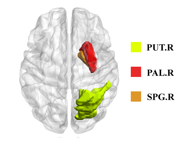
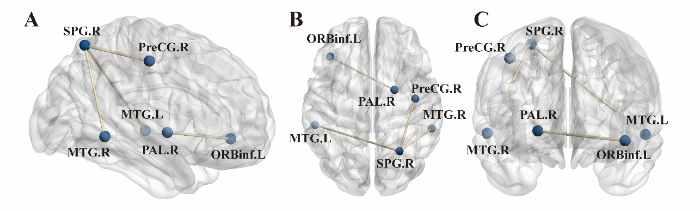
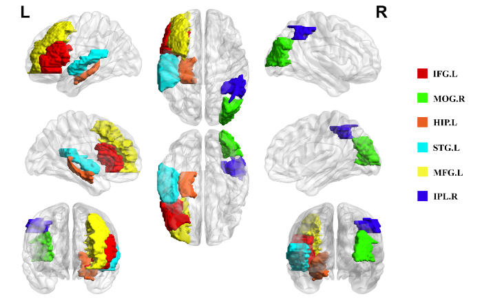

