B超和MRI在胎儿骨骼异常中的诊断价值分析
Diagnostic Efficacy Comparison of B-scan Ultrasonography and MRI in Fetal Skeletal Abnormalities
B超和MRI在胎儿骨骼异常中的诊断价值分析 |
| 舒炜 |
|
Diagnostic Efficacy Comparison of B-scan Ultrasonography and MRI in Fetal Skeletal Abnormalities |
| SHU Wei |
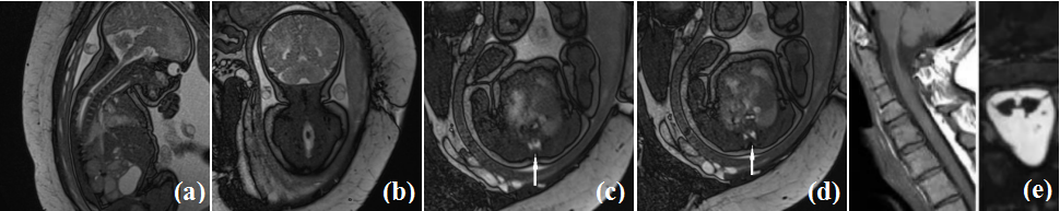
| 图3 胎儿MRI扫描脊髓纵裂畸形及出生后图像. (a)胎儿矢状位;(b)胎儿冠状位图像见椎管局部扩张、脊髓膨大,内见T2低信号灶;(c)胎儿轴位;(d)胎儿轴位见脊髓被分成左右两部分;(e)胎儿出生后图像 |
| Figure 3 Fetal MRI Scans of Diastematomyelia and Postnatal Images. (a) Fetal sagittal view; (b) Fetal coronal view showing local dilatation of the spinal canal, spinal cord enlargement, and a T2-weighted hypointense lesion inside; (c) Fetal axial view; (d) Fetal axial view showing the spinal cord divided into left and right parts; (e) Postnatal image |
|
|