


引言
磁共振波谱(Magnetic Resonance Spectroscopy,MRS)能够特异性地探究代谢物的浓度信息,评估组织的代谢功能[1-
当采用门控技术时,脉冲序列的重复时间(Repetition Time,TR)是心电或呼吸周期的整数倍. 在扫描过程中,若心电或呼吸周期出现波动,TR将随之改变,导致谱线受到T1加权的程度各异,信号幅度产生波动,最终引入ADC计算偏差[7]. 在DW-MRS数据采集中,为计算不同方向的ADC,除了采集不施加扩散梯度的磁共振信号外,还需采集施加不同方向扩散梯度的磁共振信号. 此外,为了提高谱线信噪比,需进行多次重复累加采集. 因此,数据采集过程包含b值循环和累加循环,将b值循环放置在累加循环的内侧或外侧,可以构成不同的循环策略. 由于受试者生理参数的变化趋势通常较为缓慢,将b值循环置于累加循环的内侧,有望减少不同b值间TR的差异,提高ADC测量的稳定性.
基于上述问题,本文开展了两项研究. 首先,分别采用心电门控、呼吸门控和不加门控的方法,对健康受试者的辐射冠和后扣带回皮层进行DW-MRS扫描,以评估不同门控方法对大脑不同区域测量结果的影响. 其次,在使用心电门控的情况下,采用不同的循环策略对健康受试者进行DW-MRS扫描,以探究循环方式对ADC可重复性的影响.
1 材料与方法
1.1 数据采集方案
本研究招募21名健康受试者,其中6名(5名男性,1名女性,年龄24.7 ± 0.5岁)参与门控方法研究,15名(8名男性,7名女性,年龄26.1 ± 6.0岁)参与循环策略研究. 所有扫描均在西门子Prisma Fit 3T仪器上完成,采用64通道头颈联合线圈进行信号接收. 该项研究得到了华东师范大学人体试验伦理委员会的批准(批准文号:HR 699-2024),所有受试者均自愿参加并签署知情同意书.
本研究基于西门子的面向应用程序的集成开发环境(Integrated Development Environment for Applications,IDEA),实现了扩散加权点分解波谱(Diffusion Weighted Point Resolved Spectroscopy,DW-PRESS)序列[17],脉冲序列设计如图1所示. 为实现单体素定位,首先施加选择性90˚射频脉冲激发特定层面的核自旋,随后施加两个选择性180˚射频脉冲在另两个正交层面实现核自旋的反转,只有经受所有3个射频脉冲的核自旋才能形成所需的回波. 为实现信号的扩散加权,在180˚射频脉冲两侧施加方向相反、幅度和持续时间一致的成对扩散梯度,双极梯度可降低扩散梯度切换时产生的涡流效应.
图1
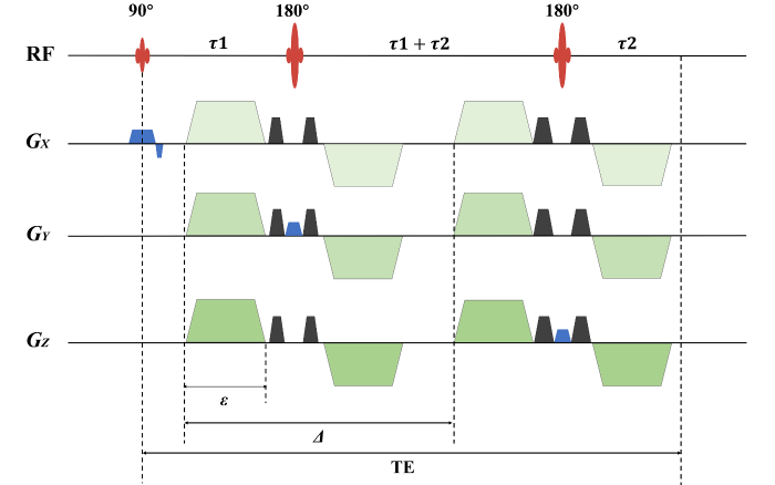
图1
DW-PRESS序列图. RF表示射频脉冲;GX、GY和GZ分别表示三个正交方向的梯度. 在本研究中,X、Y和Z分别对应受试者的左右、前后和头脚方向. 蓝色表示定位梯度;黑色表示扰相梯度;绿色表示扩散梯度. TE为回波时间,等于τ1+τ2的两倍;ε表示扩散梯度施加时长;Δ为扩散梯度对的间隔时间(即扩散时间),时长为TE的一半
Fig. 1
Sequence diagram of diffusion-weighted point resolved spectroscopy. RF denotes the radiofrequency pulse, while GX, GY, and GZ represent the gradients along the left-right, anterior-posterior, and head-foot directions, respectively. The blue color indicates the localization gradient, black represents the spoiling gradient, and green denotes the diffusion gradient. The echo time (TE) is defined as twice the sum of τ 1 and τ 2. ε denotes the duration of the applied diffusion gradient, while Δ represents the interval between the diffusion gradient pairs (i.e., the diffusion time), which is half the duration of TE
DW-MRS的回波时间(Echo Time,TE)设置为102 ms,扩散梯度持续时间ε固定为15 ms. 当不施加扩散梯度时,定位梯度和扰相梯度贡献的扩散因子b0为0.003 5 ms/μm2,此时采集到的信号记为b0信号;当施加幅度为39 mT/m的扩散梯度时,扩散因子b1为2.97 ms/μm2. 为测量三个正交方向的ADC,扩散梯度依次施加在受试者的左右(Left to Right,LR)、前后(Anterior to Posterior,AP)和头脚(Head to Foot,HF)方向,将相应条件下采集的数据分别记为b1-LR、b1-AP和b1-HF. 针对b0、b1-LR、b1-AP和b1-HF信号,均进行32次累加采集. 基于上述过程,定义了两种循环模式:一种是b值内循环模式,b值循环位于累加循环的内侧;另一种是b值外循环模式,即b值循环位于累加循环的外侧,这两种循环模式的具体情况如图2所示. 此外,将DW-MRS的采集带宽设置为2 500 Hz,采集点数为2 048,感兴趣容积(Volume of Interest,VOI)大小为20 × 20 × 20 mm³. 采用基于T1弛豫增强的水信号抑制(Water Suppression Enhanced through T1 Relaxation,WET)方法进行部分水峰抑制[18],以保留部分水峰信号用于谱线配准[7]. 并通过关闭WET模块中的射频脉冲采集不进行水峰抑制的参考信号,用于获取线圈组合所需的信息,参考水峰信号累加次数为4.
图2
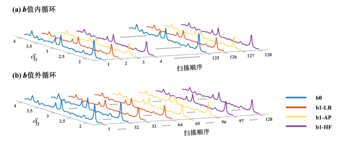
图2
两种循环模式示意图. (a) b值内循环,b值循环位于累加循环内侧,即先依次循环采集不同b值数据,再进行下一次累加采集;(b) b值外循环,b值循环位于累加循环外侧,即先累加采集完某一b值数据,再采集下一个b值数据. b0表示不施加扩散梯度时的信号;b1-LR、b1-AP和b1-HF表示扩散梯度分别施加在受试者的左右、前后或头脚方向时的信号. 蓝色、橙色、黄色和紫色分别表示b0、b1-LR、b1-AP和b1-HF
Fig. 2
Illustration of the two cycling modes. (a) Internal cycling of b-values: The b-value loop occurs within the accumulation loop, where data from different b-values are sequentially looped and collected before proceeding to the next accumulation. (b) External cycling of b-values: The b-value loop occurs outside the accumulation loop, with data for the same b-value first accumulated before moving on to the next b-value. The b0 denotes the signal when no diffusion gradient is applied. b1-LR, b1-AP, and b1-HF represent the signals when the diffusion gradient is applied in the left-right, anterior-posterior, and head-foot directions of the subject respectively. Blue, orange, yellow, and purple indicate b0, b1-LR, b1-AP, and b1-HF respectively
在门控方法的研究中,分别采用心电门控、呼吸门控和无门控三种方式对受试者进行DW-MRS扫描,将VOI定位于辐射冠和后扣带回皮层. 这两个区域位于脑室的不同方位,在神经系统疾病研究中极具临床价值[10,19
图3
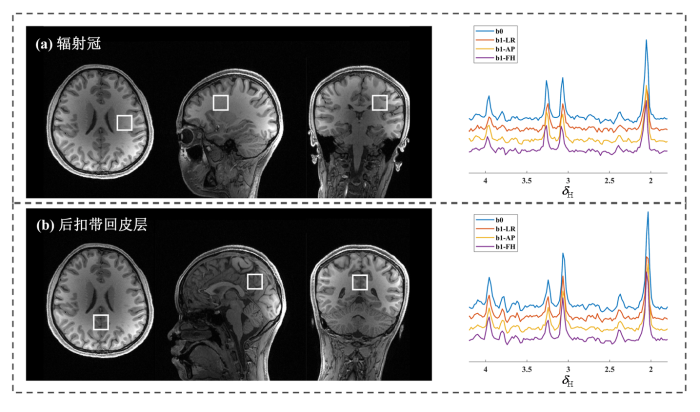
图3
DW-MRS感兴趣容积定位示意图(左列)及其对应的波谱图(右列). (a)辐射冠;(b)后扣带回皮层. b0表示不施加扩散梯度时的谱线,b1-LR、b1-AP以及b1-HF表示扩散梯度分别施加在受试者左右、前后和头脚方向时的谱线
Fig. 3
Illustration of the volume of interest localization for diffusion-weighted magnetic resonance spectroscopy (left column) and the corresponding spectra (right column). (a) Corona radiata; (b) Posterior cingulate cortex. b0 denotes the spectrum acquired without diffusion gradient application, while b1-LR, b1-AP, and b1-HF represent spectra obtained with the diffusion gradient applied in the left-right, anterior-posterior, and head-foot directions, respectively
为实现DW-MRS精准定位,每位受试者均进行了T1加权高分辨结构像扫描,序列为磁化准备快速梯度回波序列(Magnetization-Prepared Rapid Acquisition with Gradient Echo,MPRAGE),参数设置为:TR和TE分别为2 500 ms 和2.22 ms;翻转角为8˚;反转时间为1 000 ms;视野为240 × 256 × 166 mm³;采集矩阵为300 × 320 × 208;体素大小为0.8 × 0.8× 0.8 mm3;采集时长6 min 54 s.
1.2 数据后处理
本研究采用FID-A软件包对数据进行预处理[23]. 首先,使用未进行水峰抑制的b0数据获取线圈的相位和权重信息,并进行线圈组合. 随后,对不同暂态(即未经平均的单次谱线)进行配准操作[24],在此过程中,先选取与平均谱最为相似的暂态作为参考,对该参考谱进行0阶相位校正,而后调整其余暂态的频率和0阶相位,使其向参考谱配准. 在门控方法的研究中,为了评估不同门控策略下谱线受生理运动影响的程度,对配准后的谱线直接进行谱线平均. 在循环策略的研究中,对配准后的谱线进行进一步筛选,选取与平均谱最为相似的暂态作为参考,剔除偏离参考谱三倍标准偏差及以上的暂态,由于使用了心电门控,相同扩散梯度施加情况下暂态谱线间基本一致,剔除的暂态数目最多为2条,进行谱线筛选后,再进行谱线平均. 此外,为避免残余水峰的影响,采用奇异值分解法(Singular Value Decomposition,SVD)对平均后的谱线进行水峰抑制处理[25],从而得到预处理后的谱线. 最后,使用LCModel(版本为6.3-1R)对预处理后的谱线进行拟合,得到代谢物信号强度[26],代谢物的基础集由MRSCloud仿真生成[27].
计算总胆碱(total Choline,tCho)、总肌酸(total Creatine,tCr)和总N-乙酰天冬氨酸(total N-acetyl aspartate,tNAA)三种代谢物的ADC,计算公式如(1)式所示:
其中,Sb1表示施加特定方向扩散梯度时的代谢物信号强度;Sb0为未施加扩散梯度时代谢物的信号强度;b0和b1分别代表未施加或施加扩散梯度时的b值. 对于每种代谢物,分别计算其在受试者左右、前后和头脚方向上的ADC,依次记为ADCLR、ADCAP和ADCHF,并计算这三个方向ADC的平均值,记为ADCmean.
此外,为评估ADC的可重复性,本研究针对tCho、tCr和tNAA的各类ADC值,即ADCLR、ADCAP、ADCHF与ADCmean,计算受试者采用同种循环模式重复扫描两次的ADC变异系数(Coefficient of Variation,Cv)[28],计算公式如(2)式所示:
其中,σ 为两次重复扫描所获ADC的标准差,μ 为两次重复扫描所获ADC的平均值.
本研究采用线性混合效应模型(Linear Mixed-Effects Model,LMM)分析门控方法和循环模式对ADC值的影响以及循环模式对ADC测量可重复性的影响. 在门控方法研究中,通过建立以ADC为因变量、门控方法(心电门控/呼吸门控/无门控)为固定效应、被试编号为随机截距的LMM模型,分析不同门控方式对ADC值的影响. 在循环模式研究中,构建以ADC值为因变量、循环模式(b值内循环/b值外循环)为固定效应、被试编号为随机截距的LMM模型,评估循环模式对ADC值的影响;构建以ADC变异系数为因变量、循环模式(b值内循环/b值外循环)为固定效应、被试编号为随机截距的LMM模型,评估循环模式对ADC的可重复性的影响. 所有统计检验均采用P < 0.05作为显著性判断标准.
2 结果与讨论
2.1 门控方法研究结果
图4显示了一名受试者采用不同门控方式采集得到的每个暂态波谱水峰信号强度的变化情况. 当未施加扩散梯度时(b0),不同门控方式得到的暂态波谱水峰信号强度一致性高. 当施加扩散梯度后(b1-LR、b1-AP和b1-HF),水峰信号强度由于扩散效应明显下降;采用呼吸门控或无门控方式得到的暂态波谱水峰强度出现震荡,而采用心电门控时得到的暂态水峰信号强度较为一致. 此外,与后扣带回皮层的波谱数据相比,辐射冠区域的波谱水峰信号震荡更明显. 其他受试者的波谱水峰信号强度的变化情况与该受试者类似.
图4
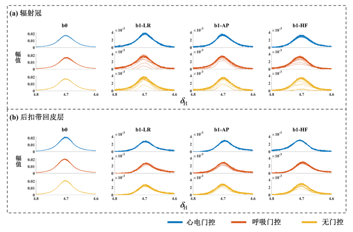
图4
一名受试者采用不同门控方式采集得到的每个暂态波谱的水峰信号强度变化情况. (a)感兴趣容积定位于辐射冠区域; (b)感兴趣容积定位于后扣带回皮层区域. b0表示不施加扩散梯度时的信号,b1-LR、b1-AP以及b1-HF表示扩散梯度分别施加在受试者左右、前后和头脚方向时的信号. 蓝色、橙色和黄色分别表示使用心电门控、呼吸门控和无门控
Fig. 4
Variations in signal intensity of the water peak in each transient spectrum acquired using different gating methods for a subject. (a) The volume of interest is located in the corona radiata region; (b) The volume of interest is located in the posterior cingulate cortex region. b0 represents the signal without diffusion gradient application, while b1-LR, b1-AP, and b1-HF denote the signals with the diffusion gradient applied in the left-right, anterior-posterior, and head-foot directions, respectively. Blue, orange, and yellow indicate the use of ECG gating, respiratory gating, and no gating, respectively
表1和表2分别展示了辐射冠区域和后扣带回皮层区域在不同门控条件下的ADC值及LMM统计结果. 结果显示,辐射冠区域所有代谢物(tNAA、tCr、tCho)的ADCLR、ADCAP、ADCHF和ADCmean的组内平均值均呈现心电门控组 < 呼吸门控组 < 无门控组的趋势,其中以心电门控组与无门控组间部分ADC指标的差异最为显著(P < 0.05). 后扣带回皮层区域的ADC值则显示更复杂的模式,虽然所有代谢物各ADC指标在心电门控组和呼吸门控组间仍保持心电门控组 < 呼吸门控组的趋势,尤其以tCr和tCho部分ADC指标变化较为显著(P < 0.05),但无门控组的ADC值变化趋势不一致;尽管如此,具有统计学意义的组间比较仍显示门控组的ADC值普遍低于无门控组.
表1 辐射冠区域不同门控条件下的表观扩散系数对比及线性混合效应模型分析结果
Table 1
| ADC/(μm2/ms) | P12 | P13 | P23 | ||||
|---|---|---|---|---|---|---|---|
| Cardiac gating | Respiratory gating | No gating | |||||
| tCho | LR | 0.144 ± 0.043 | 0.147 ± 0.017 | 0.164 ± 0.030 | 0.850 | 0.287 | 0.377 |
| AP | 0.116 ± 0.030 | 0.118 ± 0.026 | 0.135 ± 0.016 | 0.842 | 0.073 | 0.105 | |
| HF | 0.134 ± 0.013 | 0.152 ± 0.027 | 0.178 ± 0.036 | 0.193 | 0.005** | 0.077 | |
| mean | 0.131 ± 0.023 | 0.139 ± 0.018 | 0.159 ± 0.025 | 0.477 | 0.023* | 0.091 | |
| tCr | LR | 0.166 ± 0.045 | 0.191 ± 0.023 | 0.193 ± 0.034 | 0.151 | 0.204 | 0.858 |
| AP | 0.141 ± 0.026 | 0.167 ± 0.024 | 0.170 ± 0.020 | 0.072 | 0.049* | 0.839 | |
| HF | 0.162 ± 0.018 | 0.185 ± 0.015 | 0.195 ± 0.026 | 0.059 | 0.012* | 0.428 | |
| mean | 0.156 ± 0.025 | 0.181 ± 0.012 | 0.186 ± 0.024 | 0.044* | 0.028* | 0.817 | |
| tNAA | LR | 0.171 ± 0.041 | 0.186 ± 0.017 | 0.200 ± 0.038 | 0.432 | 0.125 | 0.427 |
| AP | 0.161 ± 0.020 | 0.179 ± 0.027 | 0.182 ± 0.023 | 0.076 | 0.046* | 0.794 | |
| HF | 0.153 ± 0.020 | 0.171 ± 0.016 | 0.193 ± 0.027 | 0.054 | <0.001*** | 0.016* | |
| mean | 0.162 ± 0.018 | 0.178 ± 0.013 | 0.192 ± 0.022 | 0.081 | 0.004** | 0.157 | |
注:Cardiac gating、Respiratory gating以及No gating分别是指采用心电门控、呼吸门控以及无门控;LR、AP和HF分别对应扩散梯度施加在受试者的左右、前后以及头脚方向时获取的ADC,mean对应三个方向ADC的平均值;P12:心电门控组与呼吸门控组ADC对比的差异显著性;P13:心电门控组与无门控组ADC对比的差异显著性;P23:呼吸门控组与无门控组ADC对比的差异显著性. *P < 0.05;**P < 0.01;***P<0.001
表2 后扣带回皮层区域不同门控条件下的表观扩散系数对比及线性混合效应模型分析结果
Table 2
| ADC/(μm2/ms) | P12 | P13 | P23 | ||||
|---|---|---|---|---|---|---|---|
| Cardiac gating | Respiratory gating | No gating | |||||
| tCho | LR | 0.106 ± 0.024 | 0.112 ± 0.020 | 0.111 ± 0.023 | 0.674 | 0.721 | 0.949 |
| AP | 0.097 ± 0.027 | 0.099 ± 0.011 | 0.108 ± 0.018 | 0.876 | 0.346 | 0.428 | |
| HF | 0.092 ± 0.019 | 0.111 ± 0.023 | 0.111 ± 0.021 | 0.003** | 0.003** | 0.955 | |
| mean | 0.098 ± 0.019 | 0.107 ± 0.014 | 0.110 ± 0.014 | 0.303 | 0.178 | 0.734 | |
| tCr | LR | 0.130 ± 0.017 | 0.151 ± 0.015 | 0.139 ± 0.015 | 0.034* | 0.364 | 0.185 |
| AP | 0.114 ± 0.015 | 0.129 ± 0.013 | 0.131 ± 0.013 | 0.041* | 0.022* | 0.747 | |
| HF | 0.105 ± 0.009 | 0.119 ± 0.014 | 0.127 ± 0.015 | 0.055 | 0.004** | 0.215 | |
| mean | 0.116 ± 0.008 | 0.133 ± 0.012 | 0.132 ± 0.014 | 0.019* | 0.023* | 0.928 | |
| tNAA | LR | 0.144 ± 0.030 | 0.148 ± 0.009 | 0.142 ± 0.012 | 0.750 | 0.826 | 0.592 |
| AP | 0.120 ± 0.016 | 0.125 ± 0.008 | 0.128 ± 0.006 | 0.431 | 0.205 | 0.614 | |
| HF | 0.113 ± 0.013 | 0.124 ± 0.016 | 0.130 ± 0.017 | 0.219 | 0.073 | 0.529 | |
| mean | 0.126 ± 0.018 | 0.132 ± 0.008 | 0.133 ± 0.009 | 0.368 | 0.303 | 0.893 | |
注:Cardiac gating、Respiratory gating以及No gating分别是指采用心电门控、呼吸门控以及无门控;LR、AP和HF分别对应扩散梯度施加在受试者的左右、前后以及头脚方向时获取的ADC,mean对应三个方向ADC的平均值;P12:心电门控组与呼吸门控组ADC对比的差异显著性;P13:心电门控组与无门控组ADC对比的差异显著性;P23:呼吸门控组与无门控组ADC对比的差异显著性. *P < 0.05;**P < 0.01
2.2 循环模式研究结果
表3展示了采用两种循环模式得到的15名受试者三种代谢物ADC的对比,b值内循环和外循环得到的ADC无显著性差异. 图5显示了采用两种循环模式时三种代谢物各ADC的变异系数分布情况. 采用b值内循环模式得到的三种代谢物各ADC变异系数的组内平均值均低于b值外循环模式. 并且,在以下指标存在显著差异:tCho的ADCLR变异系数(0.054 ± 0.031 vs 0.099 ± 0.056,P = 0.011)、tCho的ADCmean(0.064 ± 0.033 vs 0.102 ± 0.071,P = 0.036)、tCr的ADCLR变异系数(0.038 ± 0.038 vs 0.086 ± 0.055,P = 0.004)以及tNAA的ADCAP变异系数(0.037 ± 0.026 vs 0.063 ± 0.038,P = 0.017).
表3 b值内、外循环模式获取的表观扩散系数对比及线性混合效应模型分析结果
Table 3
| ADC/(μm2/ms) | P values | |||
|---|---|---|---|---|
| Internal b-value cycling | External b-value cycling | |||
| tCho | LR | 0.109 ± 0.014 | 0.109 ± 0.019 | 0.826 |
| AP | 0.112 ± 0.018 | 0.104 ± 0.024 | 0.158 | |
| HF | 0.097 ± 0.016 | 0.099 ± 0.021 | 0.614 | |
| mean | 0.106 ± 0.011 | 0.104 ± 0.018 | 0.601 | |
| tCr | LR | 0.133 ± 0.014 | 0.134 ± 0.019 | 0.680 |
| AP | 0.117 ± 0.016 | 0.120 ± 0.020 | 0.542 | |
| HF | 0.110 ± 0.011 | 0.109 ± 0.014 | 0.749 | |
| mean | 0.120 ± 0.010 | 0.121 ± 0.015 | 0.883 | |
| tNAA | LR | 0.144 ± 0.017 | 0.143 ± 0.021 | 0.806 |
| AP | 0.121 ± 0.008 | 0.122 ± 0.013 | 0.486 | |
| HF | 0.117 ± 0.014 | 0.116 ± 0.016 | 0.637 | |
| mean | 0.127 ± 0.011 | 0.127 ± 0.013 | 0.780 | |
注:Internal b-value cycling和External b-value cycling分别是指b值内循环和b值外循环;LR、AP和HF分别对应扩散梯度施加在受试者的左右、前后以及头脚方向时获取的ADC,mean对应三个方向ADC的平均值;P values指不同循环模式组ADC值的差异显著性
图5

图5
采用b值内循环和外循环模式时三种代谢物各ADC的变异系数分布情况. (a) tCho;(b) tCr;(c) tNAA. LR、AP和HF分别表示扩散梯度施加在受试者左右、前后方向和头脚方向时ADC的变异系数,mean表示在三个正交方向ADC的平均值的变异系数. 蓝色表示b值内循环;橙色表示b值外循环. * P <0.05;** P < 0.01
Fig. 5
The distribution of the coefficient of variation of the apparent diffusion coefficient (ADC) for three metabolites when using the internal and external b-value cycling modes. (a) tCho; (b) tCr; (c) tNAA. The LR, AP, and HF represent the coefficient of variation of the ADC when the diffusion gradient is applied in the left-right, anterior-posterior, and head-foot directions, respectively, while "mean" denotes the coefficient of variation of the averaged ADC across the three orthogonal directions. Blue indicates the internal b-value cycling mode; orange represents the external b-value cycling mode. * P < 0.05; ** P < 0.01
2.3 讨论
本文探究了不同门控技术在抑制DW-MRS生理运动伪影方面的有效性,并对比了循环模式对代谢物ADC可重复性的影响. 研究结果显示,心电门控能有效抑制生理运动导致的信号衰减,减少ADC的高估,在辐射冠区域效果更显著,而呼吸门控对改善生理运动影响的效果相对有限. 在使用心电门控的条件下,b值内循环模式采集的ADC可重复性更高.
2.3.1 门控方法研究
与未添加扩散梯度的谱线相比,添加扩散梯度谱线中出现了信号的震荡,表明谱线采集对生理运动的敏感性与扩散梯度的施加有关. 与未使用门控技术或采用呼吸门控相比,心电门控技术能够显著抑制水峰信号强度的振荡. 对辐射冠区域和后扣带回皮层区域的ADC值进行对比,心电门控组ADC组内平均值整体上低于呼吸门控组与无门控组,且部分指标的组间差异达到统计学显著水平. 这一结果充分表明,心电门控技术能够有效削弱生理运动干扰,避免 ADC 值高估,有望提高ADC测量的稳定性和准确性,与文献[15]的研究发现相符. 但心电门控组受试者间ADC值的标准差较大,推测可能是由于心电门控采用的 TR 时间相对较短,致使谱线信噪比下降所致. 此外,辐射冠区域的呼吸门控组对应各ADC组内平均值均小于无门控组,部分指标差异显著,表明呼吸门控也能一定程度上抑制生理运动的影响,并间接证实了脑脊液搏动受到呼吸和心跳的协同作用[16]. 但其效果仍弱于心电门控,这一差异可能源于两方面原因:其一,本研究采用呼吸垫采集呼吸波形,受限于其监测精度,导致门控触发的精准度不足;其二,心脏搏动在脑脊液动力学中占据主导地位,其对脑脊液流动的影响强度远超呼吸运动,从而削弱了呼吸门控的优化效能.
区域对比分析显示,当VOI位于辐射冠区域时,采用呼吸门控技术和无门控技术时谱线的震荡大于后扣带回皮层区域,这表明靠近脑室的区域受到生理运动的影响更大. 并且,在后扣带回皮层区域,呼吸门控组与无门控组变化的ADC值组间未呈现统计学差异,并且呼吸门控组与无门控组ADC值组内平均值变化趋势不一,提示在该区域呼吸门控效果不佳,进一步表明生理运动对数据采集的影响存在区域特异性.此外,在两区域不同门控方法组间ADC的统计检验结果在不同方向不完全一致,这表明生理运动的影响存在方向性差异.
2.3.2 循环模式研究
b值内循环模式下,tCho、tCr和tNAA的各ADC变异系数的组内平均值普遍小于外循环,且部分代谢物ADC变异系数在不同循环模式间的差异有统计学意义. 这一结果表明,内循环在提升DW-MRS测量ADC可重复性方面具有积极意义. 这种优势可能源于受试者的心电周期变化较为缓慢,b值内循环模式可使不同b值谱线的TR更趋一致,从而降低因T1加权程度不同导致的ADC测量偏差. 未来研究可考虑通过系统性的水模实验进一步探究内循环模式的优势机制,如配置具有不同T1值的水模,并模拟心电周期缓慢变化导致的TR变化,对比评估两种循环模式下ADC稳定性随T1值的变化规律,以明确门控条件下内循环模式的优势来源.
2.3.3 研究局限性
本研究存在一定局限性. 首先,本研究受试者样本量较小,且性别分布不均衡,可能导致统计结果存在偶然性,后续需纳入更多样本,并优化性别比例,以增强结论可靠性. 其次,心电门控虽能抑制信号震荡,提升ADC可靠性,但TR无法灵活设置,过少的心电周期次数会导致TR比较短,从而降低波谱信噪比,而过多的心电周期次数则会延长采集时间,因此针对 TR 设置方案的优化仍需深入探索,以实现生理运动抑制与测量精度的平衡.
2.3.4 应用展望
DW-MRS技术可以通过测量代谢物的扩散特性实现对特定细胞病理过程的特异性表征,为研究细胞微环境变化提供了新视角. 值得注意的是,疾病的发生和发展伴随着氧化应激、脱髓鞘、慢性神经炎症和神经元细胞死亡等过程,且与这些过程相关的脑内代谢物具有细胞群特异性分布特征:肌醇的ADC升高可能反映星形胶质细胞活化[36,37],而tCho的ADC增加与小胶质细胞激活相关[10],另外, tCr的扩散改变与胶质病理过程或能量代谢率的变化有关[38],tNAA和谷氨酸主要存在于神经元中,其扩散特性可作为轴突完整性的标志[39]. 相较于常规MRI技术,DW-MRS基于细胞特异性代谢信息的特点使其在疾病诊断和机制研究方面展现出显著的优势. 因此,优化DW-MRS技术方案,提高其稳定性和可重复性,对于推动中枢神经系统疾病的精准诊疗具有重要意义. 未来研究可进一步优化采集序列,并开发标准化分析流程,以促进该技术的临床转化应用.
3 结论
心电门控有效抑制生理运动引起的信号衰减,尤其在靠近脑室的辐射冠区域,而呼吸门控的效果相对有限. b值内循环模式在心电门控条件下可提高ADC的可重复性. 因此,建议在DW-MRS扫描中采用心电门控结合内循环方案,以提高数据采集的稳定性和准确性,推动其临床应用.
利益冲突
无
参考文献
Studies of human tumors by MRS: A review
[J].The literature describing 31P, 1H, 13C, 23Na and 19F MRS in vivo in human cancers is reviewed. Cancers have typical metabolic characteristics in 31P and 1H MRS including high levels of phospholipid metabolites and a cellular pH more alkaline than normal. These alone are not specific for cancer but are diagnostic in appropriate clinical settings. Some metabolic characteristics appear to be prognostic indices and correlation with treatment response is emerging as an important potentially cost-effective use of MRS in oncology. 19F MRS examines pharmacokinetics of 5-fluorouracil and by demonstrating its retention predicts response of a cancer to treatment. Current needs include improvement of diagnostic specificity by use of techniques like multivoxel MRS, proton decoupling of 31P, short echo time and fat-suppressed 1H MRS, 13C MRS direct or via 1H-observe, and statistical analysis of multiple spectral features. Trials in large populations in well defined clinical settings are needed to determine if MRS can provide independent prognostic indices useful in cancer management.
Progress of magnetic resonance spectroscopy in the study of the effects of smoking on the brain
[J].
磁共振波谱技术在吸烟对大脑影响的研究进展
[J].吸烟是导致过早发病和死亡的主要风险因素之一.随着磁共振技术的不断进步,磁共振波谱已成为临床诊断的重要手段之一.与磁共振成像的其他模态一样,磁共振波谱具有无创性的优点,能够反映大脑的神经递质及代谢物的浓度变化情况.吸烟是一个全球性的健康问题,近些年已有一些研究利用磁共振波谱观察吸烟对大脑的影响.针对这一广泛关注的社会问题,本文对吸烟人群的磁共振波谱研究进展进行了综述,总结了烟草对大脑代谢的影响.本综述为深入研究烟草依赖的神经生物学机制提供了新的视角,从技术层面为早期诊断、治疗及预防吸烟对大脑的负面影响提供了支持.
In vivo glutathione molecular MRS signal selection based on nuclear spin singlet states
[J].
基于核自旋单重态的活体谷胱甘肽分子MRS信号选择
[J].
DOI:10.11938/cjmr20243105
[本文引用: 1]

谷胱甘肽(GSH)是一种由谷氨酸、半胱氨酸和甘氨酸聚合而成的三肽,是人体还原驱动力的主要来源,能维持人体内部氧化还原平衡.谷胱甘肽还与生物体内自由基淬灭、肿瘤治疗密切相关.利用活体磁共振波谱(MRS)监测生物体中谷胱甘肽的含量变化对于理解其生物学作用具有重要意义.由于生物体内环境复杂,活体组织的磁共振信号往往重叠严重,难以实现特定分子的选择性检测及定量.本文介绍了一种利用核自旋单重态选择性检测活体谷胱甘肽分子的方法,通过在谷胱甘肽中选择合适的自旋体系,将其制备为单重态,借助梯度脉冲消除其它分子的信号,进而实现谷胱甘肽信号的选择性检测.该方法在健康人体大脑中得到了实验验证,理论和实验结果表明,与文献中报道的其它方法相比,本文的方法具有检测效率高、选择性强的特点.
In vivo1H NMR spectroscopy of the human brain at high magnetic fields: Metabolite quantification at 4T vs. 7T
[J].DOI:10.1002/mrm.v62:4 URL [本文引用: 1]
Diffusion NMR spectroscopy
[J].
PMID:11320536
MR offers unique tools for measuring molecular diffusion. This review focuses on the use of diffusion-weighted MR spectroscopy (DW-MRS) to non-invasively quantitate the translational displacement of endogenous metabolites in intact mammalian tissues. Most of the metabolites that are observed by in vivo MRS are predominantly located in the intracellular compartment. DW-MRS is of fundamental interest because it enables one to probe the in situ status of the intracellular space from the diffusion characteristics of the metabolites, while at the same time providing information on the intrinsic diffusion properties of the metabolites themselves. Alternative techniques require the introduction of exogenous probe molecules, which involves invasive procedures, and are also unable to measure molecular diffusion in and throughout intact tissues. The length scale of the process(es) probed by MR is in the micrometer range which is of the same order as the dimensions of many intracellular entities. DW-MRS has been used to estimate the dimensions of the cellular elements that restrict intracellular metabolite diffusion in muscle and nerve tissue. In addition, it has been shown that DW-MRS can provide novel information on the cellular response to pathophysiological changes in relation to a range of disorders, including ischemia and excitotoxicity of the brain and cancer.Copyright 2001 John Wiley & Sons, Ltd.
In vivo diffusion MRS investigation of non-water molecules in biological tissues
[J].DOI:10.1002/nbm.v30.3 URL
Diffusion-weighted MR spectroscopy: Consensus, recommendations, and resources from acquisition to modeling
[J].DOI:10.1002/mrm.v91.3 URL [本文引用: 3]
Insights into brain microstructure from in vivo DW-MRS
[J].
DOI:S1053-8119(17)30942-4
PMID:29155183
[本文引用: 1]
Many developmental processes, such as plasticity and aging, or pathological processes such as neurological diseases are characterized by modulations of specific cellular types and their microstructures. Diffusion-weighted Magnetic Resonance Imaging (DW-MRI) is a powerful technique for probing microstructure, yet its information arises from the ubiquitous, non-specific water signal. By contrast, diffusion-weighted Magnetic Resonance Spectroscopy (DW-MRS) allows specific characterizations of tissues such as brain and muscle in vivo by quantifying the diffusion properties of MR-observable metabolites. Many brain metabolites are predominantly intracellular, and some of them are preferentially localized in specific brain cell populations, e.g., neurons and glia. Given the microstructural sensitivity of diffusion-encoding filters, investigation of metabolite diffusion properties using DW-MRS can thus provide exclusive cell and compartment-specific information. Furthermore, since many models and assumptions are used for quantification of water diffusion, metabolite diffusion may serve to generate a-priori information for model selection in DW-MRI. However, DW-MRS measurements are extremely challenging, from the acquisition to the accurate and correct analysis and quantification stages. In this review, we survey the state-of-the-art methods that have been developed for the robust acquisition, quantification and analysis of DW-MRS data and discuss the potential relevance of DW-MRS for elucidating brain microstructure in vivo. The review highlights that when accurate data on the diffusion of multiple metabolites is combined with accurate computational and geometrical modeling, DW-MRS can provide unique cell-specific information on the intracellular structure of brain tissue, in health and disease, which could serve as incentives for further application in vivo in human research and clinical MRI.Copyright © 2017 Elsevier Inc. All rights reserved.
Diffusion weighted magnetic resonance spectroscopy revealed neuronal specific microstructural alterations in Alzheimer's disease
[J].
Longitudinal monitoring of microstructural alterations in cerebral ischemia with in vivo diffusion-weighted MR spectroscopy
[J].DOI:10.1148/radiol.220430 URL [本文引用: 3]
Thalamic energy dysfunction is associated with thalamo-cortical tract damage in multiple sclerosis: A diffusion spectroscopy study
[J].
DOI:10.1177/1352458520921362
URL
[本文引用: 1]
Diffusion-weighted 1H magnetic resonance spectroscopy (DW-MRS) allows to quantify creatine-phosphocreatine brain diffusivity (ADC(tCr)), whose reduction in multiple sclerosis (MS) has been proposed as a proxy of energy dysfunction.
Dysregulation of energy metabolism in multiple sclerosis measured in vivo with diffusion-weighted spectroscopy
[J].
DOI:10.1177/1352458517698249
URL
[本文引用: 1]
We employed diffusion-weighted magnetic resonance spectroscopy (DW-MRS), which allows to measure in vivo the diffusion properties of metabolites, to explore the functional neuro-axonal damage and the ongoing energetic dysregulation in multiple sclerosis (MS).
Cerebrospinal fluid flow waveforms-MR analysis in chronic adult hydrocephalus
[J].DOI:10.1097/00004424-200103000-00003 URL [本文引用: 1]
Phase contrast MRI quantification of pulsatile volumes of brain arteries, veins, and cerebrospinal fluids compartments: Repeatability and physiological interactions
[J].
DOI:10.1002/jmri.23527
PMID:22170792
[本文引用: 1]
To study measurement repeatability and physiological determinants on measurement stability for phase contrast MRI (PC-MRI) measurements of cyclic volume changes (ΔV) of brain arteries, veins, and cerebrospinal fluid (CSF) compartments.Total cerebral blood flow (tCBF), total internal jugular flow (tJBF) and spinal CSF flow at C2-C3 level and CSF in the aqueduct was measured using five repetitions in 20 healthy subjects. After subtracting net flow, waveforms were integrated to calculate ΔV of arterial, venous, and cerebrospinal fluid compartments. The intraclass correlation coefficient (ICC) was used to measure repeatability. Systematic errors were investigated by a series of phantom measurements.For ΔV calculated from tCBF, tJBF and both CSF waveforms, the ICC was ≥0.85. ΔV from the tCBF waveform decreased linearly between repetitions (P = 0.012). Summed CSF and venous volume being shifted out from the cranium was correlated with ΔV calculated from the tCBF waveform (r = 0.75; P < 0.001). Systematic errors increased at resolutions <4 pixels per diameter.Repeatability of ΔV calculated from tCBF, tJBF, and CSF waveforms allows useful interpretations. The subject's time in the MR system and imaging resolution should be considered when interpreting volume changes. Summed CSF and venous volume changes was associated with arterial volume changes.Copyright © 2011 Wiley Periodicals, Inc.
In vivo diffusion-weighted MRS using semi-LASER in the human brain at 3 T: Methodological aspects and clinical feasibility
[J].DOI:10.1002/nbm.v34.5 URL [本文引用: 3]
Heart rate and respiration influence on macroscopic blood and CSF flows
[J].
DOI:10.1177/0284185116676655
PMID:28273732
[本文引用: 2]
Background Changes in blood volume in the intracranial arteries and the resulting oscillations of brain parenchyma have been presumed as main initiating factors of cerebrospinal fluid (CSF) pulsations. However, respiration has been recently supposed to influence CSF dynamics via thoracic pressure changes. Purpose To measure blood and CSF cervical flow and quantify the contribution of cardiac and respiratory cycles on the subsequent signal evolution. Material and Methods Sixteen volunteers were enrolled. All participant underwent two-dimensional fast field echo echo planar imaging (FFE-EPI). Regions of interest were placed on internal carotids, jugular veins, and rachidian canal to extract temporal profiles. Spectral analysis was performed to extract respiratory and cardiac frequencies. The contribution of respiration and cardiac activity was assessed to signal evolution by applying a multiple linear model. Results Mean respiratory frequency was 14.6 ± 3.9 cycles per min and mean heart rate was 66.8 ± 9 cycles per min. Cardiac contribution was higher than breathing for internal carotids, explaining 74.68% and 10.27% of the signal variance, respectively. For the jugular veins, respiratory component was higher than the cardiac one contributing 44.28% and 6.53% of the signal variance, respectively. For CSF, breathing and cardiac component contributed less than half of signal variance (12.61% and 23.23%, respectively). Conclusion Respiration and cardiac activity both influence fluid flow at the cervical level. Arterial inflow is driven by the cardiac pool whereas venous blood aspiration seems more due to thoracic pressure changes. CSF dynamics acts as a buffer between these two blood compartments.
Differences in apparent diffusion coefficients of brain metabolites between grey and white matter in the human brain measured at 7 T
[J].
DOI:10.1002/mrm.23129
PMID:22083562
[本文引用: 1]
Diffusion weighted spectroscopy can provide microstructural information that is specific to compartmental geometry. So far, in human brain, apparent diffusion coefficients (ADCs) of only the metabolites N-acetyl aspartate, creatine (tCr) and choline (tCho) have been assessed. High field MR at 7 T allows the collection and analysis of diffusion weighted spectroscopy data of additional metabolites of interest such as glutamate (Glu), N-acetyl aspartyl glutamate, and glutamine (Gln), which are of interest due to their different compartmentalization and role in brain physiology. In this study, we performed (1)H diffusion weighted spectroscopy at 7 T using a diffusion-weighted PRESS sequence in parietal white matter (n = 6) and occipital grey matter (n = 7). Data were analyzed using the LCmodel. ADCs could reliably be obtained of N-acetyl aspartate, tCr, tCho, Glu, Gln in grey and white matter, and N-acetyl aspartyl glutamate in white matter. Significant differences in ADC values were observed between grey and white matter for all metabolites. ADCs in grey matter were consistently lower than in white matter. These differences can probably be attributed to different compartmentalization as well as to the differential impact of diffusion time on ADC of different molecules under conditions of restricted diffusion.Copyright © 2011 Wiley Periodicals, Inc.
WET, a T1-and B1-insensitive water-suppression method for in vivo localized 1H NMR Spectroscopy
[J].
Clinical and magnetic resonance imaging features in acute ischemic stroke with early wallerian degeneration: a case-control study
[J].DOI:10.1186/s12883-025-04179-4 [本文引用: 1]
Acquisition and voxelwise analysis of multi-subject diffusion data with tract-based spatial statistics
[J].
DOI:10.1038/nprot.2007.45
PMID:17406613
There is much interest in using magnetic resonance diffusion imaging to provide information on anatomical connectivity in the brain by measuring the diffusion of water in white matter tracts. Among the measures, the most commonly derived from diffusion data is fractional anisotropy (FA), which quantifies local tract directionality and integrity. Many multi-subject imaging studies are using FA images to localize brain changes related to development, degeneration and disease. In a recent paper, we presented a new approach, tract-based spatial statistics (TBSS), which aims to solve crucial issues of cross-subject data alignment, allowing localized cross-subject statistical analysis. This works by transforming the data from the centers of the tracts that are consistent across a study's subjects into a common space. In this protocol, we describe the MRI data acquisition and analysis protocols required for TBSS studies of localized change in brain connectivity across multiple subjects.
Regional dynamics of amyloid-β deposition in healthy elderly, mild cognitive impairment and Alzheimer's disease: a voxelwise PiB-PET longitudinal study
[J].
DOI:10.1093/brain/aws125
PMID:22628162
Amyloid-β deposition in Alzheimer's disease is thought to start while individuals are still cognitively unimpaired and it is hypothesized that after an early phase of fast accumulation, a plateau is reached by the time of cognitive decline. However, few longitudinal Pittsburgh compound B-positron emission tomography studies have tested this hypothesis, and with conflicting results. The purpose of this work is to further our understanding of the dynamics of amyloid-β deposition in a large longitudinal cohort. A total of 32 patients with Alzheimer's disease, 49 subjects with mild cognitive impairment and 103 healthy controls underwent two Pittsburgh compound B-positron emission tomography scans 18 months apart. For each participant, a parametric map of Pittsburgh compound B-positron emission tomography rate of change was created [(follow-up scan - baseline scan)/follow-up duration] and entered in a voxelwise three-way analysis of covariance, with clinical status (healthy controls, mild cognitive impairment or Alzheimer's disease), disease progression (clinical conversion from healthy controls to mild cognitive impairment or Alzheimer's disease, or from mild cognitive impairment to Alzheimer's disease) and Pittsburgh compound B status (positive versus negative) as independent factors. Only a significant effect of the Pittsburgh compound B status was found: both Pittsburgh compound B-positive and -negative subjects showed a significant increase in amyloid-β deposition, with this increase being significantly higher in Pittsburgh compound B-positive individuals. This finding suggests either that Pittsburgh compound B-negative individuals have slower rates of amyloid-β accumulation than positive, or that the proportion of individuals showing significant increase in amyloid-β deposition, termed 'Pittsburgh compound B accumulators', is higher within the Pittsburgh compound B-positive group than within the Pittsburgh compound B-negative group. The bimodal distribution of the individual rates of neocortical amyloid-β accumulation observed support the existence of 'Pittsburgh compound B non-accumulators' and 'Pittsburgh compound B accumulators' and different clustering analyses led to a consistent threshold to separate these two subgroups (0.014-0.022 standardized uptake value ratio(pons)/year). The voxelwise three-way analysis of covariance was thus recomputed with the 'Pittsburgh compound B accumulators' only and the results were almost unchanged, with the Pittsburgh compound B-positive group showing higher accumulation than the Pittsburgh compound B-negative group. Finally, a significant negative correlation was found between Pittsburgh compound B rate of change and Pittsburgh compound B baseline burden, but only in the Pittsburgh compound B-positive group (r= -0.24; P=0.025). Higher rates of amyloid-β deposition are associated with higher amyloid-β burden suggesting that amyloid-β deposition does not reach a plateau when cognitive impairments manifest but is instead an ongoing process present even at the Alzheimer's disease stage. amyloid-β accumulation also seems to slow down at the latest stages of the process, i.e. in participants with the highest amyloid burden. Furthermore, this study identified the existence of Pittsburgh compound 'accumulators' and 'non-accumulators', notably within the Pittsburgh compound B-negative group, which may be a relevant concept for future studies.
Resting-state functional connectivity in major depression: Abnormally increased contributions from subgenual cingulate cortex and thalamus
[J].DOI:10.1016/j.biopsych.2006.09.020 URL [本文引用: 1]
Advanced processing and simulation of MRS data using the FID appliance (FID-A)-An open source, MATLAB-based toolkit
[J].
DOI:10.1002/mrm.26091
PMID:26715192
[本文引用: 1]
To introduce a new toolkit for simulation and processing of magnetic resonance spectroscopy (MRS) data, and to demonstrate some of its novel features.The FID appliance (FID-A) is an open-source, MATLAB-based software toolkit for simulation and processing of MRS data. The software is designed specifically for processing data with multiple dimensions (eg, multiple radiofrequency channels, averages, spectral editing dimensions). It is equipped with functions for importing data in the formats of most major MRI vendors (eg, Siemens, Philips, GE, Agilent) and for exporting data into the formats of several common processing software packages (eg, LCModel, jMRUI, Tarquin). This paper introduces the FID-A software toolkit and uses examples to demonstrate its novel features, namely 1) the use of a spectral registration algorithm to carry out useful processing routines automatically, 2) automatic detection and removal of motion-corrupted scans, and 3) the ability to perform several major aspects of the MRS computational workflow from a single piece of software. This latter feature is illustrated through both high-level processing of in vivo GABA-edited MEGA-PRESS MRS data, as well as detailed quantum mechanical simulations to generate an accurate LCModel basis set for analysis of the same data.All of the described processing steps resulted in a marked improvement in spectral quality compared with unprocessed data. Fitting of MEGA-PRESS data using a customized basis set resulted in improved fitting accuracy compared with a generic MEGA-PRESS basis set.The FID-A software toolkit enables high-level processing of MRS data and accurate simulation of in vivo MRS experiments. Magn Reson Med 77:23-33, 2017. © 2015 Wiley Periodicals, Inc.© 2015 Wiley Periodicals, Inc.
Preprocessing, analysis and quantification in single-voxel magnetic resonance spectroscopy: Experts' consensus recommendations
[J].DOI:10.1002/nbm.v34.5 URL [本文引用: 1]
Improved Lanczos algorithms for blackbox MRS data quantitation
[J].Magnetic resonance spectroscopy (MRS) has been shown to be a potentially important medical diagnostic tool. The success of MRS depends on the quantitative data analysis, i.e., the interpretation of the signal in terms of relevant physical parameters, such as frequencies, decay constants, and amplitudes. A variety of time-domain algorithms to extract parameters have been developed. On the one hand, there are so-called blackbox methods. Minimal user interaction and limited incorporation of prior knowledge are inherent to this type of method. On the other hand, interactive methods exist that are iterative, require user involvement, and allow inclusion of prior knowledge. We focus on blackbox methods. The computationally most intensive part of these blackbox methods is the computation of the singular value decomposition (SVD) of a Hankel matrix. Our goal is to reduce the needed computational time without affecting the accuracy of the parameters of interest. To this end, algorithms based on the Lanczos method are suitable because the main computation at each step, a matrix-vector product, can be efficiently performed by means of the fast Fourier transform exploiting the structure of the involved matrix. We compare the performance in terms of accuracy and efficiency of four algorithms: the classical SVD algorithm based on the QR decomposition, the Lanczos algorithm, the Lanczos algorithm with partial reorthogonalization, and the implicitly restarted Lanczos algorithm. Extensive simulation studies show that the latter two algorithms perform best.
Automatic quantitation of localized in vivo 1H spectra with LCModel
[J].DOI:10.1002/nbm.v14:4 URL [本文引用: 1]
MRSCloud: A cloud-based MRS tool for basis set simulation
[J].
DOI:10.1002/mrm.29370
PMID:35775808
[本文引用: 1]
The purpose of this study is to present a cloud-based spectral simulation tool "MRSCloud," which allows MRS users to simulate a vendor-specific and sequence-specific basis set online in a convenient and time-efficient manner. This tool can simulate basis sets for GE, Philips, and Siemens MR scanners, including conventional acquisitions and spectral editing schemes with PRESS and semi-LASER localization at 3 T.The MRSCloud tool was built on the spectral simulation functionality in the FID-A software package. We added three extensions to accelerate computation (ie, one-dimensional projection method, coherence pathways filters, and precalculation of propagators). The RF waveforms were generated based on vendors' generic pulse shapes and timings. Simulations were compared within MRSCloud using different numbers of spatial resolution (21 × 21, 41 × 41, and 101 × 101). Simulated metabolite basis functions from MRSCloud were compared with those generated by the generic FID-A and MARSS, and a phantom-acquired basis set from LCModel. Intraclass correlation coefficients were calculated to measure the agreement between individual metabolite basis functions. Statistical analysis was performed using R in RStudio.Simulation time for a full PRESS basis set is approximately 11 min on the server. The interclass correlation coefficients ICCs were at least 0.98 between MRSCloud and FID-A and were at least 0.96 between MRSCloud and MARSS. The interclass correlation coefficients between simulated MRSCloud basis spectra and acquired LCModel basis spectra were lowest for glutamine at 0.68 and highest for N-acetylaspartate at 0.96.Substantial reductions in runtime have been achieved. High ICC values indicated that the accelerating features are running correctly and produce comparable and accurate basis sets.© 2022 International Society for Magnetic Resonance in Medicine.
Test-retest reliability of the brain metabolites GABA and Glx With JPRESS, PRESS, and MEGA-PRESS MRS sequences in vivo at 3T
[J].DOI:10.1002/jmri.v51.4 URL [本文引用: 1]
Situating the default-mode network along a principal gradient of macroscale cortical organization
[J].
DOI:10.1073/pnas.1608282113
URL
[本文引用: 1]
We describe an overarching organization of large-scale connectivity that situates the default-mode network at the opposite end of a spectrum from primary sensory and motor regions. This topography, based on the differentiation of connectivity patterns, is also embedded in the spatial distance along the cortical surface between these respective systems. In addition, this connectivity gradient accounts for the respective positions of canonical networks and captures a functional spectrum from perception and action to more abstract cognitive functions. These results suggest that the default-mode network consists of regions at the top of a representational hierarchy that describe the current cognitive landscape in the most abstract terms.
The role of the posterior cingulate cortex in cognition and disease
[J].DOI:10.1093/brain/awt162 URL [本文引用: 1]
Reproducibility and optimization of in vivo human diffusion-weighted MRS of the corpus callosum at 3 T and 7 T
[J].DOI:10.1002/nbm.v28.8 URL [本文引用: 1]
The future is 2D: Spectral-temporal fitting of dynamic MRS data provides exponential gains in precision over conventional approaches
[J].
DOI:10.1002/mrm.29456
PMID:36121336
[本文引用: 1]
Many MRS paradigms produce 2D spectral-temporal datasets, including diffusion-weighted, functional, and hyperpolarized and enriched (carbon-13, deuterium) experiments. Conventionally, temporal parameters-such as T, T, or diffusion constants-are assessed by first fitting each spectrum independently and subsequently fitting a temporal model (1D fitting). We investigated whether simultaneously fitting the entire dataset using a single spectral-temporal model (2D fitting) would improve the precision of the relevant temporal parameter.We derived a Cramer Rao lower bound for the temporal parameters for both 1D and 2D approaches for 2 experiments: a multi-echo experiment designed to estimate metabolite T s, and a functional MRS experiment designed to estimate fractional change ( ) in metabolite concentrations. We investigated the dependence of the relative standard deviation (SD) of T in multi-echo and in functional MRS.When peaks were spectrally distant, 2D fitting improved precision by approximately 20% relative to 1D fitting, regardless of the experiment and other parameter values. These gains increased exponentially as peaks drew closer. Dependence on temporal model parameters was weak to negligible.Our results strongly support a 2D approach to MRS fitting where applicable, and particularly in nuclei such as hydrogen and deuterium, which exhibit substantial spectral overlap.© 2022 The Author. Magnetic Resonance in Medicine published by Wiley Periodicals LLC on behalf of International Society for Magnetic Resonance in Medicine.
Universal dynamic fitting of magnetic resonance spectroscopy
[J].
DOI:10.1002/mrm.30001
PMID:38265152
Dynamic (2D) MRS is a collection of techniques where acquisitions of spectra are repeated under varying experimental or physiological conditions. Dynamic MRS comprises a rich set of contrasts, including diffusion-weighted, relaxation-weighted, functional, edited, or hyperpolarized spectroscopy, leading to quantitative insights into multiple physiological or microstructural processes. Conventional approaches to dynamic MRS analysis ignore the shared information between spectra, and instead proceed by independently fitting noisy individual spectra before modeling temporal changes in the parameters. Here, we propose a universal dynamic MRS toolbox which allows simultaneous fitting of dynamic spectra of arbitrary type.A simple user-interface allows information to be shared and precisely modeled across spectra to make inferences on both spectral and dynamic processes. We demonstrate and thoroughly evaluate our approach in three types of dynamic MRS techniques. Simulations of functional and edited MRS are used to demonstrate the advantages of dynamic fitting.Analysis of synthetic functional H-MRS data shows a marked decrease in parameter uncertainty as predicted by prior work. Analysis with our tool replicates the results of two previously published studies using the original in vivo functional and diffusion-weighted data. Finally, joint spectral fitting with diffusion orientation models is demonstrated in synthetic data.A toolbox for generalized and universal fitting of dynamic, interrelated MR spectra has been released and validated. The toolbox is shared as a fully open-source software with comprehensive documentation, example data, and tutorials.© 2024 The Authors. Magnetic Resonance in Medicine published by Wiley Periodicals LLC on behalf of International Society for Magnetic Resonance in Medicine.
Model-based frequency-and-phase correction of 1H MRS data with 2D linear-combination modeling
[J].DOI:10.1002/mrm.v92.5 URL [本文引用: 1]
Noise reduction of nuclear magnetic resonance spectroscopy using lightweight deep neural network
[J].
基于轻量级深度神经网络的核磁共振波谱降噪
[J].
Inflammation-driven glial alterations in the cuprizone mouse model probed with diffusion-weighted magnetic resonance spectroscopy at 11.7 T
[J].
DOI:10.1002/nbm.4480
PMID:33480101
[本文引用: 1]
Inflammation of brain tissue is a complex response of the immune system to the presence of toxic compounds or to cell injury, leading to a cascade of pathological processes that include glial cell activation. Noninvasive MRI markers of glial reactivity would be very useful for in vivo detection and monitoring of inflammation processes in the brain, as well as for evaluating the efficacy of personalized treatments. Due to their specific location in glial cells, myo-inositol (mIns) and choline compounds (tCho) seem to be the best candidates for probing glial-specific intra-cellular compartments. However, their concentrations quantified using conventional proton MRS are not specific for inflammation. In contrast, it has been recently suggested that mIns intra-cellular diffusion, measured using diffusion-weighted MRS (DW-MRS) in a mouse model of reactive astrocytes, could be a specific marker of astrocytic hypertrophy. In order to evaluate the specificity of both mIns and tCho diffusion to inflammation-driven glial alterations, we performed DW-MRS in a volume of interest containing the corpus callosum and surrounding tissue of cuprizone-fed mice after 6 weeks of intoxication, and evaluated the extent of astrocytic and microglial alterations using immunohistochemistry. Both mIns and tCho apparent diffusion coefficients were significantly elevated in cuprizone-fed mice compared with control mice, and histologic evaluation confirmed the presence of severe inflammation. Additionally, mIns and tCho diffusion showed, respectively, strong and moderate correlations with histological measures of astrocytic and microglial area fractions, confirming DW-MRS as a promising tool for specific detection of glial changes under pathological conditions.© 2021 John Wiley & Sons, Ltd.
Diffusion-weighted MR spectroscopy (DW-MRS) is sensitive to LPS-induced changes in human glial morphometry: A preliminary study
[J].DOI:10.1016/j.bbi.2021.10.005 URL [本文引用: 1]
Glial and axonal changes in systemic lupus erythematosus measured with diffusion of intracellular metabolites
[J].
DOI:10.1093/brain/aww031
PMID:26969685
[本文引用: 1]
Systemic lupus erythematosus is an inflammatory autoimmune disease with multi-organ involvement. Central nervous system involvement in systemic lupus erythematosus is common and results in several neurological and psychiatric symptoms that are poorly linked to standard magnetic resonance imaging outcome. Magnetic resonance imaging methods sensitive to tissue microstructural changes, such as diffusion tensor imaging and magnetization transfer imaging, show some correlation with neuropsychiatric systemic lupus erythematosus (NPSLE) symptoms. Histological examination of NPSLE brains reveals presence of cerebral oedema, loss of neurons and myelinated axons, microglial proliferation and reactive astrocytosis, microinfacrts and diffuse ischaemic changes, all of which can affect both diffusion tensor imaging and magnetization transfer imaging in a non-specific manner. Here we investigated the underlying cell-type specific microstructural alterations in the brain of patients with systemic lupus erythematosus with and without a history of central nervous system involvement. We did so combining diffusion tensor imaging with diffusion-weighted magnetic resonance spectroscopy, a powerful tool capable of characterizing cell-specific cytomorphological changes based on diffusion of intracellular metabolites. We used a 7 T magnetic resonance imaging scanner to acquire T1-weighted images, diffusion tensor imaging datasets, and single volume diffusion-weighted magnetic resonance spectroscopy data from the anterior body of the corpus callosum of 13 patients with systemic lupus erythematosus with past NPSLE, 16 patients with systemic lupus erythematosus without past NPSLE, and 19 healthy control subjects. Group comparisons were made between patients with systemic lupus erythematosus with/without past NPSLE and healthy controls on diffusion tensor imaging metrics and on diffusion coefficients of three brain metabolites: the exclusively neuronal/axonal N-acetylaspartate, and the predominantly glial creatine + phosphocreatine and choline compounds. In patients with systemic lupus erythematosus with past NPSLE, significantly higher diffusion tensor imaging mean and radial diffusivities were accompanied by a significantly higher intracellular diffusion of total creatine (0.202 ± 0.032 μm(2)/ms, P = 0.018) and total choline (0.142 ± 0.031 μm(2)/ms, P = 0.044) compared to healthy controls (0.171 ± 0.024 μm(2)/ms, 0.124 ± 0.018 μm(2)/ms, respectively). Total N-acetylaspartate, total creatine and total choline diffusion values from all patients with systemic lupus erythematosus correlated positively with systemic lupus erythematosus disease activity index score (P = 0.033, P = 0.040, P = 0.008, respectively). Our results indicate that intracellular alterations, and in particular changes in glia, as evidenced by increase in the average diffusivities of total choline and total creatine, correlate with systemic lupus erythematosus activity. The higher diffusivity of total creatine and total choline in patients with NPSLE, as well as the positive correlation of these diffusivities with the systemic lupus erythematosus disease activity index are in line with cytomorphological changes in reactive glia, suggesting that the diffusivities of choline compounds and of total creatine are potentially unique markers for glial reactivity in response to inflammation.© The Author (2016). Published by Oxford University Press on behalf of the Guarantors of Brain. All rights reserved. For Permissions, please email: journals.permissions@oup.com.
Investigating axonal damage in multiple sclerosis by diffusion tensor spectroscopy
[J].
DOI:10.1523/JNEUROSCI.0044-12.2012
PMID:22573688
[本文引用: 1]
Sensitive and specific in vivo measures of axonal damage, an important determinant of clinical status in multiple sclerosis (MS), might greatly benefit prognostication and therapy assessment. Diffusion tensor spectroscopy (DTS) combines features of diffusion tensor imaging and magnetic resonance spectroscopy, allowing measurement of the diffusion properties of intracellular, cell-type-specific metabolites. As such, it may be sensitive to disruption of tissue microstructure within neurons. In this cross-sectional pilot study, diffusion of the neuronal metabolite N-acetylaspartate (NAA) was measured in the human normal-appearing corpus callosum on a 7 tesla MRI scanner, comparing 15 MS patients and 14 healthy controls. We found that NAA parallel diffusivity is lower in MS (p = 0.030) and inversely correlated with both water parallel diffusivity (p = 0.020) and clinical severity (p = 0.015). Interpreted in the context of previous experiments, our findings provide preliminary evidence that DTS can distinguish axonopathy from other processes such as inflammation, edema, demyelination, and gliosis. By detecting reduced diffusion of NAA parallel to axons in white matter, DTS may thus be capable of distinguishing axonal disruption in MS in the setting of increased parallel diffusion of water, which is commonly observed in MS but pathologically nonspecific.
