蛋白冠原位调控增强肿瘤靶向19F MRI和联合治疗
In Situ Regulation of Protein Corona for Enhanced Tumor-targeted 19F MRI and Combined Therapy
蛋白冠原位调控增强肿瘤靶向19F MRI和联合治疗 |
| 李睿鹥, 李莎, 徐秋怡, 隋美菊, 陈世桢 |
|
In Situ Regulation of Protein Corona for Enhanced Tumor-targeted 19F MRI and Combined Therapy |
| LI Ruiyi, LI Sha, XU Qiuyi, SUI Meiju, CHEN Shizhen |
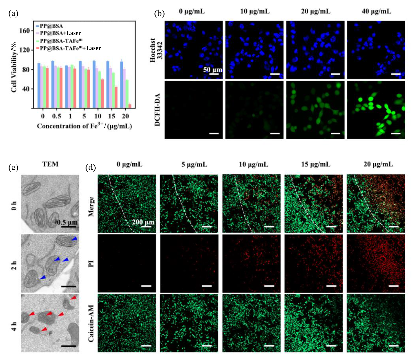
| 图5 PP@BSA-TAFeIII NPs的体外抗肿瘤性能. (a)不同纳米颗粒处理后4T1细胞的存活率;(b)不同Fe3+浓度的PP@BSA-TAFeIII NPs处理后细胞内ROS水平的共聚焦荧光图像(细胞核使用Hoechst 33342进行染色);(c) PP@BSA-TAFeIII NPs处理不同时间后4T1细胞的TEM图像;(d)不同Fe3+浓度的PP@BSA-TAFeIII NPs处理并用808 nm激光(1 W·cm-2)照射10 min后,对4T1细胞进行Calcein-AM/PI染色的共聚焦荧光图像(绿色荧光为活细胞,红色荧光为死细胞) |
| Fig. 5 In vitro antitumor efficacy of PP@BSA-TAFeIII NPs. (a) Viabilities of 4T1 cells treated with different nanoparticles; (b) CLSM images of ROS levels in 4Tl cells treated with different concentrations of Fe3+ in PP@BSA-TAFeIII NPs (cell nuclei was stained by Hoechst 33342); (c) TEM images of 4T1 cells treated with PP@BSA-TAFeIII NPs for different times; (d) CLSM images of 4T1 cells stained with Calcein-AM/PI (green fluorescence for live cells, red fluorescence for dead cells) after treatment with different concentrations of Fe3+ in PP@BSA-TAFeIII NPs and irradiation with 808 nm laser (1 W·cm-2) for 10 min |
|
|