蛋白冠原位调控增强肿瘤靶向19F MRI和联合治疗
In Situ Regulation of Protein Corona for Enhanced Tumor-targeted 19F MRI and Combined Therapy
蛋白冠原位调控增强肿瘤靶向19F MRI和联合治疗 |
| 李睿鹥, 李莎, 徐秋怡, 隋美菊, 陈世桢 |
|
In Situ Regulation of Protein Corona for Enhanced Tumor-targeted 19F MRI and Combined Therapy |
| LI Ruiyi, LI Sha, XU Qiuyi, SUI Meiju, CHEN Shizhen |
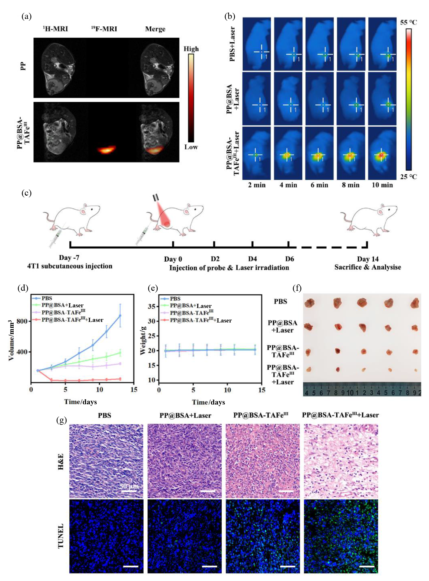
| 图6 PP@BSA-TAFeIII NPs的活体成像与抗肿瘤性能. (a)静脉注射PP@BSA-TAFeIII NPs和PP NPs 12 h后4T1荷瘤小鼠的19F MRI图像;(b)静脉注射PBS、PP@BSA NPs、PP@BSA-TAFeIII NPs至4T1荷瘤小鼠体内,经808 nm激光(1 W·cm-2)照射后肿瘤部位的光热成像;(c)活体抗肿瘤治疗示意图;不同治疗组小鼠的(d)肿瘤体积生长曲线、(e)体重变化曲线和(f)肿瘤照片;(g)不同治疗组小鼠肿瘤组织的H&E染色和TUNEL染色图像 |
| Fig. 6 In Vivo tumor MR imaging and antitumor effect of PP@BSA-TAFeIII NPs. (a) 19F MRI images of 4T1 tumor-bearing mice after intravenous injection of PP@BSA-TAFeIII NPs or PP NPs for 12 h; (b) Infrared thermal photographs of the 4T1 tumor-bearing mice after intravenous injection of PBS, PP@BSA NPs or PP@BSA-TAFeIII NPs, followed by irradiation with 808 nm laser (1 W·cm-2); (c) Schematic illustration of the in vivo antitumor therapy; (d) Tumor volume growth curves, (e) body weight change curves and (f) tumor photographs of mice in different treatment groups; (g) H&E staining and TUNEL staining images of tumor tissues from mice in different treatment groups |
|
|