

HdeA在细菌外膜囊泡环境下的原位NMR研究
收稿日期: 2023-05-16
网络出版日期: 2023-06-30
基金资助
国家自然科学基金资助项目(22174151);国家自然科学基金资助项目(21904138);国家自然科学基金资助项目(21991080);国家自然科学基金资助项目(22078280);国家重点基础研究发展计划(973计划)资助项目(2018YFE0202301);国家重点基础研究发展计划(973计划)资助项目(2018YFE0202300)
In situ Investigation of HdeA in Bacterial Outer Membrane Vesicles Using NMR Spectroscopy
Received date: 2023-05-16
Online published: 2023-06-30
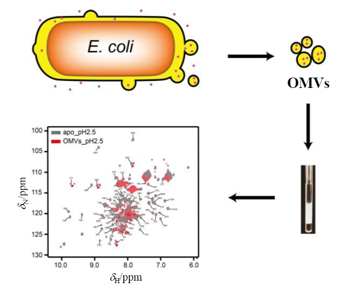
HdeA是一种定位于细菌周质的分子伴侣,在维持蛋白质稳态中起着重要的作用.以往对HdeA的研究主要是在体外条件下进行,限制了人们对HdeA在天然环境下发挥作用机制的理解.细菌外膜囊泡是细菌自发分泌到胞外环境的外膜囊泡,其内容物与周质环境相似.本研究将HdeA富集到细菌外膜囊泡(OMVs)中,通过核磁共振波谱研究HdeA在OMVs中的构象变化.结果表明,HdeA在其原位环境中表现出酸依赖性的构象变化.在低pH条件下HdeA主要通过S15、W16、T17、S27、T32、E36、G54、T57、C66、Q71、F74及D83等残基启动其分子伴侣功能.此外本研究也为原位研究其它周质分子伴侣的功能提供了新方法.
王欢 , 陶志清 , 姜国胜 , 张许 , 王冠 , 禾立春 , 刘买利 . HdeA在细菌外膜囊泡环境下的原位NMR研究[J]. 波谱学杂志, 2024 , 41(1) : 1 -8 . DOI: 10.11938/cjmr20233069
HdeA, a molecular chaperone localized in the bacterial periplasm, plays a vital role in maintaining protein homeostasis. Previous studies of HdeA were mainly carried out in in vitro conditions, limiting our understanding of the mechanism of HdeA in its native environment. Outer membrane vesicles of bacteria are extracellular vesicles released by cells into the extracellular milieu in a controlled manner, with contents almost identical to the periplasmic environment. Thus, by enriching HdeA into the bacterial outer membrane vesicles (OMVs), we investigated the conformational changes of HdeA within OMVs via nuclear magnetic resonance (NMR) spectroscopy. The results reveal that HdeA exhibits an acid-dependent conformational change in its native environment. The high-resolution spectrum of HdeA in OMVs indicates that under low pH conditions the function of HdeA is activated through residues S15, W16, T17, S27, T32, E36, G54, T57, C66, Q71, F74 and D83. Moreover, the NMR measurement of HdeA within OMVs provides a promising way for in situ investigation of mechanisms of other periplasmic molecular chaperones via NMR spectroscopy.
Key words: in situ NMR; HdeA; outer membrane vesicles; chaperone
| [1] | CHRISTOPH W, ANDREAS P. Protein folding in the periplasm of Escherichia coli[J]. Mol Microbiol, 1994, 12: 685-692. |
| [2] | HONG W Z, WU Y E, FU X M, et al. Chaperone-dependent mechanisms for acid resistance in enteric bacteria[J]. Trends Microbiol, 2012, 20(7): 328-335. |
| [3] | YU X C, HU Y F, DING J, et al. Structural basis and mechanism of the unfolding-induced activation of HdeA, a bacterial acid response chaperone[J]. J Biol Chem, 2019, 294(9): 3192-3206. |
| [4] | GARRISON M A, CROWHURST K A. NMR-monitored titration of acid-stress bacterial chaperone HdeA reveals that Asp and Glu charge neutralization produces a loosened dimer structure in preparation for protein unfolding and chaperone activation[J]. Protein Sci, 2014, 23(2): 167-178. |
| [5] | ZHAN J H, HU Q, ZHU Q J, et al. Track the conformational change of unlabeled yeast cytochrome c in cell homogenate using NMR[J]. Chinese J Magn Reson, 2023, 40(1): 22-29. |
| [5] | 占建华, 胡琴, 朱勤俊, 等. 基于磁共振的胞浆中无标记酵母细胞色素c构象变化追踪[J]. 波谱学杂志, 2023, 40(1): 22-29. |
| [6] | XING C Y, CHENGFENG Y, JIENV D, et al. Characterizations of the interactions between Escherichia coli periplasmic chaperone HdeA and its native substrates during acid stress[J]. Biochemistry, 2017, 56 (43): 5748-5757 |
| [7] | SALMON L, STULL F, SAYLE S, et al. The mechanism of HdeA unfolding and chaperone activation[J]. J Mol Biol, 2018, 430(1): 33-40. |
| [8] | ZHAI Z N, WU Q, ZHENG W W, et al. Roles of structural plasticity in chaperone HdeA activity are revealed by 19F NMR[J]. Chem Sci, 2016, 7: 2222. |
| [9] | WANG G, YU G J, GAO D W. Protein conformational exchanges modulated by the environment of outer membrane vesicles[J]. J Phys Chem Lett, 2023, 14 (11): 2772-2777. |
| [10] | THOMA J, MANIOGLU S, KALBERMATTER D, et al. Protein-enriched outer membrane vesicles as a native platform for outer membrane protein studies[J]. Comm Biol, 2018, 1: 23. |
| [11] | NIKAIDO H, VAARA M. Molecular basis of bacterial outer membrane permeability[J]. Microbiol Rev, 1985, 49(1): 1-32. |
| [12] | SCHIRMER T. General and specific porins from bacterial outer membranes[J]. J Struct Biol, 1998, 121(2): 101-109. |
| [13] | KOEBNIK R, LOCHER K P, VAN GELDER P. Structure and function of bacterial outer membrane proteins: barrels in a nutshell[J]. Mol Microbiol, 2000, 37(2): 239-253. |
| [14] | JOHANNES T, BJ?RN M B. High-resolution in situ NMR spectroscopy of bacterial envelope proteins in outer membrane vesicles[J]. Biochemistry, 2020, 59(17): 1656-1660. |
| [15] | VRANKEN W F, BOUCHER W, STEVENS T J, et al. The CCPN data model for NMR spectroscopy: development of a software pipeline[J]. Proteins, 2005, 59(4): 687-696. |
| [16] | HOEKSTRA D, VAN DER LAAN J W, DE LEIJ L, et al. Release of outer membrane fragments from normally growing Escherichia coli[J]. Biochim Biophys Acta, 1976, 455(3): 889-899. |
| [17] | MUG-OPSTELTEN D, WITHOLT B. Preferential release of new outer membrane fragments by exponentially growing Escherichia coli[J]. Biochim Biophys Acta, 1978, 508(2): 287-295. |
| [18] | KESTY N C, KUEHN M J. Incorporation of heterologous outer membrane and periplasmic proteins into Escherichia coli outer membrane vesicles[J]. J Biol Chem, 2004, 279(3): 2069-2076. |
| [19] | HAURAT M F, ADUSE-OPOKU J, RANGARAJAN M, et al. Selective sorting of cargo proteins into bacterial membrane vesicles[J]. J Biol Chem, 2011, 286(2): 1269-1276. |
| [20] | BONNINGTON K E, KUEHN M J. Protein selection and export via outer membrane vesicles[J]. Biochim Biophys Acta, 2014, 1843(8): 1612-1619. |
| [21] | SONG X, LV T, CHEN J, et al. Characterization of residue specific protein folding and unfolding dynamics in cells[J]. J Am Chem Soc, 2019, 141(29): 11363-11366. |
| [22] | TAKAOKA Y, KIOI Y, MORITO A, et al. Quantitative comparison of protein dynamics in live cells and in vitro by in-cell 19F NMR[J]. Chem Comm, 2013, 49(27): 2801-2803. |
| [23] | WILLIAMSON M P. Using chemical shift perturbation to characterize ligand binding[J]. Prog Nucl Magn Reson Spectrosc, 2013, 73: 1-16. |
/
| 〈 |
|
〉 |