

二型糖尿病大鼠纹状体白质微观结构异常的磁共振影像研究
收稿日期: 2025-07-23
网络出版日期: 2025-09-08
基金资助
湖北省实验动物研究计划资助项目(T23H1401)
Magnetic Resonance Imaging Study on the Microstructure Abnormalities of Striatal White Matter in Type 2 Diabetic Mellitus Rats
Received date: 2025-07-23
Online published: 2025-09-08
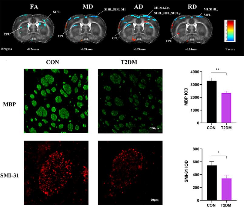
本文旨在通过磁共振成像(MRI)技术探索二型糖尿病(type 2 diabetes mellitus,T2DM)大鼠脑白质结构改变及其病理机制.本研究采用8周高脂饮食加单次腹腔注射30 mg/kg链脲佐菌素(STZ)建立T2DM模型,造模4周后,通过MRI技术对大鼠进行脑白质可视化分析,通过免疫组化进一步解释MRI指标变化的生理机制.结果发现,T2DM大鼠纹状体白质体积减小,各向异性分数(FA)、平均扩散率(MD)、轴向扩散速率(AD)值降低,径向扩散率(RD)值升高,磷酸化纤维丝(SMI-31)和髓鞘碱性蛋白(MBP)免疫组化提示T2DM组大鼠纹状体存在轴突受损、脱髓鞘.综上所述,弥散张量成像(DTI)结果显示T2DM大鼠纹状体损伤,其参数异常可能是纹状体轴突受损和脱髓鞘的表现,DTI指标有望作为评估糖尿病脑损伤的潜在指标.
蔡悦 , 王旭霞 , 刘思婕 , 郭浩东 , 陈茜 , 程琳琳 , 康彦 , 林富春 . 二型糖尿病大鼠纹状体白质微观结构异常的磁共振影像研究[J]. 波谱学杂志, 2026 , 43(2) : 175 -185 . DOI: 10.11938/cjmr20253177
This study aims to explore the structural changes in white matter and the physiological mechanisms in type 2 diabetes mellitus (T2DM) rats by magnetic resonance imaging (MRI) technology. A T2DM model was established by 8 weeks of high-fat diet combined with intraperitoneal injection of a dose of 30 mg/kg streptozotocin (STZ). 4 weeks after modeling, visual analysis of white matter in rats was performed by MRI, and the physiological mechanism of MRI index changes was further explained by immunohistology. The results showed that the striatal white matter volume decreased, the fractional anisotropy (FA), mean diffusivity (MD) and axial diffusivity (AD) decreased, and radial diffusivity (RD) increased in T2DM rats. Immunostain of phosphorylated neurofilament (SMI-31) and myelin basic protein (MBP) indicated axonal damage and demyelination of striatum in T2DM rats. In conclusion, the striatal injury in T2DM rats was observed by diffusion tensor imaging (DTI), and abnormal DTI index may be a manifestation of striatal axonal damage and demyelination, which can potentially be used as surrogates for evaluating diabetic brain injuries.
| [1] | AHMAD E, LIM S, LAMPTEY R, et al. Type 2 diabetes[J]. Lancet, 2022, 400(10365): 1803-1820. |
| [2] | OGURTSOVA K, GUARIGUATA L, BARENGO N C, et al. IDF diabetes atlas: Global estimates of undiagnosed diabetes in adults for 2021[J]. Diabetes Res Clin Pract, 2022, 183: 109118. |
| [3] | ZHENG Y, LEY S H, HU F B. Global aetiology and epidemiology of type 2 diabetes mellitus and its complications[J]. Nat Rev Endocrinol, 2018, 14(2): 88-98. |
| [4] | TOMIC D, SHAW J E, MAGLIANO D J. The burden and risks of emerging complications of diabetes mellitus[J]. Nat Rev Endocrinol, 2022, 18(9): 525-539. |
| [5] | HUANG M, GAO L, YANG L, et al. Abnormalities in the brain of streptozotocin-induced type 1 diabetic rats revealed by diffusion tensor imaging[J]. Neuroimage Clin, 2012, 1(1): 57-65. |
| [6] | ZHANG T, SHAW M, CHERBUIN N. Association between type 2 diabetes mellitus and brain atrophy: a meta-analysis[J]. Diabetes Metab J, 2022, 46(5): 781-802. |
| [7] | MA T, LI Z Y, YU Y, et al. Gray and white matter abnormality in patients with T2DM-related cognitive dysfunction: a systemic review and meta-analysis[J]. Nutr Diabetes, 2022, 12(1): 39. |
| [8] | QIU W, YUE X, HUANG H, et al. Structural characteristics of amygdala subregions in type 2 diabetes mellitus[J]. Behav Brain Res, 2024, 466: 114992. |
| [9] | TANG Q, LI S, YANG Z, et al. A narrative review of multimodal imaging of white matter lesions in type-2 diabetes mellitus[J]. Ann Palliat Med, 2021, 10(12): 12867-12876. |
| [10] | TOURNIER J D, MORI S, LEEMANS A. Diffusion tensor imaging and beyond[J]. Magn Reson Med, 2011, 65(6): 1532-1556. |
| [11] | ALOTAIBI A, TENCH C, STEVENSON R, et al. Investigating brain microstructural alterations in type 1 and type 2 diabetes using diffusion tensor imaging: a systematic review[J]. Brain Sci, 2021, 11(2): 140. |
| [12] | XIONG Y, SUI Y, ZHANG S, et al. Brain microstructural alterations in type 2 diabetes: diffusion kurtosis imaging provides added value to diffusion tensor imaging[J]. Eur Radiol, 2019, 29(4): 1997-2008. |
| [13] | HUANG L, ZHANG Q, TANG T, et al. Abnormalities of brain white matter in type 2 diabetes mellitus: a meta-analysis of diffusion tensor imaging[J]. Front Aging Neurosci, 2021, 13: 693890. |
| [14] | TAN X, FANG P, AN J, et al. Micro-structural white matter abnormalities in type 2 diabetic patients: a DTI study using TBSS analysis[J]. Neuroradiology, 2016, 58(12): 1209-1216. |
| [15] | WU C Y, HUANG S M, LIN Y H, et al. Reproducibility of diffusion tensor imaging-derived parameters: implications for the streptozotocin-induced type 1 diabetic rats[J]. Magn Reson Mater Phy, 2023, 36(4): 631-639. |
| [16] | LI J, GUO Y, LI Q, et al. Presence of white matter lesions associated with diabetes-associated cognitive decline in male rat models of pre-type 2 diabetes[J]. Med Sci Monit, 2019, 25: 9679-9689. |
| [17] | LI M Z, ZHANG L, SHI Z Y, et al. Magnetic resonance imaging detects cerebral gray and white matter injury correlated with cognitive impairments in diabetic db/db mice[J]. Behav Brain Res, 2023, 451: 114510. |
| [18] | HU Y D, CAI Y, WANG X X, et al. Magnetic resonance imaging the brain structures involved in nicotine susceptibility in rats[J]. Chinese J Magn Reson, 2021, 38(3): 345-355. |
| 胡赢丹, 蔡悦, 王旭霞, 等. 尼古丁易感的脑结构特征的磁共振成像研究[J]. 波谱学杂志, 2021, 38(3): 345-355. | |
| [19] | CHEN X, LIU S J, CAI Y, et al. Effects of seizure-inducing doses nicotine on hippocampal structure in adolescent female rats[J]. Chinese J Magn Reson, 2025, 42(4): 345-354. |
| 陈茜, 刘思婕, 蔡悦, 等. 致痫剂量尼古丁对青少年雌性大鼠海马结构的影响[J]. 波谱学杂志, 2025, 42(4): 345-354. | |
| [20] | HUANG W, CAO Z Y. STZ-induced progressive brain atrophy studied by magnetic resonance imaging and histochemical staining[J]. Chinese J Magn Reson, 2015, 32(3): 439-449. |
| 黄微, 曹子玉. STZ诱导大鼠1型糖尿病进行性脑萎缩的磁共振成像及组织化学研究[J]. 波谱学杂志, 2015, 32(3): 439-449. | |
| [21] | WANG X, LIN F, KANG Y, et al. Brain structural plasticity in rats subjected to early binocular enucleation characterized by high resolution anatomical magnetic resonance imaging and diffusion tensor imaging[J]. Magn Reson Lett, 2023, 3(1): 14-21. |
| [22] | TANAKA S, HAYASHI T, TOYODA T, et al. High-fat diet impairs the effects of a single bout of endurance exercise on glucose transport and insulin sensitivity in rat skeletal muscle[J]. Metabolism, 2007, 56(12): 1719-1728. |
| [23] | SRINIVASAN K, VISWANAD B, ASRAT L, et al. Combination of high-fat diet-fed and low-dose streptozotocin-treated rat: a model for type 2 diabetes and pharmacological screening[J]. Pharmacol Res, 2005, 52(4): 313-320. |
| [24] | KOWLURU R A. Retinopathy in a diet-induced type 2 diabetic rat model and role of epigenetic modifications[J]. Diabetes, 2020, 69(4): 689-698. |
| [25] | MORAN C, PHAN T G, CHEN J, et al. Brain atrophy in type 2 diabetes: regional distribution and influence on cognition[J]. Diabetes Care, 2013, 36(12): 4036-4042. |
| [26] | YAU P L, JAVIER D C, RYAN C M, et al. Preliminary evidence for brain complications in obese adolescents with type 2 diabetes mellitus[J]. Diabetologia, 2010, 53(11): 2298-2306. |
| [27] | LI C, JIN R, LIU K, et al. White matter atrophy in type 2 diabetes mellitus patients with mild cognitive impairment[J]. Front Neurosci, 2020, 14: 602501. |
| [28] | ZHOU Y, LI X L, XIE H L, et al. Voxel-based morphology analysis of STZ-induced type 1 diabetes mellitus rats with and without cognitive impairment[J]. Neurosci Lett, 2018, 684: 210-217. |
| [29] | CHEN J, ZHANG J, LIU X, et al. Abnormal subcortical nuclei shapes in patients with type 2 diabetes mellitus[J]. Eur Radiol, 2017, 27(10): 4247-4256. |
| [30] | SUN Q, CHEN G Q, WANG X B, et al. Alterations of white matter integrity and hippocampal functional connectivity in type 2 diabetes without mild cognitive impairment[J]. Front Neuroanat, 2018, 12: 00021. |
| [31] | ABE Y, YAMAMOTO T, SOEDA T, et al. Diabetic striatal disease: clinical presentation, neuroimaging, and pathology[J]. Intern Med, 2009, 48(13): 1135-1141. |
| [32] | LEI H, DOOLEY P, PEELING J, et al. Temporal profile of magnetic resonance imaging changes following forebrain ischemia in the gerbil[J]. Neurosci Lett, 1998, 257(2): 105-108. |
| [33] | WANG H, WANG Z, GAO Y, et al. STZ-induced diabetes exacerbates neurons ferroptosis after ischemic stroke by upregulating LCN2 in neutrophils[J]. Exp Neurol, 2024, 377: 114797. |
| [34] | DE BRESSER J, KUIJF H J, ZAANEN K, et al. White matter hyperintensity shape and location feature analysis on brain MRI; proof of principle study in patients with diabetes[J]. Sci Rep, 2018, 8(1): 1893. |
| [35] | ZHANG J H, XU H Z, SHEN Q F, et al. Nepsilon-(carboxymethyl)-lysine, white matter, and cognitive function in diabetes patients[J]. Can J Neurol Sci, 2016, 43(4): 518-522. |
| [36] | YOON S, CHO H, KIM J, et al. Brain changes in overweight/obese and normal-weight adults with type 2 diabetes mellitus[J]. Diabetologia, 2017, 60(7): 1207-1217. |
| [37] | ZHOU C, LI J, DONG M, et al. Altered white matter microstructures in type 2 diabetes mellitus: a coordinate-based meta-analysis of diffusion tensor imaging studies[J]. Front Endocrinol (Lausanne), 2021, 12: 658198. |
| [38] | VAN DE VONDERVOORT I, AMIRI H, BRUCHHAGE M M K, et al. Converging evidence points towards a role of insulin signaling in regulating compulsive behavior[J]. Transl Psychiatry, 2019, 9(1): 225. |
| [39] | ZHANG J, CHEN S, SHI W, et al. Effects of Xiaoshuan enteric-coated capsule on white and gray matter injury evaluated by diffusion tensor imaging in ischemic stroke[J]. Cell Transplant, 2019, 28(6): 671-683. |
| [40] | GAO J, PAN P, LI J, et al. Analysis of white matter tract integrity using diffusion kurtosis imaging reveals the correlation of white matter microstructural abnormalities with cognitive impairment in type 2 diabetes mellitus[J]. Front Endocrinol (Lausanne), 2024, 15: 1327339. |
| [41] | PORTER A, LECKIE R, VERSTYNEN T. White matter pathways as both a target and mediator of health behaviors[J]. Ann N Y Acad Sci, 2018, 1428(1): 71-88. |
| [42] | SONG S K, SUN S W, JU W K, et al. Diffusion tensor imaging detects and differentiates axon and myelin degeneration in mouse optic nerve after retinal ischemia[J]. NeuroImage, 2003, 20(3): 1714-1722. |
| [43] | SOUSTELLE L, ANTAL M C, LAMY J, et al. Correlations of quantitative MRI metrics with myelin basic protein (MBP) staining in a murine model of demyelination[J]. NMR Biomed, 2019, 32(9): e4116. |
| [44] | MUKHERJEE P, MILLER J H, SHIMONY J S, et al. Diffusion-tensor MR imaging of gray and white matter development during normal human brain maturation[J]. AJNR Am J Neuroradiol, 2002, 23(9): 1445-1456. |
| [45] | ZHU Y S, XIONG K L, ZHANG Y L, et al. Establishment of an acute diffuse axonal injury model and early diffusion tensor imaging manifestations[J]. Chin J Trauma, 2014, 30(5): 460-463. |
| 朱永山, 熊坤林, 张玉龙, 等. 急性弥漫性轴索损伤模型的建立与早期弥散张量成像表现[J]. 中华创伤杂志, 2014, 30(5): 460-463. | |
| [46] | WANG S, WU E X, QIU D, et al. Longitudinal diffusion tensor magnetic resonance imaging study of radiation-induced white matter damage in a rat model[J]. Cancer Res, 2009, 69(3): 1190-1198. |
/
| 〈 |
|
〉 |