
波谱学杂志 ›› 2026, Vol. 43 ›› Issue (2): 175-185.doi: 10.11938/cjmr20253177cstr: 32225.14.cjmr20253177
蔡悦1,2,*( ), 王旭霞1,2, 刘思婕1,2, 郭浩东1,2, 陈茜1,2, 程琳琳1,3, 康彦1,2, 林富春1,2
收稿日期:2025-07-23
出版日期:2026-06-05
在线发表日期:2025-09-08
通讯作者:
蔡悦
E-mail:caiyue192@mails.ucas.ac.cn
基金资助:
CAI Yue1,2,*(), WANG Xuxia1,2, LIU Sijie1,2, GUO Haodong1,2, CHEN Xi1,2, CHENG Linlin1,3, KANG Yan1,2, LIN Fuchun1,2
Received:2025-07-23
Published:2026-06-05
Online:2025-09-08
Contact:
CAI Yue
E-mail:caiyue192@mails.ucas.ac.cn
摘要:
本文旨在通过磁共振成像(MRI)技术探索二型糖尿病(type 2 diabetes mellitus,T2DM)大鼠脑白质结构改变及其病理机制.本研究采用8周高脂饮食加单次腹腔注射30 mg/kg链脲佐菌素(STZ)建立T2DM模型,造模4周后,通过MRI技术对大鼠进行脑白质可视化分析,通过免疫组化进一步解释MRI指标变化的生理机制.结果发现,T2DM大鼠纹状体白质体积减小,各向异性分数(FA)、平均扩散率(MD)、轴向扩散速率(AD)值降低,径向扩散率(RD)值升高,磷酸化纤维丝(SMI-31)和髓鞘碱性蛋白(MBP)免疫组化提示T2DM组大鼠纹状体存在轴突受损、脱髓鞘.综上所述,弥散张量成像(DTI)结果显示T2DM大鼠纹状体损伤,其参数异常可能是纹状体轴突受损和脱髓鞘的表现,DTI指标有望作为评估糖尿病脑损伤的潜在指标.
中图分类号:
蔡悦, 王旭霞, 刘思婕, 郭浩东, 陈茜, 程琳琳, 康彦, 林富春. 二型糖尿病大鼠纹状体白质微观结构异常的磁共振影像研究[J]. 波谱学杂志, 2026, 43(2): 175-185.
CAI Yue, WANG Xuxia, LIU Sijie, GUO Haodong, CHEN Xi, CHENG Linlin, KANG Yan, LIN Fuchun. Magnetic Resonance Imaging Study on the Microstructure Abnormalities of Striatal White Matter in Type 2 Diabetic Mellitus Rats[J]. Chinese Journal of Magnetic Resonance, 2026, 43(2): 175-185.
表1
实验药品与试剂
| 试剂名称 | 纯度 | 供应商 |
|---|---|---|
| 链脲佐菌素(STZ) | ≥99% | S0130, Sigma |
| 戊巴比妥钠 | ≥99% | 57-33-0, Sigma |
| 柠檬酸-柠檬酸钠缓冲液 | 0.1 mol/L, pH=4.5 | pH1716,飞净 |
| 0.9%生理盐水 | 0.9% (w/v) | 武汉滨湖双鹤药业有限公司 |
| 丙三醇 | ≥99% | 国药集团化学试剂有限公司 |
| 乙二醇 | ≥99.5% | 国药集团化学试剂有限公司 |
| 多聚甲醛 | ≥95% | 国药集团化学试剂有限公司 |
| 氯化钠 | ≥99.5% | 国药集团化学试剂有限公司 |
| 氯化钾 | ≥99.5% | 国药集团化学试剂有限公司 |
| 十二水合磷酸氢二钾 | ≥99% | 国药集团化学试剂有限公司 |
| 磷酸二氢钾 | ≥99.5% | 国药集团化学试剂有限公司 |
| 氢氧化钠 | ≥99.8% | 国药集团化学试剂有限公司 |
| 羊血清 | ≥99% | 武汉飞弈科技有限公司 |
| 异氟烷 | ≥99% | 深圳市瑞沃德生命科技股份有限公司 |
| 葡萄糖 | ≥99.5% | 国药集团化学试剂有限公司 |
| 蔗糖 | ≥99% | 国药集团化学试剂有限公司 |
| 髓鞘碱性蛋白(MBP)一抗 | ≥95% | Ab218011, sigma |
| 磷酸化神经丝(SMI-31)一抗 | 纯化型 | SMI-31P-100, Covance |
| 二抗羊抗小鼠 Alexa Fluor 594 | 免疫原亲和纯化型 | ab150116, abcam |
| 二抗羊抗兔 Alexa Fluor 488 | 免疫原亲和纯化型 | ab150077, abcam |
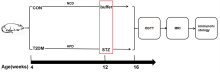
图1
实验流程图. CON:对照组大鼠;T2DM:二型糖尿病组大鼠;NCD:标准维持饲料;HFD:高脂饮食;STZ:链脲佐菌素;OGTT:口服葡萄糖耐量测试

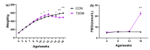
图2
大鼠体重和血糖的变化. (a) 4~16周龄两组大鼠体重变化;(b) 4~16周龄两组大鼠空腹血糖变化. *p < 0.05;***p < 0.001

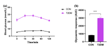
图3
大鼠口服糖耐量测试. (a)两组大鼠OGTT测试血糖动态变化曲线;(b)两组大鼠OGTT变化曲线的曲线下面积,***p < 0.001
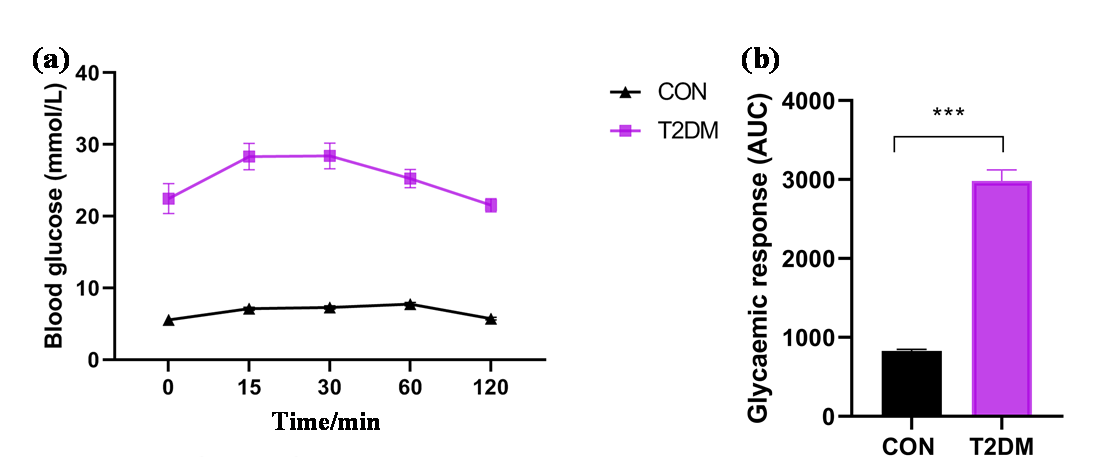

图4
二型糖尿病对大鼠脑白质微观结构的影响. CPU:尾状壳核(纹状体);M1:初级运动皮层;S1FL:初级体感皮层,前肢区域;S1HL:初级体感皮层,后肢区域. p<0.005(FDR校正),RARE的聚类大小为100,DTI的聚类大小为50
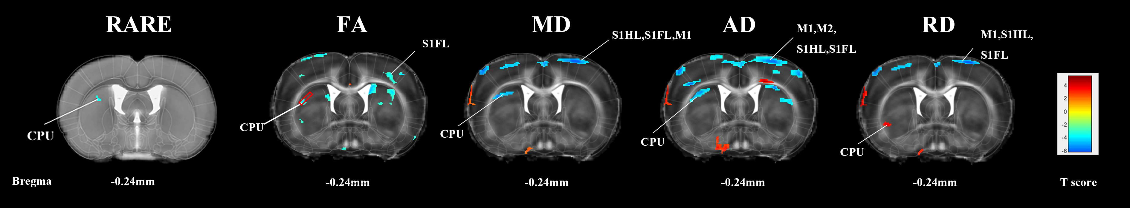
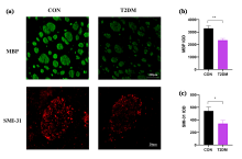
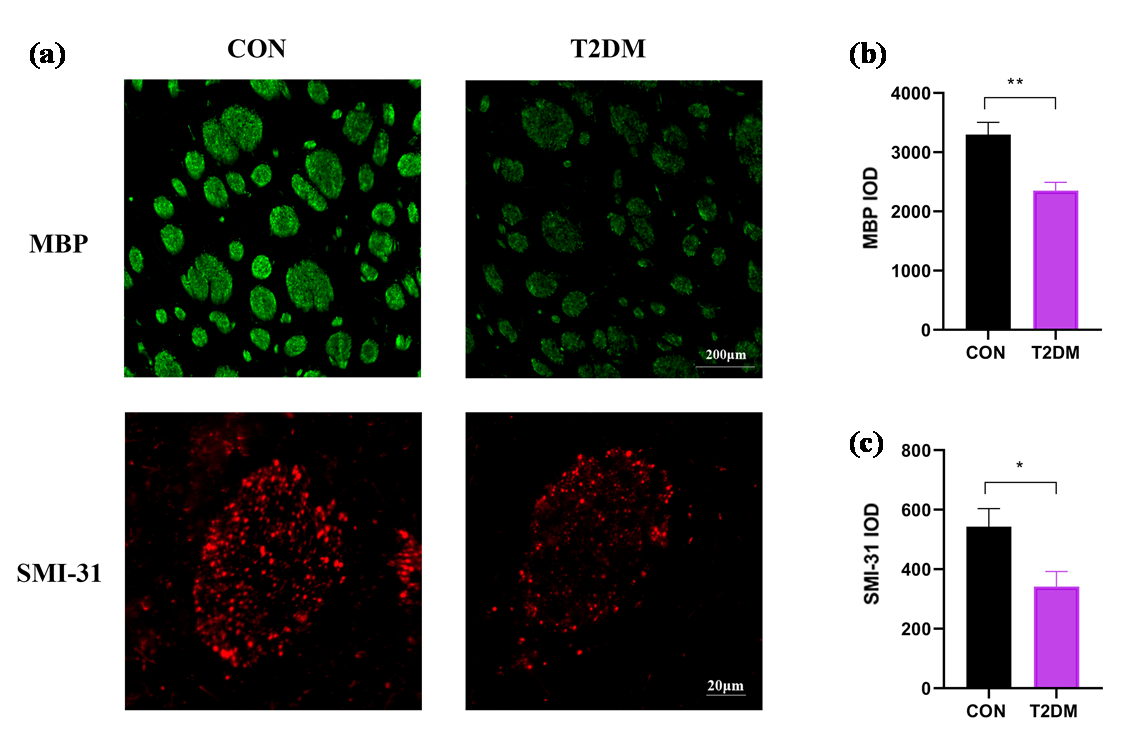
图5
大鼠纹状体免疫组化. (a)两组大鼠纹状体区域的MBP和SMI-31免疫组化染色,MBP免疫反应染色的比例尺为200 μm,SMI-31免疫反应染色的比例尺为20 μm;(b)纹状体MBP的IOD值;(c)纹状体SMI-31的IOD值. * p < 0.05;** p < 0.01
| [1] |
AHMAD E, LIM S, LAMPTEY R, et al. Type 2 diabetes[J]. Lancet, 2022, 400(10365): 1803-1820.
doi: 10.1016/S0140-6736(22)01655-5 pmid: 36332637 |
| [2] |
OGURTSOVA K, GUARIGUATA L, BARENGO N C, et al. IDF diabetes atlas: Global estimates of undiagnosed diabetes in adults for 2021[J]. Diabetes Res Clin Pract, 2022, 183: 109118.
doi: 10.1016/j.diabres.2021.109118 |
| [3] |
ZHENG Y, LEY S H, HU F B. Global aetiology and epidemiology of type 2 diabetes mellitus and its complications[J]. Nat Rev Endocrinol, 2018, 14(2): 88-98.
doi: 10.1038/nrendo.2017.151 pmid: 29219149 |
| [4] |
TOMIC D, SHAW J E, MAGLIANO D J. The burden and risks of emerging complications of diabetes mellitus[J]. Nat Rev Endocrinol, 2022, 18(9): 525-539.
doi: 10.1038/s41574-022-00690-7 |
| [5] |
HUANG M, GAO L, YANG L, et al. Abnormalities in the brain of streptozotocin-induced type 1 diabetic rats revealed by diffusion tensor imaging[J]. Neuroimage Clin, 2012, 1(1): 57-65.
doi: 10.1016/j.nicl.2012.09.004 |
| [6] |
ZHANG T, SHAW M, CHERBUIN N. Association between type 2 diabetes mellitus and brain atrophy: a meta-analysis[J]. Diabetes Metab J, 2022, 46(5): 781-802.
doi: 10.4093/dmj.2021.0189 pmid: 35255549 |
| [7] |
MA T, LI Z Y, YU Y, et al. Gray and white matter abnormality in patients with T2DM-related cognitive dysfunction: a systemic review and meta-analysis[J]. Nutr Diabetes, 2022, 12(1): 39.
doi: 10.1038/s41387-022-00214-2 pmid: 35970833 |
| [8] |
QIU W, YUE X, HUANG H, et al. Structural characteristics of amygdala subregions in type 2 diabetes mellitus[J]. Behav Brain Res, 2024, 466: 114992.
doi: 10.1016/j.bbr.2024.114992 |
| [9] |
TANG Q, LI S, YANG Z, et al. A narrative review of multimodal imaging of white matter lesions in type-2 diabetes mellitus[J]. Ann Palliat Med, 2021, 10(12): 12867-12876.
doi: 10.21037/apm-21-3299 pmid: 35016461 |
| [10] |
TOURNIER J D, MORI S, LEEMANS A. Diffusion tensor imaging and beyond[J]. Magn Reson Med, 2011, 65(6): 1532-1556.
doi: 10.1002/mrm.v65.6 |
| [11] |
ALOTAIBI A, TENCH C, STEVENSON R, et al. Investigating brain microstructural alterations in type 1 and type 2 diabetes using diffusion tensor imaging: a systematic review[J]. Brain Sci, 2021, 11(2): 140.
doi: 10.3390/brainsci11020140 |
| [12] |
XIONG Y, SUI Y, ZHANG S, et al. Brain microstructural alterations in type 2 diabetes: diffusion kurtosis imaging provides added value to diffusion tensor imaging[J]. Eur Radiol, 2019, 29(4): 1997-2008.
doi: 10.1007/s00330-018-5746-y pmid: 30338363 |
| [13] |
HUANG L, ZHANG Q, TANG T, et al. Abnormalities of brain white matter in type 2 diabetes mellitus: a meta-analysis of diffusion tensor imaging[J]. Front Aging Neurosci, 2021, 13: 693890.
doi: 10.3389/fnagi.2021.693890 |
| [14] |
TAN X, FANG P, AN J, et al. Micro-structural white matter abnormalities in type 2 diabetic patients: a DTI study using TBSS analysis[J]. Neuroradiology, 2016, 58(12): 1209-1216.
pmid: 27783100 |
| [15] |
WU C Y, HUANG S M, LIN Y H, et al. Reproducibility of diffusion tensor imaging-derived parameters: implications for the streptozotocin-induced type 1 diabetic rats[J]. Magn Reson Mater Phy, 2023, 36(4): 631-639.
doi: 10.1007/s10334-022-01048-w |
| [16] |
LI J, GUO Y, LI Q, et al. Presence of white matter lesions associated with diabetes-associated cognitive decline in male rat models of pre-type 2 diabetes[J]. Med Sci Monit, 2019, 25: 9679-9689.
doi: 10.12659/MSM.918557 |
| [17] |
LI M Z, ZHANG L, SHI Z Y, et al. Magnetic resonance imaging detects cerebral gray and white matter injury correlated with cognitive impairments in diabetic db/db mice[J]. Behav Brain Res, 2023, 451: 114510.
doi: 10.1016/j.bbr.2023.114510 |
| [18] | HU Y D, CAI Y, WANG X X, et al. Magnetic resonance imaging the brain structures involved in nicotine susceptibility in rats[J]. Chinese J Magn Reson, 2021, 38(3): 345-355. |
|
胡赢丹, 蔡悦, 王旭霞, 等. 尼古丁易感的脑结构特征的磁共振成像研究[J]. 波谱学杂志, 2021, 38(3): 345-355.
doi: 10.11938/cjmr20212890 |
|
| [19] | CHEN X, LIU S J, CAI Y, et al. Effects of seizure-inducing doses nicotine on hippocampal structure in adolescent female rats[J]. Chinese J Magn Reson, 2025, 42(4): 345-354. |
| 陈茜, 刘思婕, 蔡悦, 等. 致痫剂量尼古丁对青少年雌性大鼠海马结构的影响[J]. 波谱学杂志, 2025, 42(4): 345-354. | |
| [20] | HUANG W, CAO Z Y. STZ-induced progressive brain atrophy studied by magnetic resonance imaging and histochemical staining[J]. Chinese J Magn Reson, 2015, 32(3): 439-449. |
|
黄微, 曹子玉. STZ诱导大鼠1型糖尿病进行性脑萎缩的磁共振成像及组织化学研究[J]. 波谱学杂志, 2015, 32(3): 439-449.
doi: 10.11938/cjmr20150305 |
|
| [21] |
WANG X, LIN F, KANG Y, et al. Brain structural plasticity in rats subjected to early binocular enucleation characterized by high resolution anatomical magnetic resonance imaging and diffusion tensor imaging[J]. Magn Reson Lett, 2023, 3(1): 14-21.
doi: 10.1016/j.mrl.2022.10.001 pmid: 40919275 |
| [22] |
TANAKA S, HAYASHI T, TOYODA T, et al. High-fat diet impairs the effects of a single bout of endurance exercise on glucose transport and insulin sensitivity in rat skeletal muscle[J]. Metabolism, 2007, 56(12): 1719-1728.
doi: 10.1016/j.metabol.2007.07.017 |
| [23] |
SRINIVASAN K, VISWANAD B, ASRAT L, et al. Combination of high-fat diet-fed and low-dose streptozotocin-treated rat: a model for type 2 diabetes and pharmacological screening[J]. Pharmacol Res, 2005, 52(4): 313-320.
doi: 10.1016/j.phrs.2005.05.004 pmid: 15979893 |
| [24] |
KOWLURU R A. Retinopathy in a diet-induced type 2 diabetic rat model and role of epigenetic modifications[J]. Diabetes, 2020, 69(4): 689-698.
doi: 10.2337/db19-1009 pmid: 31949005 |
| [25] |
MORAN C, PHAN T G, CHEN J, et al. Brain atrophy in type 2 diabetes: regional distribution and influence on cognition[J]. Diabetes Care, 2013, 36(12): 4036-4042.
doi: 10.2337/dc13-0143 pmid: 23939539 |
| [26] |
YAU P L, JAVIER D C, RYAN C M, et al. Preliminary evidence for brain complications in obese adolescents with type 2 diabetes mellitus[J]. Diabetologia, 2010, 53(11): 2298-2306.
doi: 10.1007/s00125-010-1857-y pmid: 20668831 |
| [27] |
LI C, JIN R, LIU K, et al. White matter atrophy in type 2 diabetes mellitus patients with mild cognitive impairment[J]. Front Neurosci, 2020, 14: 602501.
doi: 10.3389/fnins.2020.602501 |
| [28] |
ZHOU Y, LI X L, XIE H L, et al. Voxel-based morphology analysis of STZ-induced type 1 diabetes mellitus rats with and without cognitive impairment[J]. Neurosci Lett, 2018, 684: 210-217.
doi: S0304-3940(18)30555-X pmid: 30125641 |
| [29] |
CHEN J, ZHANG J, LIU X, et al. Abnormal subcortical nuclei shapes in patients with type 2 diabetes mellitus[J]. Eur Radiol, 2017, 27(10): 4247-4256.
doi: 10.1007/s00330-017-4790-3 pmid: 28374074 |
| [30] |
SUN Q, CHEN G Q, WANG X B, et al. Alterations of white matter integrity and hippocampal functional connectivity in type 2 diabetes without mild cognitive impairment[J]. Front Neuroanat, 2018, 12: 00021.
doi: 10.3389/fnana.2018.00021 |
| [31] |
ABE Y, YAMAMOTO T, SOEDA T, et al. Diabetic striatal disease: clinical presentation, neuroimaging, and pathology[J]. Intern Med, 2009, 48(13): 1135-1141.
doi: 10.2169/internalmedicine.48.1996 |
| [32] |
LEI H, DOOLEY P, PEELING J, et al. Temporal profile of magnetic resonance imaging changes following forebrain ischemia in the gerbil[J]. Neurosci Lett, 1998, 257(2): 105-108.
pmid: 9865938 |
| [33] |
WANG H, WANG Z, GAO Y, et al. STZ-induced diabetes exacerbates neurons ferroptosis after ischemic stroke by upregulating LCN2 in neutrophils[J]. Exp Neurol, 2024, 377: 114797.
doi: 10.1016/j.expneurol.2024.114797 |
| [34] |
DE BRESSER J, KUIJF H J, ZAANEN K, et al. White matter hyperintensity shape and location feature analysis on brain MRI; proof of principle study in patients with diabetes[J]. Sci Rep, 2018, 8(1): 1893.
doi: 10.1038/s41598-018-20084-y pmid: 29382936 |
| [35] |
ZHANG J H, XU H Z, SHEN Q F, et al. Nepsilon-(carboxymethyl)-lysine, white matter, and cognitive function in diabetes patients[J]. Can J Neurol Sci, 2016, 43(4): 518-522.
doi: 10.1017/cjn.2015.398 |
| [36] |
YOON S, CHO H, KIM J, et al. Brain changes in overweight/obese and normal-weight adults with type 2 diabetes mellitus[J]. Diabetologia, 2017, 60(7): 1207-1217.
doi: 10.1007/s00125-017-4266-7 pmid: 28447116 |
| [37] |
ZHOU C, LI J, DONG M, et al. Altered white matter microstructures in type 2 diabetes mellitus: a coordinate-based meta-analysis of diffusion tensor imaging studies[J]. Front Endocrinol (Lausanne), 2021, 12: 658198.
doi: 10.3389/fendo.2021.658198 |
| [38] |
VAN DE VONDERVOORT I, AMIRI H, BRUCHHAGE M M K, et al. Converging evidence points towards a role of insulin signaling in regulating compulsive behavior[J]. Transl Psychiatry, 2019, 9(1): 225.
doi: 10.1038/s41398-019-0559-6 |
| [39] |
ZHANG J, CHEN S, SHI W, et al. Effects of Xiaoshuan enteric-coated capsule on white and gray matter injury evaluated by diffusion tensor imaging in ischemic stroke[J]. Cell Transplant, 2019, 28(6): 671-683.
doi: 10.1177/0963689718802755 |
| [40] |
GAO J, PAN P, LI J, et al. Analysis of white matter tract integrity using diffusion kurtosis imaging reveals the correlation of white matter microstructural abnormalities with cognitive impairment in type 2 diabetes mellitus[J]. Front Endocrinol (Lausanne), 2024, 15: 1327339.
doi: 10.3389/fendo.2024.1327339 |
| [41] |
PORTER A, LECKIE R, VERSTYNEN T. White matter pathways as both a target and mediator of health behaviors[J]. Ann N Y Acad Sci, 2018, 1428(1): 71-88.
doi: 10.1111/nyas.2018.1428.issue-1 |
| [42] |
SONG S K, SUN S W, JU W K, et al. Diffusion tensor imaging detects and differentiates axon and myelin degeneration in mouse optic nerve after retinal ischemia[J]. NeuroImage, 2003, 20(3): 1714-1722.
doi: 10.1016/j.neuroimage.2003.07.005 |
| [43] |
SOUSTELLE L, ANTAL M C, LAMY J, et al. Correlations of quantitative MRI metrics with myelin basic protein (MBP) staining in a murine model of demyelination[J]. NMR Biomed, 2019, 32(9): e4116.
doi: 10.1002/nbm.v32.9 |
| [44] | MUKHERJEE P, MILLER J H, SHIMONY J S, et al. Diffusion-tensor MR imaging of gray and white matter development during normal human brain maturation[J]. AJNR Am J Neuroradiol, 2002, 23(9): 1445-1456. |
| [45] | ZHU Y S, XIONG K L, ZHANG Y L, et al. Establishment of an acute diffuse axonal injury model and early diffusion tensor imaging manifestations[J]. Chin J Trauma, 2014, 30(5): 460-463. |
| 朱永山, 熊坤林, 张玉龙, 等. 急性弥漫性轴索损伤模型的建立与早期弥散张量成像表现[J]. 中华创伤杂志, 2014, 30(5): 460-463. | |
| [46] |
WANG S, WU E X, QIU D, et al. Longitudinal diffusion tensor magnetic resonance imaging study of radiation-induced white matter damage in a rat model[J]. Cancer Res, 2009, 69(3): 1190-1198.
doi: 10.1158/0008-5472.CAN-08-2661 pmid: 19155304 |
| [1] | 李睿鹥, 李莎, 徐秋怡, 隋美菊, 陈世桢. 蛋白冠原位调控增强肿瘤靶向19F MRI和联合治疗[J]. 波谱学杂志, 2026, 43(2): 200-213. |
| [2] | 谢心怡, 王远军. 脑部扩散磁共振成像超分辨率重建研究进展[J]. 波谱学杂志, 2026, 43(2): 223-240. |
| [3] | 傅芬芳, 林国兵, 李梅芳. 磁共振成像技术在轻度创伤性脑损伤诊断与预后中的应用[J]. 波谱学杂志, 2026, 43(2): 214-222. |
| [4] | 王奕雯, 吴光耀, 文之, 林富春. 抗逆转录病毒治疗相关的HIV感染者脑功能动态改变[J]. 波谱学杂志, 2026, 43(1): 61-70. |
| [5] | 倪广茂, 李玉伟, 侯文轩, 刘彩云, 董鹏, 张艳辉. 基于MR-DWI的急性脑梗塞复发影响因素的研究[J]. 波谱学杂志, 2026, 43(1): 87-93. |
| [6] | 陈茜, 刘思婕, 蔡悦, 程琳琳, 王旭霞, 康彦, 林富春, 雷皓. 致痫剂量尼古丁对青少年雌性大鼠海马结构的影响[J]. 波谱学杂志, 2025, 42(4): 345-354. |
| [7] | 李英豪, 王丽辉, 王苏成, 朱中旗, 黄长栋, 李仁峰, 曹开明, 胡海洋, 贾一鸣, 梁松涛, 杨光, 路青, 汪红志. 胰腺自动分割与区域定量及糖尿病评估研究[J]. 波谱学杂志, 2025, 42(4): 378-389. |
| [8] | 高照耀, 张展, 胡亮亮, 许光宇, 周胜, 胡雨欣, 林子捷, 周超. 基于虚拟线圈和GRAPPA增强网络的PMRI方法[J]. 波谱学杂志, 2025, 42(4): 390-401. |
| [9] | 马滢雪, 赵晏强, 杨晓冬, 蒋滨, 陶诚. 我国高场及超高场磁共振成像设备研制和市场化的机遇与挑战[J]. 波谱学杂志, 2025, 42(3): 334-344. |
| [10] | 李鹏, 纪雨萍, 胡悦. 基于流形结构正则化的快速高质量磁共振指纹定量成像[J]. 波谱学杂志, 2025, 42(3): 249-264. |
| [11] | 舒炜. B超和MRI在胎儿骨骼异常中的诊断价值分析[J]. 波谱学杂志, 2025, 42(3): 265-274. |
| [12] | 隋美菊, 张磊, 王瑞芳, 骆盈盈, 李莎, 丘茂松, 徐秋怡, 陈代钦, 陈世桢, 周欣. MRI示踪的纳米酶用于级联反应增强的免疫治疗[J]. 波谱学杂志, 2025, 42(3): 231-248. |
| [13] | 孟靖欣, 王远军. 基于扩散磁共振的大脑浅表白质纤维束研究进展[J]. 波谱学杂志, 2025, 42(2): 205-220. |
| [14] | 孙灏芸, 王丽嘉. 融合注意力机制和空洞卷积的3D ELD_MobileNetV2在肝结节分类中的应用[J]. 波谱学杂志, 2025, 42(2): 130-142. |
| [15] | 曹飞, 徐芊芊, 陈浩, 祖洁, 李晓文, 田锦, 鲍磊. 基于交叉自监督和DWI的NIID智能诊断方法[J]. 波谱学杂志, 2025, 42(2): 154-163. |
| 阅读次数 | ||||||
|
全文 |
|
|||||
|
摘要 |
|
|||||