

脉冲偶极耦合EPR技术在生物大分子结构变化与互作研究中的应用
收稿日期: 2025-12-11
网络出版日期: 2026-08-11
基金资助
国家自然科学基金资助项目(22177056);国家自然科学基金资助项目(22574083);天津市自然科学基金资助项目(22JCYBJC01490)
Applications of Pulsed Dipolar EPR Spectroscopy in Characterizing Interactions and Structural Changes of Biomacromolecules
Received date: 2025-12-11
Online published: 2026-08-11
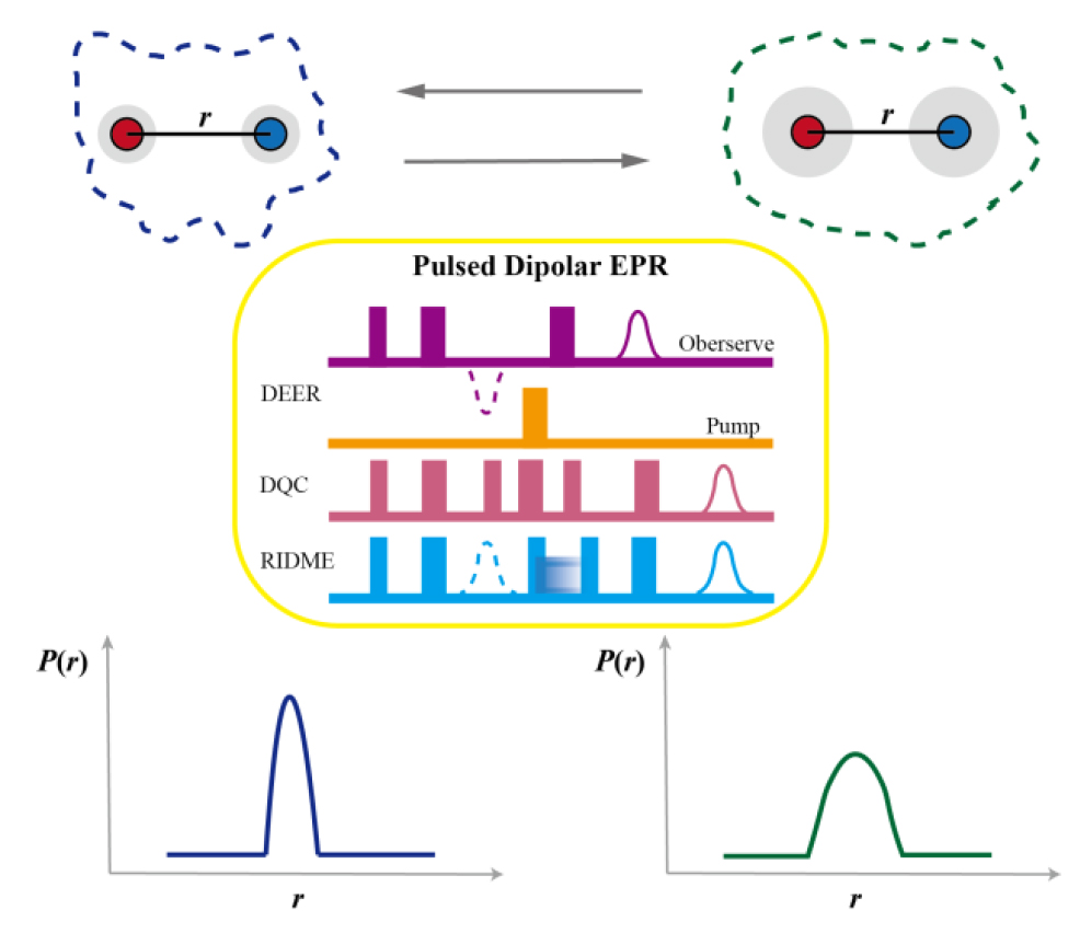
脉冲偶极耦合电子顺磁共振(PD-EPR)技术是一种研究蛋白质等生物大分子结构变化与相互作用的重要生物物理技术.该技术通过测量定点引入到目标分子上的自旋探针之间未成对电子的偶极相互作用,直接获取纳米尺度下电子自旋之间的距离分布信息,并通过比较不同实验条件下自旋对间的距离分布,解析生物大分子的结构变化、互作与动态行为.本文系统综述了三种主要的PD-EPR技术包括双电子-电子共振(DEER)、双量子相干(DQC)和弛豫诱导偶极调制增强(RIDME)的基本原理、技术特点及相关应用,总结了PD-EPR研究中常用的自旋探针类型及蛋白质标记策略,重点介绍了近年来该技术在生物大分子及其复合物研究中的最新进展.
谢雅欣 , 杨茵 , 苏循成 . 脉冲偶极耦合EPR技术在生物大分子结构变化与互作研究中的应用[J]. 波谱学杂志, 2026 , 43(3) : 350 -366 . DOI: 10.11938/cjmr20253191
Pulsed dipolar electron paramagnetic resonance (PD-EPR) is a powerful biophysical technique for probing the dynamic structures and interactions of biomacromolecules such as proteins. By measuring dipolar interactions between unpaired electrons, PD-EPR directly yields nanometer-scale distance distributions between electron spins, from which conformational transitions can be inferred by comparison under different experimental conditions. This review summarizes three major PD-EPR techniques, including double electron-electron resonance, double quantum coherence, and relaxation-induced dipolar modulation enhancement, together with commonly used spin labels and protein spin-labeling strategies, highlighting recent advances in the study of biomacromolecules and complexes.
| [1] | DRENTH J. Principles of protein X-ray crystallography[M]. 3rd ed. New York: Springer, 2007. |
| [2] | SHI Y A. Glimpse of structural biology through X-ray crystallography[J]. Cell, 2014, 159(5): 995-1014. |
| [3] | BAI X-C, MCMULLAN G, SCHERES S H W. How cryo-EM is revolutionizing structural biology[J]. Trends Biochem Sci, 2015, 40(1): 49-57. |
| [4] | FERNANDEZ-LEIRO R, SCHERES S H W. Unravelling biological macromolecules with cryo-electron microscopy[J]. Nature, 2016, 537(7620): 339-346. |
| [5] | PALMER A G. NMR characterization of the dynamics of biomacromolecules[J]. Chem Rev, 2004, 104(8): 3623-3640. |
| [6] | BANARI A, SAMANTA A K, MUNKE A, et al. Advancing time-resolved structural biology: latest strategies in cryo-EM and X-ray crystallography[J]. Nat Methods, 2025, 22(7): 1420-1435. |
| [7] | RüDIGER S, FREUND S M V, VEPRINTSEV D B, et al. CRINEPT-TROSY NMR reveals p53 core domain bound in an unfolded form to the chaperone Hsp90[J]. Proc Natl Acad Sci USA, 2002, 99(17): 11085-11090. |
| [8] | SCHIEMANN O, PRISNER T F. Long-range distance determinations in biomacromolecules by EPR spectroscopy[J]. Q Rev Biophys, 2007, 40(1): 1-53. |
| [9] | SCHIEMANN O, HEUBACH C A, ABDULLIN D, et al. Benchmark test and guidelines for DEER/PELDOR experiments on nitroxide-labeled biomolecules[J]. J Am Chem Soc, 2021, 143(43): 17875-17890. |
| [10] | JESCHKE G. DEER distance measurements on proteins[J]. Annu Rev Phys Chem, 2012, 63: 419-446. |
| [11] | BORDIGNON E, SEEGER M A, GALAZZO L, et al. From in vitro towards in situ: structure-based investigation of ABC exporters by electron paramagnetic resonance spectroscopy[J]. FEBS Lett, 2020, 594(23): 3839-3856. |
| [12] | SAHU I D, LORIGAN G A. Role of membrane mimetics on biophysical EPR studies of membrane proteins[J]. BBA-Biomembranes, 2023, 1865(4): 184138. |
| [13] | FACCHETTI D, DANG Y F, SEIF-EDDINE M, et al. Film-electrochemical EPR spectroscopy to investigate electron transfer in membrane proteins in their native environment[J]. Chem Commun, 2024, 60(87): 12690-12693. |
| [14] | BALO A R, LEE J, ERNST O P. Stationary phase EPR spectroscopy for monitoring membrane protein refolding by conformational response[J]. Anal Chem, 2019, 91(1): 1071-1079. |
| [15] | VALERA S, ACKERMANN K, PLIOTAS C, et al. Accurate extraction of nanometer distances in multimers by pulse EPR[J]. Chem-Eur J, 2016, 22(14): 4700-4703. |
| [16] | ABRAGAM A, BLEANEY B. Electron paramagnetic resonance of transition ions[M]. Oxford: Oxford University Press, 1970. |
| [17] | ABDULLIN D, SCHIEMANN O. Pulsed dipolar EPR spectroscopy and metal ions: Methodology and biological applications[J]. ChemPlusChem, 2020, 85(2): 353-372. |
| [18] | BEDILO A F, MARYASOV A G. Electron spin resonance of dipole-coupled anisotropic pairs in disordered systems[J]. J Magn Reson, 1995, 116(1): 87-96. |
| [19] | KLARE J P. Site-directed spin labeling EPR spectroscopy in protein research[J]. Biol Chem, 2013, 394(10): 1281-1300. |
| [20] | GARCíA-RUBIO I. EPR of site-directed spin-labeled proteins: A powerful tool to study structural flexibility[J]. Arch Biochem Biophys, 2020, 684: 108323. |
| [21] | MILOV A D, MARYASOV A G, TSVETKOV Y D. Pulsed electron double resonance (PELDOR) and its applications in free-radicals research[J]. Appl Magn Reson, 1998, 15(1): 107-143. |
| [22] | STEIN R A, BETH A H, HUSTEDT E J. A straightforward approach to the analysis of double electron-electron resonance data[J]. Methods Enzymol, 2015, 563: 531-567. |
| [23] | MILOV A D, SALIKHOV K M, SHIROV M D. Application of ELDOR in electron-spin echo for paramagnetic center space distribution in solids[J]. Fiz Tverd Tela, 1981, 23(4): 975-982. |
| [24] | JESCHKE G, POLYHACH Y. Distance measurements on spin-labelled biomacromolecules by pulsed electron paramagnetic resonance[J]. Phys Chem Chem Phys, 2007, 9(16): 1895-1910. |
| [25] | BORBAT P P, FREED J H. Multiple-quantum ESR and distance measurements[J]. Chem Phys Lett, 1999, 313(1-2): 145-154. |
| [26] | SUNIL S, FREED J H. Double quantum two-dimensional Fourier transform electron spin resonance: Distance measurements[J]. Chem Phys Lett, 1996, 251(1-2): 102-110. |
| [27] | SUNIL S, FREED J H. Theory of double quantum two-dimensional electron spin resonance with application to distance measurements[J]. J Chem Phys, 1997, 107(5): 1317-1340. |
| [28] | KUZIN S, YULIKOV M. RIDME spectroscopy: New topics beyond the determination of electron spin-spin distances[J]. J Phys Chem Lett, 2025, 16(4): 1024-1037. |
| [29] | COLLAUTO A, FRYDMAN V, LEE M D, et al. RIDME distance measurements using Gd(III) tags with a narrow central transition[J]. Phys Chem Chem Phys, 2016, 18(28): 19037-19049. |
| [30] | MEYER A, SCHIEMANN O. PELDOR and RIDME measurements on a high-spin manganese(II)bisnitroxide model complex[J]. J Phys Chem A, 2016, 120(20): 3463-3472. |
| [31] | AKHMETZYANOV D, CHING H Y V, DENYSENKOV V, et al. RIDME spectroscopy on high-spin Mn2+ centers[J]. Phys Chem Chem Phys, 2016, 18(44): 30857-30866. |
| [32] | RITSCH I, HINTZ H, JESCHKE G, et al. Improving the accuracy of Cu(II)-nitroxide RIDME in the presence of orientation correlation in water-soluble Cu(II)-nitroxide rulers[J]. Phys Chem Chem Phys, 2019, 21(19): 9810-9830. |
| [33] | WORT J L, ACKERMANN K, GIANNOULIS A, et al. Sub-micromolar pulse dipolar EPR spectroscopy reveals increasing CuII-labelling of double-histidine motifs with lower temperature[J]. Angew Chem Int Ed, 2019, 58(34): 11681-11685. |
| [34] | ABDULLIN D, DUTHIE F, MEYER A, et al. Comparison of PELDOR and RIDME for distance measurements between nitroxides and low-spin Fe(III) ions[J]. J Phys Chem B, 2015, 119(43): 13534-13542. |
| [35] | ASTASHKIN A V. Mapping the structure of metalloproteins with RIDME[J]. Methods Enzymol, 2015, 563: 251-284. |
| [36] | ORANGES M, WORT J L, FUKUSHIMA M, et al. Pulse dipolar electron paramagnetic resonance spectroscopy reveals buffer-modulated cooperativity of metal-templated protein dimerization[J]. J Phys Chem Lett, 2022, 13(33): 7847-7852. |
| [37] | KUZHELEV A A, KRUMKACHEVA O A, SHEVELEV G Y, et al. Room-temperature distance measurements using RIDME and the orthogonal spin labels trityl/nitroxide[J]. Phys Chem Chem Phys, 2018, 20(15): 10224-10230. |
| [38] | KULIK L V, DZUBA S A, GRIGORYEV I A, et al. Electron dipole-dipole interaction in ESEEM of nitroxide biradicals[J]. Chem Phys Lett, 2001, 343(3-4): 315-324. |
| [39] | JESCHKE G, CHECHIK V, IONITA P, et al. DeerAnalysis2006—a comprehensive software package for analyzing pulsed ELDOR data[J]. Appl Magn Reson, 2006, 30(3): 473-498. |
| [40] | AMEY J L, KEELEY J, CHOUDHURY T, et al. Neural network interpretation using descrambler groups[J]. Proc Natl Acad Sci USA, 2021, 118(5): e2016917118. |
| [41] | FáBREGAS IBá?EZ L, JESCHKE G, STOLL S. DeerLab: a comprehensive software package for analyzing dipolar electron paramagnetic resonance spectroscopy data[J]. Magn Reson, 2020, 1: 209-224. |
| [42] | HAUGLAND M M, LOVETT J E, ANDERSON E A. Advances in the synthesis of nitroxide radicals for use in biomolecule spin labelling[J]. Chem Soc Rev, 2018, 47(3): 668-680. |
| [43] | SAMUNI A, GOLDSTEIN S, RUSSO A, et al. Kinetics and mechanism of hydroxyl radical and OH-adduct radical reactions with nitroxides and with their hydroxylamines[J]. J Am Chem Soc, 2002, 124(29): 8719-8724. |
| [44] | KIRILYUK I A, POLIENKO Y F, KRUMKACHEVA O A, et al. Synthesis of 2,5-Bis(spirocyclohexane)-substituted nitroxides of pyrroline and pyrrolidine series, including thiol-specific spin label: An analogue of MTSSL with long relaxation time[J]. J Org Chem, 2012, 77(18): 8016-8027. |
| [45] | PALETTA J T, PINK M, FOLEY B, et al. Synthesis and reduction kinetics of sterically shielded pyrrolidine nitroxides[J]. Org Lett, 2012, 14(20): 5322-5325. |
| [46] | JAGTAP A P, KRSTIC I, KUNJIR N C, et al. Sterically shielded spin labels for in-cell EPR spectroscopy: Analysis of stability in reducing environment[J]. Free Radic Res, 2015, 49(1): 78-85. |
| [47] | KUZHELEV A A, TRUKHIN D V, KRUMKACHEVA O A, et al. Room-temperature electron spin relaxation of triarylmethyl radicals at the X- and Q-bands[J]. J Phys Chem B, 2015, 119(43): 13630-13640. |
| [48] | MILIKISYANTS S, SCARPELLI F, FINIGUERRA M G, et al. A pulsed EPR method to determine distances between paramagnetic centers with strong spectral anisotropy and radicals: The dead-time free RIDME sequence[J]. J Magn Reson, 2009, 201(1): 48-56. |
| [49] | HUNTER H R, KANKATI S, HASANBASRI Z, et al. Endogenous Cu(II) labeling for distance measurements on proteins by EPR[J]. Chem-Eur J, 2024, 30(72): e202403160. |
| [50] | HEUBACH C A, HASANBASRI Z, ABDULLIN D, et al. Differentiating between label and protein conformers in pulsed dipolar EPR spectroscopy with the dHis-Cu2+(NTA) motif[J]. Chem-Eur J, 2023, 29(72): e202302541. |
| [51] | BOGETTI X W, HASANBASRI Z, HUNTER H R, et al. An optimal acquisition scheme for Q-band EPR distance measurements using Cu2+-based protein labels[J]. Phys Chem Chem Phys, 2022, 24(24): 14727-14739. |
| [52] | KISGEROPOULOS E C, GAN Y J, GREER S M, et al. Pulsed multifrequency electron paramagnetic resonance spectroscopy reveals key branch points for one- vs two-electron reactivity in Mn/Fe proteins[J]. J Am Chem Soc, 2022, 144(27): 11991-12006. |
| [53] | CASTO J, MANDATO A, HOFMANN L, et al. Cu(II)-based DNA labeling identifies the structural link between transcriptional activation and termination in a metalloregulator[J]. Chem Sci, 2022, 13(6): 1693-1697. |
| [54] | YANG Y, YANG F, GONG Y J, et al. High sensitivity in-cell EPR distance measurements on proteins using an optimized Gd(III) spin label[J]. J Phys Chem Lett, 2018, 9(20): 6119-6123. |
| [55] | PROKOPIOU G, LEE M D, COLLAUTO A, et al. Small Gd(III) tags for Gd(III)-Gd(III) distance measurements in proteins by EPR spectroscopy[J]. Inorg Chem, 2018, 57(9): 5048-5059. |
| [56] | KUCHER S, KORNEEV S, KLARE J P, et al. In cell Gd3+-based site-directed spin labeling and EPR spectroscopy of eGFP[J]. Phys Chem Chem Phys, 2020, 22(24): 13358-13362. |
| [57] | BEN-ISHAY Y, BARAK Y, FEINTUCH A, et al. Exploring the dynamics and structure of PpiB in living Escherichia coli cells using electron paramagnetic resonance spectroscopy[J]. Protein Sci, 2024, 33(3): e4903. |
| [58] | FIELDING A J, CONCILIO M G, HEAVEN G, et al. New developments in spin labels for pulsed dipolar EPR[J]. Molecules, 2014, 19(10): 16998-17025. |
| [59] | AZARKH M, BIEBER A, QI M, et al. Gd(III)-Gd(III) relaxation-induced dipolar modulation enhancement for in-cell electron paramagnetic resonance distance determination[J]. J Phys Chem Lett, 2019, 10(7): 1477-1481. |
| [60] | MILEO E, ETIENNE E, MARTINHO M, et al. Enlarging the panoply of site-directed spin labeling electron paramagnetic resonance (SDSL-EPR): Sensitive and selective spin-labeling of tyrosine using an isoindoline-based nitroxide[J]. Bioconjugate Chem, 2013, 24(6): 1110-1117. |
| [61] | GMEINER C, KLOSE D, MILEO E, et al. Orthogonal tyrosine and cysteine site-directed spin labeling for dipolar pulse EPR spectroscopy on proteins[J]. J Phys Chem Lett, 2017, 8(19): 4852-4857. |
| [62] | LORENZI M, PUPPO C, LEBRUN R, et al. Tyrosine-targeted spin labeling and EPR spectroscopy: An alternative strategy for studying structural transitions in proteins[J]. Angew Chem Int Ed, 2011, 50(39): 9108-9111. |
| [63] | NOREN C J, ANTHONY-CAHILL S J, GRIFFITH M C, et al. A general method for site-specific incorporation of unnatural amino acids into proteins[J]. Science, 1989, 244(4901): 182-188. |
| [64] | CORNISH V W, BENSON D R, ALTENBACH C A, et al. Site-specific incorporation of biophysical probes into proteins[J]. Proc Natl Acad Sci USA, 1994, 91(8): 2910-2914. |
| [65] | SCHMIDT M J, BORBAS J, DRESCHER M, et al. A genetically encoded spin label for electron paramagnetic resonance distance measurements[J]. J Am Chem Soc, 2014, 136(4): 1238-1241. |
| [66] | WANG L, ZHANG Z W, BROCK A, et al. Addition of the keto functional group to the genetic code of Escherichia coli[J]. Proc Natl Acad Sci U S A, 2003, 100(1): 56-61. |
| [67] | KALAI T, FLEISSNER M R, JEKO J, et al. Synthesis of new spin labels for Cu-free click conjugation[J]. Tetrahedron Lett, 2011, 52(21): 2747-2749. |
| [68] | FLEISSNER M R, BRUSTAD E M, KALAI T, et al. Site-directed spin labeling of a genetically encoded unnatural amino acid[J]. Proc Natl Acad Sci USA, 2009, 106(51): 21637-21642. |
| [69] | WORT J L, ARYA S, ACKERMANN K, et al. Pulse dipolar EPR reveals double-histidine motif CuII-NTA spin-labeling robustness against competitor ions[J]. J Phys Chem Lett, 2021, 12(11): 2815-2819. |
| [70] | CHING H Y V, MASCALI F C, BERTRAND H C, et al. The use of Mn(II) bound to His-tags as genetically encodable spin-label for nanometric distance determination in proteins[J]. J Phys Chem Lett, 2016, 7(6): 1072-1076. |
| [71] | JARVI A G, BOGETTI X, SINGEWALD K, et al. Going the dHis-tance: Site-directed Cu2+ labeling of proteins and nucleic acids[J]. Acc Chem Res, 2021, 54(6): 1481-1491. |
| [72] | STOLLER S, SICOLI G, BARANOVA T Y, et al. TOPP: A novel nitroxide-labeled amino acid for EPR distance measurements[J]. Angew Chem Int Ed, 2011, 50(41): 9743-9746. |
| [73] | KARIM C B, KIRBY T L, ZHANG Z W, et al. Phospholamban structural dynamics in lipid bilayers probed by a spin label rigidly coupled to the peptide backbone[J]. Proc Natl Acad Sci USA, 2004, 101(40): 14437-14442. |
| [74] | BECKER C F W, LAUSECKER K, BALOG M, et al. Incorporation of spin-labelled amino acids into proteins[J]. Magn Reson Chem, 2005, 43: S34-S39. |
| [75] | TESSMER M H, STOLL S. Protein modeling with DEER spectroscopy[J]. Annu Rev Biophys, 2025, 54: 35-57. |
| [76] | KONG L W, KUANG G L, WU X Y. Research progress of EPR spectrometer under high frequency and high field[J]. Chinese J Magn Reson, 2023, 40(3): 341-364. |
| 孔令文, 匡光力, 吴向阳. 高频高场下EPR谱仪的研究进展[J]. 波谱学杂志, 2023, 40(3): 341-364. | |
| [77] | PETER M F, GEBHARDT C, MAECHTEL R, et al. Cross-validation of distance measurements in proteins by PELDOR/DEER and single-molecule FRET[J]. Nat Commun, 2022, 13(1): 4396. |
| [78] | JOSEPH B, JAUMANN E A, SIKORA A, et al. In situ observation of conformational dynamics and protein ligand-substrate interactions in outer-membrane proteins with DEER/PELDOR spectroscopy[J]. Nature Protocols, 2019, 14(8): 2344-2369. |
| [79] | JOSEPH B, SIKORA A, CAFISO D S. Ligand induced conformational changes of a membrane transporter in E. coli cells observed with DEER/PELDOR[J]. J Am Chem Soc, 2016, 138(6): 1844-1847. |
| [80] | WU T, STEIN R A, KAO T-Y, et al. Modeling protein conformational ensembles by guiding AlphaFold2 with double electron electron resonance (DEER) distance distributions[J]. Nat Commun, 2025, 16(1): 7107. |
| [81] | TANG Q, SINCLAIR M, HASDEMIR H S, et al. Asymmetric conformations and lipid interactions shape the ATP-coupled cycle of a heterodimeric ABC transporter[J]. Nat Commun, 2023, 14(1): 7184. |
| [82] | KARTHIKEYAN G, BONUCCI A, CASANO G, et al. A bioresistant nitroxide spin label for in-cell EPR spectroscopy: In vitro and in oocytes protein structural dynamics studies[J]. Angew Chem Int Ed, 2018, 57(5): 1366-1370. |
| [83] | HAYSOM S F, MACHIN J, WHITEHOUSE J M, et al. Darobactin B stabilises a lateral-closed conformation of the bam complex in E. coli cells[J]. Angew Chem Int Ed, 2023, 62(34): e202218783. |
| [84] | JOSEPH B, TORMYSHEV V M, ROGOZHNIKOVA O Y, et al. Selective high-resolution detection of membrane protein-ligand interaction in native membranes using trityl-nitroxide PELDOR[J]. Angew Chem Int Ed, 2016, 55(38): 11538-11542. |
| [85] | HALBMAIR K, SEIKOWSKI J, TKACH I, et al. High-resolution measurement of long-range distances in RNA: pulse EPR spectroscopy with TEMPO-labeled nucleotides[J]. Chem Sci, 2016, 7(5): 3172-3180. |
| [86] | HEINZ M, ERLENBACH N, STELZL L S, et al. High-resolution EPR distance measurements on RNA and DNA with the non-covalent ? spin label[J]. Nucleic Acids Res, 2020, 48(2): 924-933. |
| [87] | ZHANG J, FANG X. Empowering the molecular ruler techniques with unnatural base pair system to explore conformational dynamics of flaviviral RNAs[J]. Curr Opin Struct Biol, 2024, 89: 102944. |
| [88] | DUSS O, YULIKOV M, JESCHKE G, et al. EPR-aided approach for solution structure determination of large RNAs or protein-RNA complexes[J]. Nat Commun, 2014, 5: 3669. |
| [89] | WARD R, KEEBLE D J, EL-MKAMI H, et al. Distance determination in heterogeneous DNA model systems by pulsed EPR[J]. ChemBioChem, 2007, 8(16): 1957-1964. |
| [90] | SCHMIDT T, JEON J, OKUNO Y, et al. Submillisecond freezing permits cryoprotectant-free EPR double electron-electron resonance spectroscopy[J]. ChemPhysChem, 2020, 21: 1224-1229. |
| [91] | CHEN Y T, ZHANG X, CHEN J L, et al. Rigid and stable nitroxide spin label for high-resolution distance measurements on proteins by DEER experiments[J]. Magn Reson Lett, 2025, 5(3): 200194. |
| [92] | GRAENZ M, ERLENBACH N, SPINDLER P, et al. Dynamics of nucleic acids at room temperature revealed by pulsed EPR spectroscopy[J]. Angew Chem Int Ed, 2018, 57(33): 10540-10543. |
| [93] | QIN Z, WANG Z, KONG F, et al. In situ electron paramagnetic resonance spectroscopy using single nanodiamond sensors[J]. Nat Commun, 2023, 14(1): 6278. |
| [94] | SHI F, KONG F, ZHAO P, et al. Single-DNA electron spin resonance spectroscopy in aqueous solutions[J]. Nat Methods, 2018, 15(9):?697-699. |
/
| 〈 |
|
〉 |